Inhaling Fentanyl Could Cause Irreparable Brain Damage


A man found unconscious in his hotel room in February 2023 became the first recorded case of brain disease caused by fentanyl inhalation.

Inhaling Fentanyl Could Cause Irreparable Brain Damage
Evidence bags containing fentanyl are displayed during a news conference at Surrey RCMP Headquarters, in Surrey, B.C., on Sept. 3, 2020.

A 47-year-old Seattle man who had been visiting Oregon on business became the first recorded case of brain disease as a result of fentanyl inhalation. Although the incident happened in February 2023, the report of toxic leukoencephalopathy (disease of white brain matter) was just published in BMJ Case Reports.

Reports Due to Heroin Inhalation, but Not Fentanyl

On Feb. 25, 2023, the man was found unresponsive in his hotel room. While he had no known previous medical issues, the man had been unconscious for an unknown period before he was found. He was discovered close to unidentified crushed pills and white residue on a nearby table.

When he arrived at the Oregon Health & Science University (OHSU) emergency department, he could not follow commands or answer questions. He could respond to pain stimuli in his legs but not his arms.

A hospital brain scan revealed that the white matter in his brain was inflamed and swollen, and his cerebellum—responsible for gait and balance—was injured.

While an initial drug screening produced negative results, a subsequent urinalysis showed a very high level of fentanyl, prompting a diagnosis of toxic leukoencephalopathy due to fentanyl inhalation.

Medical experts have previously documented cases of brain disease caused by heroin inhalation, but this is the first case involving inhalation of illicit fentanyl.

Alleged 1st-Time Use

Fentanyl is a synthetic opioid known to be 50 to 100 times more potent than morphine, according to the U.S. Centers for Disease Control and Prevention (CDC). While it can be prescribed for pain, it is often sold through illegal drug markets for its heroin-like effect. As a result, rates of overdose deaths involving synthetic opioids, including fentanyl and fentanyl analogs, increased by more than 22 percent between 2021 and 2022. The overdose death rate was nearly 22 times higher in 2021 than it was in 2013, the CDC reports.

“Opioid use, especially fentanyl, has become very stigmatized,” Dr. Chris Eden, lead author of the case study, said in a news release. “This is a case of a middle-class man, in his late 40s, with kids, who used fentanyl for the first time. It demonstrates that fentanyl can affect everyone in our society.”

Over 1 Million Fentanyl Pills Seized Near San Diego Border

Eighteen days after he was brought to the hospital, the man was still bedridden and required a feeding tube. He received multiple medications to treat urinary incontinence, kidney injury, cognitive impairment, suspected opioid withdrawal, pain and agitation, and pneumonia.

“We know very well the classic opiate side effects: respiratory depression, loss of consciousness, disorientation,” Dr. Eden said. “But we don’t classically think of it causing possibly irreversible brain damage and affecting the brain, as it did in this case.”

‘Miraculous’ Recovery

After 26 days, the man was discharged from the hospital to a rehabilitation facility, where he spent another month. He returned home with the support of an outpatient physiotherapist and occupational therapist. He fully recovered and returned to work after less than a year.

“This case involved internal medicine, neurology, neuroradiology and palliative care physicians, in addition to nurses, social workers, discharge planners, physical therapists, dieticians and pharmacists,” Dr. Eden said in the press release. “I’m proud of these multidisciplinary teams at OHSU working together to take care of complex patients, both from a medical and social perspective.”

Today, the man has no recollection of the episode but expressed gratitude for his “miraculous” recovery.

“Early on it was looking like I would need 24 hour care after being discharged, but I focused and worked hard in my therapy session and was determined not to leave the hospital only to be checked into a group facility for ongoing care,” he said in a news release. “I have regrets often about what I did to myself, my wife, and my family.”

Cannabis-Induced Brain Damage Identified


From this month, adults in Germany can legally possess and consume certain amounts of cannabis. While opinions on this development vary, it’s widely acknowledged that cannabis can pose potential health risks. Hajar Zebbakh and colleagues at the Ibn Sina University Hospital in Rabat, Morocco, have highlighted a potential complication of recreational cannabis use based on the case history of a young man.

The Patient and His Story 

According to the authors, the 24-year-old man was admitted to the clinic in Rabat experiencing status epilepticus. He reported a daily consumption of 8-10 cannabis joints since the age of 18 years, with no use of alcohol or tobacco.

The Findings

  • Glasgow Coma Scale: 8 out of 15 
  • Temperature: 36.8 °C 
  • Stable cardiovascular status; SpO2: 91% 
  • Leukocytosis: 25,000/mm≥ 
  • Raised C-reactive protein and procalcitonin levels: 45 and 2.65 mg/L, respectively 
  • Normal kidney function, and lactate and serum carboxyhaemoglobin levels 
  • Electroencephalogram within normal limits 
  • Cerebral computed tomography revealed no lesions 
  • Cerebrospinal fluid analysis was normal 
  • Toxicological blood test positive for cannabis, but negative for alcohol or other drugs 
  • Cerebral MRI showed bilaterally symmetrical swelling of the hippocampi with high signal anomalies in T2-FLAIR and T2 sequences, hyperintense diffusion, and no enhancement after contrast injection 

Diagnosis: Acute hippocampal encephalopathy

Treatment and Course 

The patient received treatment that included intravenous acyclovir (10 mg/kg every 8 hours) despite negative PCR tests for herpes simplex viruses in the cerebrospinal fluid. Additionally, a corticosteroid bolus (500 mg/day), antibiotics, and immunoglobulins were administered due to suspected autoimmune encephalitis.

After initial intensive care, the patient was transferred to a general neurological ward. According to Zebbakh and her colleagues, the patient showed clinical improvement and was discharged from the clinic after 10 days.

Discussion

The authors explain that cannabis contains two psychoactive components, Δ9-tetrahydrocannabinol and cannabidiol, both of which target type 1 cannabinoid receptors found in the neural endings of several brain regions, including the hippocampus. This clarifies the involvement of the hippocampus in the case of the young patient. Additionally, these psychoactive components interact with type 2 receptors in immune cells.

Excessive cannabis use can lead to acute bilateral hippocampal lesions, as observed on FLAIR and diffusion sequences of MRI. In the case of the 24-year-old patient, other potential causes of bilateral hippocampal lesions, such as herpes encephalitis and autoimmune encephalitis, were ruled out.

A few years ago, French neurologists led by Laurent Cleret de Langavant from Assistance Publique-Hôpitaux de Paris, CHU Henri Mondor, France, highlighted the risk of acute hippocampal encephalopathy in heavy cannabis users in The American Journal of Medicine.

They reported similar findings in two men with heavy cannabis use (>10 joints/day), where MRI showed significant signal anomalies in the hippocampal regions. These patients also exhibited kidney dysfunction, rhabdomyolysis, and an inflammatory syndrome. However, no evidence of infectious or autoimmune encephalitis was found. Repeated electroencephalograms showed no epileptic activity. The acute clinical and magnetic resonance imaging findings improved within a few weeks. Notably, a subsequent exposure to cannabis led to a new encephalopathic episode. A few months later, both patients experienced severe and long-lasting impairments of episodic memory, along with hippocampal atrophy, as observed by the authors.

Cannabis is the most commonly used drug in Europe, according to Lars Wilhelm and Professor Jan Kramer and colleagues from LADR GmbH Medical Care Center in Geesthacht, Germany, as outlined in a journal article on the drug’s toxicology. Approximately 60% of the nearly 80 million consumers are male. Lifetime prevalence of cannabis use in Europe varies widely, ranging from 4% in Malta to 45% in France. Cannabis-related offences dominate drug-related crimes, with 57% of trafficking offences related to cannabis. The consumption patterns among today’s youth suggest a continued increase in the recreational use of cannabis as a narcotic drug.

In addition to the desired intoxicating effect of cannabis, undesirable effects also occur. These include impaired cognitive performance, particularly affecting memory, attention, and psychomotor skills. Regular use has been observed to lead to a drop of eight IQ points. However, cognitive restrictions are usually no longer detectable after prolonged abstinence. Furthermore, structural changes in brain regions with high CB1 receptor density, such as the amygdala and hippocampus, have been demonstrated, as reported by the two authors.

Unmasking the Immune System’s Secret Role in Brain Damage From Viral Infections.


Recent research has revealed that neurological damage from acute viral infections, like Zika and COVID-19, is caused by the immune system’s response, particularly by a unique population of T cells. This discovery shifts the focus from the viruses to the immune system, offering new avenues for treatment.

A new study discovered that T cells, not viruses, are responsible for neurological damage in diseases like Zika and COVID-19, suggesting new treatment strategies.

For years, there has been a long-held belief that acute viral infections like Zika or COVID-19 are directly responsible for neurological damage, but researchers from McMaster University have now discovered that it’s the immune system’s response that is behind it.

The research, published today (February 5, 2024) in Nature Communications, was led by Elizabeth Balint, a PhD student at McMaster, and Ali Ashkar, a professor with the Department of Medicine and the Canada Research Chair in Natural Immunity and NK Cell Function.

The Role of T Cells in Neurological Diseases

“We were interested in trying to understand why so many viral infections are associated with neurological diseases,” says Balint. “Our evidence suggests that it’s not the virus itself that causes the damage, but a unique population of T cells, which are part of the immune system, that are actually responsible for the damage.”

To come to this conclusion, the McMaster team focused on Zika virus. During laboratory testing, researchers, as expected, found T cells that were specific for Zika and designed to eliminate infected cells. They found something else, too.

“What was interesting in our study is that although we did find some T cells specific for Zika, we identified cells that weren’t functioning like a normal T cell and were killing lots of cells that weren’t infected with Zika.”

These cells are called NKG2D+CD8+ T cells and researchers say their aggressive response is responsible for neurological damage suffered from infections beyond just Zika, like COVID-19 and even septic shock.

Immune Response and Potential Treatments

The aggressive response is the result of the body producing large amounts of inflammatory proteins called cytokines, which in moderation help to coordinate the body’s response in battling an infection or injury by telling immune cells where to go and what to do when they arrive.

“If our body’s immune cells overreact and overproduce inflammatory cytokines, this condition will lead to non-specific activation of our immune cells which in turn leads to collateral damage. This can have severe consequences if it happens in the brain,” Ashkar says.

The discovery offers researchers and scientists a new target for treatments of neurological diseases sparked by acute viral infections. In fact, Balint has already found a treatment that holds promise.

“Elizabeth has experimented with an antibody that can completely block and treat devastating neurotoxicity in the animal model, which is already in clinical trials for different uses in humans,” says Ashkar.

Balint hopes to continue her work towards finding a treatment that would be effective in humans.

“There are a few different other viruses we’re interested in studying, which will aid us in creating the best treatment options,” Balint says.

Monitoring Cerebral Blood Flow With Ultrasound in Newborns Undergoing Surgery Prevents Brain Damage


Researchers and neonatal specialists at Norwegian University of Science and Technology (NTNU) and St. Olavs Hospital are behind the first study in which ultrasound is used to monitor cerebral blood flow in newborn babies under general anesthesia. Ultrasound technology from NTNU makes it possible to monitor cerebral blood flow in newborn babies, helping prevent brain damage in premature and sick infants who require surgery.

“We have always believed that this could revolutionize neonatal care. Everything indicates that we are closer to being able to prevent brain damage in premature infants and sick newborns,” says pediatrician Sigrid Dannheim Vik, who is doing a PhD on NeoDoppler technology.

Over a two-year period, she has used the equipment to measure cerebral blood flow in 30 newborn babies under general anesthesia. Some of them were born very prematurely. Others came into the world around their due date, but were born with congenital defects that had developed in the womb.

Most of the patients had gastrointestinal complications, such as lack of passage through the small intestine. Premature babies often have an immature intestine that is easily perforated in the neonatal period.

“These babies require surgery – often acutely. There is no other choice,” says Vik.

The longest courses of continuous ultrasound measurements lasted 10-11 hours, and some of her patients weighed less than 1000 grams.

Babies who are born prematurely are prone to brain damage in the neonatal period and are very vulnerable to blood pressure fluctuations. The risk increases if they have to undergo surgery.

The aim of the NTNU study has been to use the new ultrasound technology to see how cerebral blood flow changes while the infants are under general anesthesia.

The conclusion is that NeoDoppler provides doctors with access to critical information that has previously been unavailable.

“The equipment provides important additional information compared with today’s standard monitoring equipment,”  Vik said.

Currently, indirect factors such as blood pressure, pulse measurements and general clinical assessments are used to see if the child has adequate blood circulation.

“We don’t actually have a reliable measurement method for monitoring the most important organ, namely the brain,” says the pediatrician and researcher.

NeoDoppler measurements are taken continuously before, during and after surgery. The equipment is able to instantly detect changes in cerebral blood flow. As a result, measures that can prevent brain damage and that are adapted to the individual child can be implemented immediately.

It also helps doctors make individual assessments and tailor treatment, rather than having to rely on general recommendations.

“I am in no doubt that we are now moving closer to preventing brain damage.”

The researchers believe NeoDoppler can provide a new standard in monitoring newborn babies.

“The problem today is that we don’t know what the most favorable blood pressure in newborns is with regard to maintaining stable blood circulation in the brain, and it probably varies from one infant to the next,” says Sigrid Dannheim Vik.

What she and her colleagues saw, and which they believe gives cause for concern, was that cerebral blood flow speed was more than halved during anesthesia compared with when the patients were awake.

“Our findings indicate that newborns who are placed under general anesthesia should probably have a higher blood pressure than is currently recommended. The current reference range should probably be higher,” says Vik.

The NeoDoppler ultrasound probe is the shape of a small button and has a diameter of around 1 centimeter.  It can be attached to a cap and placed gently over the fontanelle (the soft spot at the top of a baby’s head).

The fontanelle closes towards the end of the first year of life when the bones of the skull fuse together. Ultrasound is not able to pass through bone. Therefore, the fontanelle is like a window that is only open for a limited period of time, through which doctors can directly monitor blood flow.

More clinical trials are needed before NeoDoppler becomes standard monitoring equipment in neonatal care. These will be accurate with regard to effect, but the children must be followed up over a longer period of time in order to show that it helps prevent brain damage.

Paediatric cardiologist and researcher Siri Ann Nyrnes is behind the invention, together with Professor Hans Torp. She is also Sigrid Dannheim Vik’s main academic supervisor.

“The NeoDoppler system is CE certified and can therefore be used in multiple environments. Nyrnes says that the ultrasound is now being tested by partners at Oslo University Hospital, UMC Utrecht in the Netherlands and at the Hospital for Sick Children in Toronto, Canada.

Better Blood Sugar Control in Teens May Limit Diabetes-Related Brain Damage


Summary: Better glucose control can improve brain structure and function in young people with Type 1 diabetes.

Source: Nemours

Tight control of blood sugar in teens with Type 1 diabetes may help reduce the disease’s damaging effects on the brain, effects which have been shown even in younger children, according to a study published online today in Nature Communications.

The findings indicate that better glucose control can actually improve brain structure and function in youth with Type 1 diabetes, bringing them closer to their peers without diabetes, investigators said.

This proof-of-concept pilot study – the most detailed to date on this topic – was jointly led by investigators from Nemours Children’s Health, Jacksonville and Stanford University School of Medicine.

“These results offer hope that harm to the developing brain from Type 1 diabetes might be reversible with rigorous glucose control,” said the paper’s senior author, and co-principal investigator, pediatric endocrinologist Nelly Mauras, MD, of Nemours Children’s Health Jacksonville and professor of pediatrics at Mayo Clinic College of Medicine.

“Use of an automated hybrid closed-loop system–an insulin delivery system linked to a continuous glucose monitor–was associated with better blood glucose concentrations, which translated in our study to quantifiable differences in brain structure and cognition.”

The study was conducted through the five-center Diabetes Research in Children Network (DirecNet). Funded by the National Institutes of Health (NIH), DirecNet has longitudinally followed a cohort of children with and without diabetes for nearly eight years. DirecNet studies have added to evidence that children and teens with diabetes undergo detrimental changes in the brain – with effects including below-normal IQ – and that these differences are linked to high blood sugars.

Dr. Mauras and the DirecNet investigators recruited 42 adolescents, aged 14 to 17 years, diagnosed with Type 1 diabetes before the age of eight who were receiving insulin therapy. The teens were randomized to one of two groups – one using a hybrid closed-loop insulin delivery system and the other receiving standard diabetes care.

The researchers conducted cognitive assessments and multi-modal brain imaging with all participants before and after the six-month study period.

A hybrid closed-loop insulin delivery system, used properly and continuously, can increase the amount of time when blood sugar is in a healthy range. It particularly helps stabilize blood sugar during sleep, when detection and treatment of early signs of hypoglycemia are more difficult.

The system uses a closed glucose monitor (CGM) to measure blood sugar every five minutes through a sensor under the skin. The CGM connects wirelessly to an insulin pump that adjusts the amount of insulin based on the latest CGM reading.

Participants using the closed-loop glucose control system showed significantly greater improvement than the standard care group in key brain metrics indicative of normal adolescent brain development – in other words, their results were closer to those of teens without diabetes.

The closed-loop group also showed higher cognitive (IQ) outcomes and functional brain activity, more in line with normal adolescent brain development.

“We have known for some time that better control of blood glucose levels in persons with Type 1 diabetes can prevent or reduce damage to a number of biological systems (for example, kidney, eyes, nerves, blood vessels).

This shows a brain
Type 1 diabetes is an autoimmune disease, in which the body’s immune system mistakenly destroys insulin-producing cells in the pancreas. Its causes are not fully understood, and currently no cure exists. Image is in the public domain

“Our new research joins with other studies to highlight that better control of blood glucose levels in children with Type 1 diabetes can potentially reduce injury to the maturing brain and lead to measurable improvements in brain development and function as well,” said lead author and co-principal investigator, Allan Reiss, MD, the Howard C. Robbins Professor of Psychiatry and Behavioral Sciences and a professor of radiology at Stanford.

Type 1 diabetes is an autoimmune disease, in which the body’s immune system mistakenly destroys insulin-producing cells in the pancreas. Its causes are not fully understood, and currently no cure exists.

An estimated 244,000 children and adolescents in the U.S. have this condition, which can cause sometimes serious health problems that develop quickly or that show up later in life.

This study builds on previous research showing that strict blood sugar control leads to lower rates of diabetes complications such as blindness, kidney failure, and amputations.

Funding: This research was supported by grants from the NIH Eunice Kennedy Shriver National Institute of Child Health and Human Development (Grant 5R01-HD-078463) and a grant from the Juvenile Diabetes Research Foundation. Other investigators at Nemours participated in the project (Larry Fox, MD, Allison Cato, PhD, Keisha Bird DNP and Kim Englert, RN). Researchers at Stanford University (Bruce Buckingham, MD), Washington University in St. Louis (Neil White, MD), the University of Iowa (Eva Tsalikian, MD), Yale University (Stuart Weinzimer, MD) and the Jaeb Center for Health Research (John Lum) also contributed to the project.

A Pilot randomized trial to examine effects of a hybrid closed-loop insulin delivery system on neurodevelopmental and cognitive outcomes in adolescents with type 1 diabetes

Type 1 diabetes (T1D) is associated with lower scores on tests of cognitive and neuropsychological function and alterations in brain structure and function in children.

This proof-of-concept pilot study (ClinicalTrials.gov Identifier NCT03428932) examined whether MRI-derived indices of brain development and function and standardized IQ scores in adolescents with T1D could be improved with better diabetes control using a hybrid closed-loop insulin delivery system.

Eligibility criteria for participation in the study included age between 14 and 17 years and a diagnosis of T1D before 8 years of age. Randomization to either a hybrid closed-loop or standard diabetes care group was performed after pre-qualification, consent, enrollment, and collection of medical background information.

Of 46 participants assessed for eligibility, 44 met criteria and were randomized. Two randomized participants failed to complete baseline assessments and were excluded from final analyses.

Participant data were collected across five academic medical centers in the United States.

Research staff scoring the cognitive assessments as well as those processing imaging data were blinded to group status though participants and their families were not. Forty-two adolescents, 21 per group, underwent cognitive assessment and multi-modal brain imaging before and after the six month study duration. HbA1c and sensor glucose downloads were obtained quarterly.

Primary outcomes included metrics of gray matter (total and regional volumes, cortical surface area and thickness), white matter volume, and fractional anisotropy. Estimated power to detect the predicted treatment effect was 0.83 with two-tailed, α = 0.05.

Adolescents in the hybrid closed-loop group showed significantly greater improvement in several primary outcomes indicative of neurotypical development during adolescence compared to the standard care group including cortical surface area, regional gray volumes, and fractional anisotropy.

The two groups were not significantly different on total gray and white matter volumes or cortical thickness. The hybrid closed loop group also showed higher Perceptual Reasoning Index IQ scores and functional brain activity more indicative of neurotypical development relative to the standard care group (both secondary outcomes). No adverse effects associated with study participation were observed.

These results suggest that alterations to the developing brain in T1D might be preventable or reversible with rigorous glucose control. Long term research in this area is needed.

Brain Damage Could Explain Violent Crimes, But It’s Not Always a Simple Defence


Where do we draw the line?

Four years ago, Chelsea fell head first onto the hardwood floor at her college. She had a seizure and stopped breathing, causing an anoxic injury – when the brain is damaged from not receiving enough oxygen.

Before the accident, Chelsea experienced a level of anger similar to any other person. She had certainly never been aggressive. But ever since, she has grappled with mood swings and can’t contain her rage or impulses.

“I have really bad anger problems now. I can be fine one second and when the slightest inconvenience happens I’d be throwing and breaking things,” said Chelsea, a pseudonym we are using to protect her identity.

“One time was that I was really pissed off about not being able to go back to school. I really wanted to go and try and get my old life back… I was screaming, throwing things. My mum was yelling and I start getting physical with her, and then my dad stepped in and out of nowhere I punched him in the face.”

Chelsea’s story is not unique. Behaviour changes after brain injuries have been well documented for many years.

There are cases of people recovering from a brain damage with a new talent, or even in some cases, an accent from a foreign country, like this woman who survived a stroke only to acquire a Chinese accent.

Sometimes, brain damage can create a criminal.

On 1 August 1966, Charles Whitman infamously became the “Texas Tower Sniper”. He killed his mother and wife with knives, then climbed up the tower at the University of Texas and started randomly shooting at people for 96 minutes.

He killed 14 and injured 31 others, before he was gunned down.

During his autopsy, doctors found a tumour on his brain. It is indeterminable whether the tumour caused Whitman to act the way he did, but the autopsy report concluded it was certainly a possibility.

Injuries can be linked to areas of the brain that control morals

Whitman’s story, and others like it, intrigued Ryan Darby, a professor of neurology at Vanderbilt University in Nashville, Tennessee.

“Cases like this raise a question,” he told Business Insider. “What is it about brain lesions in different areas that could lead to this behaviour?”

In a recent study, Darby and his team looked at 17 cases where people appeared to behave normally, then started committing crimes after changes to their brain like a tumour or an injury.

They wanted to see if lesions in certain areas of the brain were associated with criminal behaviour, but the results were inconsistent.

However, when they looked at whether lesions were connected to the same area of the brain, they started to see a pattern.

“Even though all the brain lesions were in different parts of the brain, they were all connected to the same brain network,” Darby said. “Our idea is that after an injury in one location, other parts of the brain become dysfunctional.”

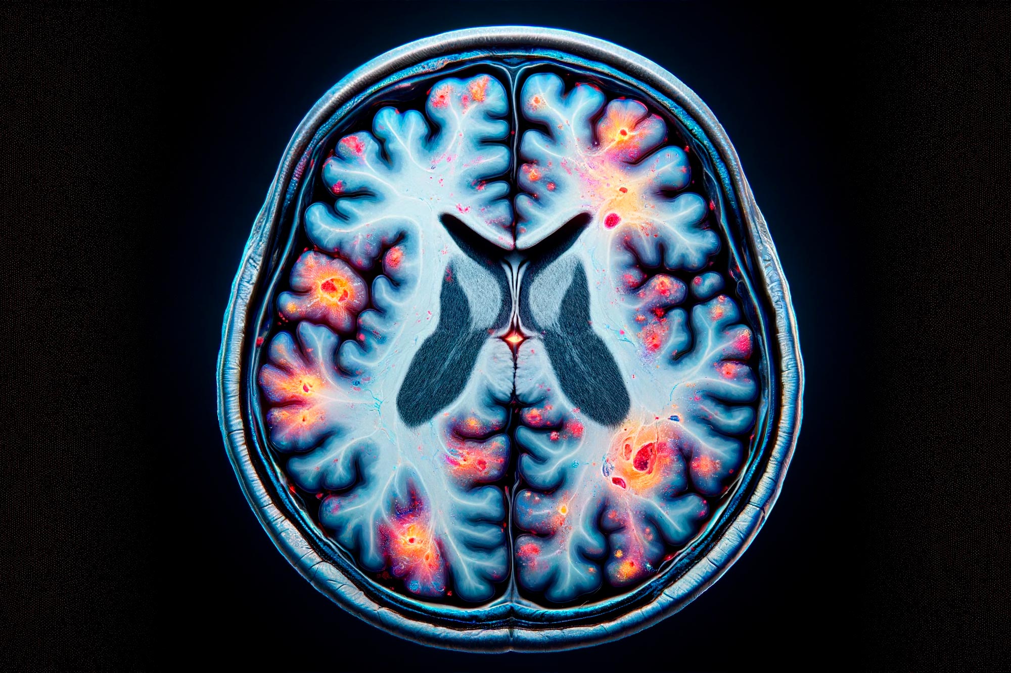
Areas they saw the lesions in were the anterior temporal lobe, the ventromedial frontal cortex, the amygdala, and the nucleus accumbens. These are all areas connected to morals, value decision making, reward and punishment.

“Those were the processes we thought would be important for what keeps a normal person from committing crimes,” Darby said.

There are studies linking brain damage to crime

Brain injuries typically result in having problems with “executive skills,” explained Huw Williams, a professor of clinical neuropsychology and co-director of the Centre for Clinical Neuropsychology Research at Exeter University in Britain.

These skills include planning ahead, thinking things through, managing impulses, and memory.

When you’re dealing with such a complicated organ like the brain, it takes decades of research to pinpoint what exactly might be happening.

There still isn’t strong research to suggest you can completely determine the way someone will behave depending on what their brain scans look like.

What is certain is that in a community sample of the UK population, analysed by Williams, about 10 percent will have had some kind of brain injury at some point.

In the prison population, this number jumps dramatically to somewhere between 50 and 70 percent. This trend is echoed in the US and South Africa

“This doesn’t mean necessarily that head injury equals crime,” Williams pointed out. “It might, but also it might be coincidental, because if you’ve got a violent life, and you crash cars, you’re likely to get a head injury at some point.”

A team at Oxford University tried to shed some light on this question in a 35-year population study in Sweden led by Psychiatrist Seena Fazel.

Results showed that overall, Swedes had a 2.5 percent chance of becoming violent offenders. If they had a head injury on their records, that rose to 9 percent.

To make allowances for the fact brain injuries could be a result of upbringing, the researchers also looked at the siblings of those with brain damage – they had a 4.5 percent chance of becoming offenders too.

In other words, the Oxford research shows that people have nearly double the chance of becoming violent offenders if they something in their upbringing, genetics, or environment predisposes it. Add a head injury on top of that, then the risk is doubled again.

Criminals might not be getting the right rehabilitation

Brain injuries are not easy to recover from. If memory is damaged, for example, patients will easily forget everything they have learned about how to behave. Or they might struggle to contain their outbursts of anger, like Chelsea does.

This is more pronounced among criminals, Williams said, partly because they are not getting the right rehabilitation.

When he switched to academia from clinical work, he was asked to supervise a student’s work looking into whether prisoners had post-traumatic stress disorder.

“I gave a workshop in a local prison, to get an idea of the nature of the environment… and one of the prisoners had an area of skull missing,” Williams said.

“This was probably because he’d had a head injury and hadn’t had follow up surgery to put in a titanium plate to replace the missing part of his skull probably removed to release pressure on the brain.

“He asked me why he had these funny feelings in his body that were quite nice, when he pressed himself on that area – and it was because he was pressing on the sensory motor strip. He was told he needed more surgery, and to take care with the area.”

After that, Williams became aware that the medical and neurological needs of the prison population probably weren’t being met.

“A lot of people who are in the system seem to have head injuries,” he said.

“That complicates their rehabilitation, which means there is a lot of re-offending. That may be because the present system isn’t necessarily well geared towards rehabilitation yet, especially neuro-rehabilitation where you have to remind people when to do things, and how to do them.”

Williams and his team started projects where they helped prison staff work with young people with brain injuries. So far the outcomes have been very promising because young people are more able to participate in their own improvement.

For example, for those with injuries to the left side of the brain, who have trouble remembering what they have been told, were given visual cues instead. “It’s about trying to find a way of helping and working with the population more effectively,” Williams said.

Neurology can be used in someone’s defence

When the law comes into it, things get even more complicated. Defence attorneys have a duty to defend their clients with rigour, and so they keep up with the latest research to help minimise their punishment.

In some cases, this means zealously claiming their client was neurologically predisposed to acting the way they did. It isn’t their fault, they may claim, because their brains are just wired that way.

Judith Edersheim is the co-founder and co-director of the Massachusetts General Hospital Centre for Law, Brain and Behaviour. It’s an organisation that is helping bring science into the courtroom, by separating the fringe ideas from the theories that are a lot more established.

“It’s very hard to tell [the difference] when neuroscience itself is so new,” Edersheim told Business Insider. “And there are some features of brain science that make it perhaps more alluring and look more probative than it should be.”

It’s important to make the distinction because law and science behave in conflicting ways. Science is evolutionary like “building blocks”, where you make assumptions and test them, then talk about the limitations of your studies and invite other scientists to do the same.

Law is not like that. In a courtroom, the outcome is final, and somebody’s life is going to change. So you have to be pretty certain of your evidence.

But when applied sparingly, looking carefully at the condition of an individual rather a collective, neuroscientific evidence can be a useful defence tool.

If a person is accused of a violent crime, there may be attributes of that person’s brain which would explain abhorrent violent behaviour, like a tumour in the brain’s frontal lobe. That would be a well-founded reason that somebody’s neurology could contribute to their defence.

One of the most successful defences along these lines was a case of an obstetrician in New York, Allan Zarkin, who started behaving peculiarly at work. He became unusually angry, and was short and provocative with the nursing staff.

Zarkin performed a cesarean section on a patient, delivering a healthy baby. He then carved his initials “AZ” into her abdomen. When asked about it, he simply said he thought he should sign it because he did such a good job.

After an investigation, doctors discovered that Zarkin had progressive Pick’s disease, a frontal lobe dementia similar to Alzheimer’s, which catastrophically disrupted his ability to plan, contain himself, and behave socially appropriately.

Zarkin wasn’t convicted, due to the medical diagnosis, but his licence was revoked.

In another case, a 40-year-old man who began looking at child pornography and propositioning prostitutes at massage parlours – behaviour that was completely out of character.

He was eventually kicked out by his wife and found guilty of child molestation.

But while awaiting jail, he complained of a terrible headache and was admitted to hospital. That’s when a large, egg-sized tumour was detected in the right lobe of the orbitofrontal cortex in his brain. Surgeons immediately removed it, and after that the pedophilic urges disappeared.

He successfully completed a Sexaholics Anonymous programme and was allowed to return home.

But it’s not possible to defend everyone

These two cases are extreme, and the cause and effect are easily identifiable. But when brain damage is less severe, it is difficult to draw definitive conclusions about how it can impact behaviour.

This becomes even more complicated when you look group data.

Research may show certain brain patterns or injuries that are loosely associated with certain types of behaviour, but then the lawyer has to correlate that with the defendant’s characteristics.

For example, you may have a whole set of brain scans of people who have committed violent crimes, and a pattern has been detected by researchers. You then have to show where the defendant fits into that group data.

“Crime is about behaviour, and the trial is about the mental state of the person who is sitting in front of you,” Edersheim explained.

“If you say someone has a propensity for violence, someone who has killed his wife for example, why is your propensity for violence only towards your wife? The individuated questions will dominate.”

Without the specifics, the behaviour is explained by motive. For now, at least, there are too many vulnerabilities in the neurological evidence for it to be used effectively.

Neuroscience’s contribution to criminal behaviour is still in its infancy, Edersheim said. Researchers are building on what they have to try and map out what makes a criminal mind, but there is still a lot they do not know.

Only when the science is a lot more concrete can it be used with confidence as a defence.

Researchers are still piecing the puzzle together

With Chelsea, she doesn’t know the next time she will lose control. Last time the police were called but no charges were brought against her.

If she ever commits a violent crime, though, it looks unlikely that the evidence of her post-injury behavioural changes will be strong enough to help her.

A neurologist and psychiatrist have both said her behaviour is a result of her brain damage, but she isn’t convinced of any of the treatment they offer her.

“When I get angry like that I literally have no control. It’s almost like the fight or flight response turns on and I choose to fight for no reason. It’s not even like I enjoy attacking people or getting worked up like that… it just happens,” she said.

“They put me on mood medication, but I don’t see the point in taking it. I see it as my brain is physically damaged, not chemically imbalanced. Then again, I could just misunderstand all of this.”

Of course, not everyone with a brain injury will become a criminal. As Darby explained, brain lesions may predispose someone to criminal behaviour, but that certainly doesn’t mean everyone becomes an offender.

As for those who suddenly acquire a propensity for violence from less serious injuries, like Chelsea, they are still awaiting answers.

Malaria drug causes brain damage that mimics PTSD: case study


The case of a service member diagnosed with post-traumatic stress disorder but found instead to have brain damage caused by a malaria drug raises questions about the origin of similar symptoms in other post-9/11 veterans.

According to the case study published online in Drug Safety Case Reports in June, a U.S. military member sought treatment at Walter Reed National Military Medical Center in Bethesda, Maryland, for uncontrolled anger, insomnia, nightmares and memory loss.

The once-active sailor, who ran marathons and deployed in 2009 to East Africa, reported stumbling frequently, arguing with his family and needing significant support from his staff while on the job due to cognitive issues.

Physicians diagnosed the service member with anxiety, PTSD and a thiamine deficiency. But after months of treatment, including medication, behavioral therapy and daily doses of vitamins, little changed.

The patient continued to be hobbled by his symptoms, eventually leaving the military on a medical discharge and questioning his abilities to function or take care of his children.

It wasn’t until physicians took a hard look at his medical history, which included vertigo that began two months after his Africa deployment, that they suspected mefloquine poisoning: The medication once used widely by the U.S. armed forces to prevent and treat malaria has been linked to brain stem lesions and psychiatric symptoms.

While no test is available to prove the sailor suffered what is called “mefloquine toxicity,” he scored high enough on an adverse drug reaction probability survey to tie his symptoms to the drug, also known as Lariam.

The sailor told his Walter Reed doctors that he began experiencing vivid dreams and disequilibrium within two months of starting the required deployment protocol.

Symptoms can last years

Case reports of mefloquine side effects have been published before, but the authors of “Prolonged Neuropsychiatric Symptoms in a Military Service Member Exposed to Mefloquine” say their example is unusual because it shows that symptoms can last years after a person stops taking the drug.

And since the symptoms are so similar to PTSD, the researchers add, they serve to “confound the diagnosis” of either condition.

“It demonstrates the difficulty in distinguishing from possible mefloquine-induced toxicity versus PTSD and raises some questions regarding possible linkages between the two diagnoses,” wrote Army Maj. Jeffrey Livezey, chief of clinical pharmacology at the Walter Reed Army Institute of Research, Silver Spring, Maryland.

Once the U.S. military’s malaria prophylactic of choice, favored for its once-a-week dosage regimen, mefloquine was designated the drug of last resort in 2013 by the Defense Department after the Food and Drug Administration slapped a boxed warning on its label, noting it can cause permanent psychiatric and neurological side effects,

50,000 prescriptions in 2003

At the peak of mefloquine’s use in 2003, nearly 50,000 prescriptions were written by military doctors.

That figure dropped to 216 prescriptions in 2015, according to data provided by the Defense Department. According to DoD policy, mefloquine is prescribed only to personnel who can’t tolerate other preventives.

But Dr. Remington Nevin, a former Army epidemiologist and researcher at the Johns Hopkins Bloomberg School of Public Health in Baltimore, said any distribution of the drug, which was developed by the Army in the late 1970s, is too much.

“This new finding should motivate the U.S. military to consider further revising its mefloquine policy to ban use of the drug altogether,” Nevin told Military Times.

While a case study is a snapshot of one patient’s experience and not an indication that everyone who took or takes mefloquine has similar issues, one randomized study conducted in 2001 — more than a decade after the medication was adopted by the military for malaria prevention — showed that 67 percent of study participants reported more than one adverse side effect, such as nightmares and hallucinations, and 6 percent needed medical treatment after taking the drug.

Yet mefloquine remains on the market while Walter Reed Army Institute of Research conducts research on medications in the same family as mefloquine, including tafenoquine, hoping to find a malarial preventive that is less toxic but as effective.

Mefloquine was developed under the Army’s malaria drug discovery program and approved for use as a malaria prophylactic in 1989. Shortly after commercial production began, stories surfaced about side effects, including hallucinations, delirium and psychoses.

Once considered ‘well-tolerated’

Military researchers maintained, however, that it was a “well-tolerated drug,” with one WRAIR scientist attributing reports of mefloquine-associated psychoses to a “herd mentality.”

“Growing controversies over neurological side effects, though, are appearing in the literature, from journal articles to traveler’s magazines and resulting legal ramifications threaten global availability,” wrote researcher Army Col. Wilbur Milhous in 2001. “As the ‘herd mentality’ of mefloquine associated psychoses continues to gain momentum, it will certainly affect operational compliance and readiness. … The need for a replacement drug for weekly prophylaxis will continue to escalate.”

Mefloquine was implicated in a series of murder-suicides at Fort Bragg, North Carolina, in 2002, and media reports also tied it to an uptick in military suicides in 2003.

A 2004 Veterans Affairs Department memo urged doctors to refrain from prescribing mefloquine, citing individual cases of hallucinations, paranoia, suicidal thoughts, psychoses and more.

The FDA black box warning nine years later led to a sharp decline in demand for the medication. But while the drug is no longer widely used, it has left damage in its wake, with an unknown number of troops and veterans affected, according to retired Navy Cmdr. Bill Manofsky, who was discharged from the military in 2004 for PTSD and later documented to have mefloquine toxicity.

He said the Defense Department and VA should do more to understand the scope of the problem and reach out to those who have been affected.

New concerns rising over antimalaria drug

“I’m kind of the patient zero for this and I now spend my life trying to help other veterans who have health problems that may have been caused by mefloquine. More needs to be done,” Manofsky said.

He said while there is no cure for the vertigo and vestibular damage or the psychiatric symptoms caused by mefloquine, treatments for such symptoms, such as behavior and vestibular therapy help.

And, he added, simply having a diagnosis is comforting.

Veterans can seek help

“Veterans need to come forward,” he said. “The VA’s War Related Illness and Injury Study Center can help.”

The patient in the case study written by Livezey continues to see a behavioral therapist weekly but takes no medications besides vitamins and fish oil.

He sleeps just three to four hours a night, has vivid dreams and nightmares and vertigo that causes him to fall frequently, and continues to report depression, restlessness and a lack of motivation.

The sailor’s experience with mefloquine has been “severely life debilitating” and Livezey notes that the case should alert physicians to the challenges of diagnosing patients with similar symptoms.

“This case documents the potential long-term and varied mefloquine-induced neuropsychiatric side effects,” he wrote.

Source:www.militarytimes.com

Statin Scam Exposed! Cholesterol Drugs Cause Rapid Aging, Brain Damage And Diabetes


Over 100 million people across the world use statin drugs. The bad news is that these drugs are considered as cellular poisons that speed up the aging process, and their use is connected with muscle fatigue, diabetes, memory loss, etc.

According to scientists at Tulane University in New Orleans, statins “prematurely age” stem cells that control the cellular repair in the body. These drugs generate tens of billions of dollars every year. Although the pharmaceutical companies do their best to sell these drugs, statins can cause disastrous and even fatal side-effects.

 Some of the most severe side-effects include “memory loss, muscle pain, diabetes, cataracts, liver dysfunction, diabetes, fatigue and memory loss”.

“Statins make regular users become older faster”, UK Express reports, “leaving them open to long-term mental and physical decline …”

UK Express added:

“Scientists have found the heart disease drugs badly affects our stem cells, the internal medical system which repairs damage to our bodies and protects us from muscle and joint pain as well as memory loss … Professor Reza Izadpanah, a stem cell biologist and lead author of the research published in the American Journal of Physiology, said: “Our study shows statins may speed up the aging process.”

Side effects

 Pleotropic Beneficial and Adverse Clinical Effects.”

According to the study, statin drugs “side effects include memory loss, myopathy, cataract formation, and increased risk of diabetes.” The study also warned that “Statins also impaired the expression of DNA repair genes including XRCC4, XRCC6, and Apex1.”

Doctors and pharmaceutical companies worked together in an effort to hide the side-effects of stating drugs, claiming that these are “miracle” drugs. Some even suggested that stating should be added to water supply.

Patients experience severe side-effects like memory loss, diabetes, and muscle fatigue, but doctors tend to give them even more medications. In other words, statins are some sort of “gateway drugs” that lead to increased use of more high-profit drugs.

High cholesterol limits lowered

The medical system tried so hard to lower the values of “high cholesterol,” and millions of people rushed to pharmacies, asking for their medications. Pharmaceutical companies bribed panelist in the American Heart Association and the American College of Cardiology.

Here’s what Natural News wrote in 2013:

Doctors claim the new guidelines will limit how many people with low heart risks are put on statins simply because of a cholesterol number. However, under the new advice, one-third of U.S. adults would meet the threshold to consider taking a statin, more than twice the 15 percent of adults who are recommended statins under current guidelines.

The justification for the panel having half its members with ties to Big Pharma: Ties between heart doctors and Big Pharma are so extensive that it is almost impossible to find a large group of doctors who have no industry ties.

Natural News has long warned about the side-effects caused by stating, noting that nerve damage is the most common. However, medical experts ignored this warning. It was all about the profits.

The stating drug industry is nothing better than the vaccine industry. But, researchers at the Tulane University revealed the truth, and helped people understand the damaging effect of statins.

Statin Scam Exposed: Cholesterol Drugs Cause Rapid Aging, Brain Damage And Diabetes


Statins are prescribed to patients who suffer from high levels of “bad” cholesterol, as it lowers the harmful levels, and reduces the risk of having heart issues.

Lately, statins have come under fire as a study concluded that they do more harm than good. A lot of people take statin drugs such as Crestor, Lipitor and Zocor. In the United States, prescription drug spending rose to $374 billion in the year 2014 (the highest level of spending since 2001). Statins make up for a huge portion of spending, and consumers who take these drugs are going to have a lot to worry over than the damage to their wallets.

The American Journal of Physiology, conducted a study that states that statins “…impact on other biologic properties of stem cells provides a novel explanation for their adverse clinical effects.” Specifically, the study states that such adverse effects include advancing the “process of aging” and also notes that “…long-term use of statins has been associated with adverse effects including myopathy, neurological side effects and an increased risk of diabetes.” Myopathy means skeletal muscle weakness.

Statins make cells unable to repair properly, create nerve problems and destroy memory

In the study, experts suggest that the health issues incurred due to statins, have been downplayed in the recent years. People who do take these drugs usually report having fatigue, cataracts, muscle pain, liver damage and loss of memory. These drugs have been proven to mess with cells in a way that heir main purpose is to reproduce and the process of body repair is blocked. Professor Reza Izadpanah, stem cell biologist and lead author of the published study, states, “Our study shows statins may speed up the aging process. People who use statins as a preventative medicine for [health] should think again as our research shows they may have general unwanted effects on the body which could include muscle pain, nerve problems and joint problems.”
Despite health problems linked to statin drugs, FDA says people shouldn’t be scared of them
While our FDA expresses on its online site that “Cognitive (brain-related) impairment, such as memory loss, forgetfulness and confusion, has been reported by some statin users” and that “People being treated with statins may have an increased risk of raised blood sugar levels and the development of Type 2 diabetes,” they also maintain its safety and effectiveness.

The site directs people’s attention to the advice of Amy G. Egan, M.D., M.P.H., who is the deputy director for safety in the FDA’s Division of Metabolism and Endocrinology Products (DMEP). She says, “This new information should not scare people off statins. Their benefit is indisputable, but they need to be taken with care and knowledge of their side effects.”

 This isn’t hard to dispute at all, especially after a previous study, we beg to differ. What’s so great and healthy about accelerated aging, muscle weakness, memory loss and cells that function improperly?
The need to continually assess prescription drugs and older studies that tout their benefits…
These findings have opened up the importance of researching the benefits of prescription drugs, which is hopefully something people will continue to do, to stay well-informed and in the best shape possible. There was a similar study that opened up many eyes, and involved the adolescent antidepressant “Paxil”, which made headlines when a re-analysis of an original study had shown incomplete information and exposed all the errors. Although, in reality, this drug wasn’t found to not be safe and efficient for its recommended demographic. Brian Nosek, a professor of psychology at the University of Virginia says “signals that the community is waking up, checking its work and doing what science is supposed to do — self-correct.”

Statin scam exposed: Cholesterol drugs cause rapid aging, brain damage and diabetes.


Sadly, many people take statin drugs, which are commonly known by brand names including Lipitor, Crestor and Zocor. Prescription drug spending in the U.S. shot up to about $374 billion in 2014, representing the highest level of spending since 2001. Statins undoubtedly made up a significant portion of this spending, and now consumers who take such drugs have much more to worry about than the dent it’s making in their wallets.

The study, which was published in the American Journal of Physiology, states that statins’ “…impact on other biologic properties of stem cells provides a novel explanation for their adverse clinical effects.” Specifically, the study states that such adverse effects include advancing the “process of aging” and also notes that “…long-term use of statins has been associated with adverse effects including myopathy, neurological side effects and an increased risk of diabetes.” Myopathy refers to skeletal muscle weakness.

cholesterol-drugs-cause-rapid-aging

Statins make cells unable to repair properly, create nerve problems and destroy memory

Experts involved in the study suggest that the health problems associated with statins have likely been downplayed through the years. In reality, those taking such cholesterol-lowering drugs have been experiencing cataracts, fatigue, liver problems, muscle pain and memory loss. Simply put, the drugs have been found to tamper with cells in such a way that their primary purpose of reproducing and helping the body repair is thwarted. With that comes the onset of terrible health issues or the worsening of existing ones.

Professor Reza Izadpanah, a stem cell biologist and lead author of the published study, says, “Our study shows statins may speed up the ageing process. People who use statins as a preventative medicine for [health] should think again as our research shows they may have general unwanted effects on the body which could include muscle pain, nerve problems and joint problems.”