HIV Drugs Show Promise in Averting Complications from Bacterial Infections


Study in mice finds a trigger for devastating condition, hints at possible treatment

Hundreds of rod-shaped bacteria, colorized magenta, strewn across a pink background.

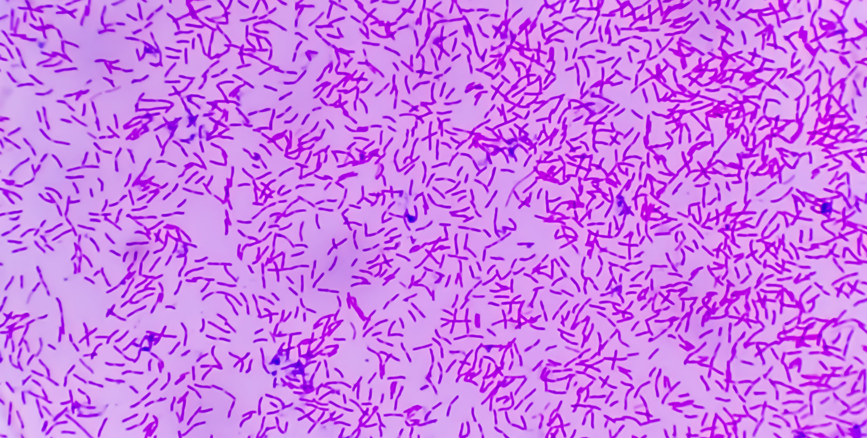
Micrograph of Escherichia coli (E. coli) bacteria.

At a glance:

  • Remnant viral genes from ancestral viral infections linked to severity of bacterial infections and formation of abscesses in mice.
  • Antiviral drugs used to fight HIV were shown to prevent these dangerous complications of infection in lab experiments.
  • Insights into the mechanisms that lead to abscess formation open the door for new lines of thought about treating bacterial infections.

Bacterial infections inside the body can lead to abscesses — pockets of dead cells and debris surrounded by inflammation-fueling immune cells. In these pockets, bacteria multiply, causing more inflammation and further damage to surrounding tissues.

For reasons that remain poorly understood, in some cases the immune reaction can spread throughout the body, resulting in organ damage and life-threatening organ failure, a condition known as sepsis, estimated to affect 1.7 million people in the U.S. each year.

But how do the initial abscesses that set off this devastating immune cascade begin? And why do some escalate to sepsis?

Research published Jan. 16 in PNAS sheds new light on this question and also hints at a possible treatment.

Using animal models, Harvard Medical School investigators at Brigham and Women’s Hospital have identified one key mechanism that may be driving abcess formation in the liver that could culminate in sepsis.

The researchers caution that sepsis could be the result of multiple factors and that other mechanisms could be at play. However, they say their findings pinpoint a previously unknown trigger that could have important implications for treatment.

“Our work to understand the mechanisms of abscess formation and sepsis points to a new way of thinking about treatment and prevention of deleterious consequences following bloodstream infection,” said first author Karthik Hullahalli, an HMS graduate student in microbiology working in the lab of Matthew Waldor at Brigham and Women’s. Waldor is the HMS Edward H. Kass Professor of Medicine at Brigham and Women’s and an affiliated faculty member of the HMS Department of Microbiology.

The researchers say that if replicated in further studies, both in larger animals and then in humans, the work could pave the way to using existing drugs in a novel way to prevent some of the most devastating complications of bacterial infections.

The surprising role of dormant viral genes in igniting bacterial infections

The researchers examined the livers of mice infected with Escherichia coli, or E. coli, a common bacterium that infects many animals, including humans.

They found that abscess formation was linked to the presence and reactivation of dormant endogenous retroviruses (ERVs), genetic remnants of viruses that integrated into the genome of the mice following infections in previous generations.

The authors hypothesized that genetic material produced by the reawakened viruses stimulate inflammatory immune responses that in turn damage surrounding cells and thus drive abscess development. If so, the researchers surmised, suppressing the activity of the ERVs might prevent abscesses from forming.

A possible new use for antiviral drugs

To test this hypothesis, the researchers treated mice with a cocktail of reverse transcriptase inhibitors — antiretroviral drugs used to curb viral replication in people infected with HIV. They found that a single dose of the antiviral medication was enough to prevent abscess formation if delivered quickly after bacterial infection.

“Our findings show that drugs used to treat HIV can be used to prevent inflammatory complications of bacterial sepsis in animals,” Waldor said.

Further work is needed to understand whether and how antiretroviral drugs might prevent complications such as bacterial sepsis in different cases, the researchers said.

For example, Hullahalli noted that in mice, abscess susceptibility varies by sex and among tissues. In addition, the researchers noted that ERVs play a complex role in the life cycle of their host.

ERVs are switched on and off at different points in the course of an animal’s life. Most ERVs are usually deactivated and go into dormancy. In fact, if ERVs aren’t successfully silenced by the host immune system, they could lead to mutations that cause cancer or to immune dysregulation that causes autoimmune diseases later on. But these ERV genes are not always harmful. In some circumstances, ERVs could play a protective role in fighting infections by stimulating defensive immune responses.

“Whether these elements of the genome are beneficial or detrimental to an individual likely depends on context,” Hullahalli noted. “But the findings of this study suggest that there may be a role for the drugs that we currently think of as antivirals in treating inflammatory responses to bacterial infection.”

AI-designed catheter could help prevent bacterial infections.


A diagram of the new catheter design. Fin-like triangular protrusions line the...

A diagram of the new catheter design. Fin-like triangular protrusions line the interior of the catheter wall, creating turbulence that hinders bacteria’s upstream progress along the tube’s wall. In this diagram, the normal flow of the catheter is to the right, while bacteria (yellow) attempt to swim upstream to the left.

One of the most common bacterial infections in a healthcare setting comes from bacteria entering the body through catheters, thin tubes inserted in the urinary tract.

Though catheters are designed to draw fluids out of a patient, bacteria are able to propel themselves upstream and into the body via catheter tubes using a unique swimming motion, causing $300 million of catheter-associated urinary infections in the U.S. annually. Now, an interdisciplinary project at Caltech has designed a new type of catheter tube that impedes the upstream mobility of bacteria, without the need for antibiotics or other chemical antimicrobial methods. With the new design, which was optimized by novel artificial intelligence (AI) technology, the number of bacteria that are able to swim upstream in laboratory experiments was reduced 100-fold. 

A paper describing the study appears in the journal Science Advances. The work was a collaboration between the laboratories of Chiara Daraio, G. Bradford Jones Professor of Mechanical Engineering and Applied Physics and Heritage Medical Research Institute Investigator; Paul Sternberg, Bren Professor of Biology; John Brady, Chevron Professor of Chemical Engineering and Mechanical Engineering; and Anima Anandkumar, Bren Professor of Computing and Mathematical Sciences.

Photo

In catheter tubes, fluid exhibits a so-called Poiseuille flow, an effect where fluid movement is faster in the center but slow near the wall, similar to the flow in a river’s current, where the velocity of the water varies from fast in the center to slow near the banks. Bacteria, as self-propelling organisms, exhibit a unique “two-step forward along the wall, one-step back in the middle” motion that produces their forward progress in tubular structures. Researchers in the Brady lab had previously modeled this phenomenon. “One day, I shared this intriguing phenomenon with Chiara Daraio, framing it simply as a ‘cool thing,’ and her response shifted the conversation toward a practical application,” says Tingtao Edmond Zhou, postdoctoral scholar in chemical engineering and a co-first author of the study. “Chiara’s research often plays with all kinds of interesting geometries, and she suggested tackling this problem with simple geometries.” 

Following that suggestion, the team designed tubes with triangular protrusions, like shark fins, along the inside of the tube’s walls. Simulations yielded promising results: These geometric structures effectively redirected bacterial movement, propelling them toward the center of the tube where the faster flow pushed them back downstream. The triangles’ fin-like curvature also generated vortices that further disrupted bacterial progress.

Zhou and his collaborators aimed to verify the design experimentally but needed additional biology expertise. For that, Zhou reached out to Olivia Xuan Wan, a postdoctoral scholar in the Sternberg laboratory. “I study nematode navigation, and this project resonated deeply with my specialized interest in motion trajectories,” says Wan, who is also a co-first author on the new paper. For years, the Sternberg laboratory has conducted research into the navigation mechanisms of the nematode Caenorhabditis elegans, a rice grain–sized soil organism commonly studied in research labs and thus had many of the tools to observe and analyze the movements of microscopic organisms. 

The team quickly transitioned from theoretical modeling to practical experimentation, using 3D printed catheter tubes and high-speed cameras to monitor bacterial progress. The tubes with triangular inclusions resulted in a reduction of upstream bacterial movement by two orders of magnitude (a 100-fold decrease). The team then continued simulations to determine the most effective triangular obstacle shape to impede bacteria’s upstream swimming. They then fabricated microfluidic channels analogous to common catheter tubes with the optimized triangular designs to observe the movement of E. coli bacteria under various flow conditions. The observed trajectories of the E. coli within these microfluidic environments aligned almost perfectly with the simulated predictions. 

The collaboration grew as the researchers aimed to continue improving the geometric tube design. Artificial intelligence experts in the Anandkumar laboratory provided the project with cutting-edge AI methods called neural operators. This technology was able to accelerate the catheter design optimization computations so they required not days but minutes. The resulting model proposed tweaks to the geometric design, further optimizing the triangle shapes to prevent even more bacteria from swimming upstream. The final design enhanced the efficacy of the initial triangular shapes by an additional 5% in simulations. 

“A collaborative spirit defines Caltech,” says Sternberg. “Caltech people help each other. This endeavor was truly an interdisciplinary journey, weaving together diverse fields of study.” 

“Our journey from theory to simulation, experiment, and, finally, to real-time monitoring within these microfluidic landscapes is a compelling demonstration of how theoretical concepts can be brought to life, offering tangible solutions to real-world challenges,” says Zhou. “I’m very lucky to be at Caltech with so many talented colleagues.” 

Vitamin D: The Pac-Man of Viruses, Bacterial Infections, and Cancer Cells


Vitamin D, also called the Sunshine Vitamin, is actually a steroid with hormone-like activity. (Shutterstock)

Vitamin D, also called the Sunshine Vitamin, is actually a steroid with hormone-like activity. (Shutterstock)

In a fascinating study conducted at the University of Copenhagen, researchers found that vitamin D is essential in order to activate the body’s immune system. Without it — or with insufficient levels — the immune system’s killer T cells can’t fight off serious infections and instead remain unheroically dormant. But with sufficient levels of vitamin D, these T cells spring into action, do what they’re designed to do, and gobble up viruses, harmful bacteria, and can even destroy cancer cells.

According to Professor Carsten Geisler (Department of International Health, Immunology and Microbiology): “When a T cell is exposed to a foreign pathogen, it extends a signaling device or ‘antenna’ known as a vitamin D receptor, with which it searches for vitamin D. This means that the T cell must have vitamin D or activation of the cell will cease. If the T cells cannot find enough vitamin D in the blood, they won’t even begin to mobilize.”

With an estimated 42% of the world’s population deficient in vitamin D, many experts consider this deficiency to be a global health problem. But it’s  a problem that can be easily remedied.

It begins with knowing what your current vitamin D levels are.

Checking Your Vitamin D Levels—How Much Is Enough?

Knowing your vitamin D levels is critical, especially if you have cancer. Fortunately, a simple blood test is all you’ll need. Your doctor can recommend the test, or you can order your own from companies such as LabCorp, Private MD Labs, and Life Extension.

You’ll want your vitamin D levels to be a minimum of 60-80 ng/mL on this test. Anything below 25 is dangerously deficient.

Risk Factors

Obesity. Because vitamin D is fat-soluble, people with higher amounts of body fat will store vitamin D in fat cells, causing lower amounts of vitamin D to circulate in the bloodstream. Those who are obese typically require higher amounts of vitamin D to correct a deficiency.

Ethnicity. African Americans are of particular risk for vitamin D deficiency. A study from the Medical University of South Carolina states that this class of people is 90% more likely to be deficient in vitamin D and that daily doses of 4,000IU may be necessary to combat the deficiency. According to a study published in the Journal of Investigative Medicine, if you’re black AND obese, you are 70% more likely to be deficient in Vitamin D.

Age. For those aged 50 and older, vitamin D deficiency can be a problem. A variety of reasons may account for this, such as excessive time spent indoors, reduced appetite and malabsorption of nutrients. In addition, our skin becomes thinner as we age, affecting the body’s ability to synthesize vitamin D.

The Ideal Form of Vitamin D

Vitamin D, also called the Sunshine Vitamin, is actually a steroid with hormone-like activity. It regulates the functions of over 200 genes and, in addition to its phenomenal immune system boosting properties, is absolutely essential for strong bones.

It only takes about 20 minutes in the midday sun (from 10AM—2PM) for the body to absorb UVB rays through the skin and, via a chemical reaction process, turn it into one of the best immune system defenses on the planet. Studies show that vitamin D derived from the sun may circulate for double the time as vitamin D from food or supplements.

You would think that since the sun shines on all of us, there wouldn’t be a lack of vitamin D, but for many reasons, this simply isn’t the case. For those who are housebound or who live at latitudes too far from the equator, getting enough natural vitamin D can be problematic. And for those who use sunscreens, they’re missing out on a chance for the body to produce this precious steroid, as sunscreen blocks the body’s ability to produce vitamin D.

The Shadow Rule: you make more vitamin D when you are taller than your shadow.

You’re fooling yourself if you think that by sitting indoors near a sunny window or driving in the car on a sunny day is enough to increase your levels of vitamin D. Window glass blocks UVB ultraviolet light. You really need to be outdoors, exposing as much skin as possible.

If you’re curious as to how much vitamin D potential you have in the area where you live, check this chart from National Oceanic and Atmospheric Administration (NOAA): https://gml.noaa.gov/grad/solcalc/azel.html

Enjoy Vitamin D-rich Foods

You can fortify your diet by increasing dietary sources of vitamin D. These include egg yolks, beef liver, salmon, herring, sardines, cod liver oil, and mushrooms.

When eating fatty fish like salmon, be sure to opt for wild-caught as wild-caught salmon (on average) contains anywhere from 988-1,300 IU of vitamin D per 3.5-ounce serving. Farmed salmon disappoints with only about one-quarter of the amount of vitamin D.

If you’re not a fish eater, consider cod liver oil. It was used for centuries as a preventative measure against vitamin D deficiency. Those who lived in northern climates may be familiar with the advantages of relying on cod liver oil as a vitamin D supplement when sunlight is scarce in wintertime.

A word about eggs — vitamins, minerals, and fat are concentrated in the yolk, and the protein in eggs is found mainly in the whites. One typical egg yolk contains approximately 37 IU of vitamin D.

Vitamin D Mushroom Hack

Mushrooms are a great source of vitamin D. Like humans, mushrooms can synthesize this vitamin when exposed to UV light. Researchers in Virginia have documented that sliced or chopped mushrooms when exposed to natural sunlight for only 15 minutes recorded a significant increase in Vitamin D levels. In some cases, daily Vitamin D requirements of 600 IU were exceeded by just three sliced white button mushrooms! (Bruce Hudson, Fitness and Health)

Simply place sliced mushrooms in a pan and place in the sunlight for about 15 minutes before adding to a fresh salad or your favorite recipe. Be sure to soak up the sunlight yourself while you’re waiting!

Tan-Through Clothing

Ideally, you’ll want to have at least 40% of bare skin exposed to the sun, with NO sunscreens or lotions. If you’re too modest to bare up to 40% of your body to the sunlight, consider tan-through clothing options like this line of swimwear and shirts: https://www.tanthrough.com/.

Vitamin D Supplements

Vitamin D supplements are available in two forms: D2 (made from plants) and D3 (found in animal foods). Vitamin D3 is the type that is naturally produced in the human body and is widely considered to be the optimal form of supplementation.

Look for quality brands—never skimp on this important vitamin. We recommend those such as Bio-D-Mulsion Forte by Biotics Research, Nordic Naturals Arctic Cod Liver Oil, and Thorne Vitamin D/K2. We also recommend that you check with your personal healthcare practitioner to determine if supplementation is right for you.

Researchers developing new tool to distinguish between viral, bacterial infections


Antibiotics are lifesaving drugs, but overuse is leading to antibiotic resistance, one of the world’s most pressing health threats. Scientists identified 11 genetic markers in blood that accurately distinguished between viral and bacterial infections 80 to 90 percent of the time. The finding is important because physicians don’t have a good way to confirm bacterial infections like pneumonia and more-often-than-not default to an antibiotic. The goal of the research is to develop a tool, such as a blood test, that physicians can use to rule out a bacterial infection with enough certainty that they are comfortable, and their patients are comfortable, foregoing an antibiotic. Credit: University of Rochester Medical Center

Antibiotics are lifesaving drugs, but overuse is leading to one of the world’s most pressing health threats: antibiotic resistance. Researchers at the University of Rochester Medical Center are developing a tool to help physicians prescribe antibiotics to patients who really need them, and avoid giving them to individuals who don’t.

Scientists from the University’s National Institutes of Health-funded Respiratory Pathogens Research Center identified 11  in blood that accurately distinguished between viral and bacterial infections (antibiotics help us fight bacterial infections, but aren’t effective and shouldn’t be used to treat viruses). The finding, published today in the journal Scientific Reports, is important because physicians don’t have a good way to confirm bacterial infections like pneumonia and more-often-than-not default to an antibiotic.

“It’s extremely difficult to interpret what’s causing a , especially in very ill patients who come to the hospital with a high fever, cough, shortness of breath and other concerning symptoms,” said Ann R. Falsey, M.D., lead study author, professor and interim chief of the Infectious Diseases Division at UR Medicine’s Strong Memorial Hospital. “My goal is to develop a tool that physicians can use to rule out a  with enough certainty that they are comfortable, and their patients are comfortable, foregoing an antibiotic.”

Falsey’s project caught the attention of the federal government; she’s one of 10 semifinalists in the Antimicrobial Resistance Diagnostic Challenge, a competition sponsored by NIH and the Biomedical Advanced Research and Development Authority to help combat the development and spread of . Selected from among 74 submissions, Falsey received $50,000 to continue her research and develop a prototype diagnostic test, such as a blood test, using the genetic markers her team identified.

A group of 94 adults hospitalized with lower respiratory tract infections were recruited to participate in Falsey’s study. The team gathered clinical data, took blood from each patient, and conducted a battery of microbiologic tests to determine which individuals had a bacterial  (41 patients) and which had a non-bacterial or viral infection (53 patients). Thomas J. Mariani, Ph.D., professor of Pediatrics and Biomedical Genetics at URMC, used complex genetic and statistical analysis to pinpoint markers in the blood that correctly classified the patients with bacterial infections 80 to 90 percent of the time.

“Our genes react differently to a virus than they do to bacteria,” said Mariani, a member of the Respiratory Pathogens Research Center (RPRC). “Rather than trying to detect the specific organism that’s making an individual sick, we’re using genetic data to help us determine what’s affecting the patient and when an antibiotic is appropriate or not.”

Falsey, co-director of the RPRC, and Mariani say that the main limitation of their study is the small sample size and that the genetic classifiers selected from the study population may not prove to be universal to all .

A patent application has been filed for their method of diagnosing bacterial infection. Edward Walsh, M.D., professor of Infectious Diseases, and Derick Peterson, Ph.D., professor of Biostatics and Computational Biology at URMC, also contributed to the research.

According to the Centers for Disease Control and Prevention, antibiotic resistant bacteria cause at least 2 million infections and 23,000 deaths each year in the United States. The use of  is the single most important factor leading to  around the world.

Scientists Develop Drug to Replace Antibiotics


New medicine effective against superbugs

A small patient trial showed that the new treatment was effective at eradicating the MRSA superbug which is resistant to most antibiotics. The drug is already available as a cream for skin infections and researchers hope to create a pill or an injectable version of it in the next five years.

Antibiotics have been one of the most important drugs since the invention of penicillin almost 90 years ago. But the World Health Organization has repeatedly warned of the threat of antimicrobial resistance, saying “a post-antibiotic era – in which common infections and minor injuries can kill” is a very real possibility in the 21st century.

But scientists say this new technology is less prone to resistance than antibiotics because the treatment attacks infections in a completely different way. The treatment uses enzymes called endolysins — naturally occurring viruses that attack certain bacterial species but leave beneficial microbes alone.

Mark Offerhaus, the Chief Executive of the Dutch biotech firm Micreos which is leading the research, said the development of the new drug marks “a new era in the fight against antibiotic-resistant bacteria”, adding that millions of people stand to benefit from this.

Newborns Born With Genetic Code Signaling Sepsis, Other Bacterial Infections; Scientists Able To Decode DNA In Search For Better Treatment.


Some newborns, after birth, can develop life-threatening infections such as sepsis due to their underdeveloped immune systems. But pioneering research published Thursday in Nature Communicationscan enable doctors to identify the presence of infection-causing bacteria in the bloodstream of newborns, and target them. And how can they identify the pathogens? By decoding a signal generated from the baby’s DNA.

The signal is like an SOS, sent by the messenger RNA of the baby’s genome. By analyzing this code, the doctors can understand if there is a sepsis present in the blood. The signal can be deciphered using just a single drop of blood. Scientists are now trying to develop tests based on this signal detection, so that fatality due to bacterial infections can be significantly reduced.

Bacterial infections in newborns are difficult to diagnose unless blood tests are performed, and this generally requires testing large amounts of blood. Babies can develop infections either during childbirth or a few days after being born. If the mother is suffering from an active infection, then pathogens can be inhaled by the newborn when passing through the birth canal. After birth, the child may catch contagious bugs on contact with someone who has cold or flu. Pathogens that cause sepsis include Group B Streptococci, E. coli, Listeria, and viruses.

Since the baby’s body cannot put up much of a fight, pathogens multiply fast and the newborn can get very sick very quickly. So it is prudent that the infection is discovered and treated soon. Also, typical symptoms like high fever may not occur, and even if they do, they are not indicative of an infection. If a blood culture is performed to identify the pathogens, it takes two to three days for the results to come. Since the infant is put on antibiotics before the results are in, even children without sepsis may end up getting antibiotics or they may be given antibiotics not suited for that particular infection. Unnecessary use of meds also increases the risk of antibiotic resistance.

Scientists from the University of Edinburgh spent years trying to find causes and methods of identification of blood infections in preterm and full-term babies. Using blood samples from newborn babies in Edinburgh, they investigated thousands of signals written in mRNAs. With meticulous code breaking, the scientists identified a signal consisting of 52 molecular characters specific to bacterial infection. This signal, with complete accuracy, can tell if the baby is suffering from sepsis.

Newborn

“Just as a Twitter user can send a 140 character message so a baby’s genome produces short messages or signals that produce code information to communicate with the infant’s immune and metabolic systems so that it can fight the infection,” said Dr. Peter Ghazal, professor of molecular genetics and biomedicine at the University of Edinburgh’s Division of Pathway Medicine, in a statement. “The 52-character ‘tweet’ or message that we have identified appears to be specific for bacterial but not viral infection.” Ghazal said researchers believe the detection method could also be used in children and adults. Essentially, researchers would use a single drop of blood to detect the vital distress signal in DNA. The findings create hope for also “tackling antibiotics resistance,” he added.

Stressing that infections lead to a number of deaths or disabilities in infants worldwide, Dr. Claire Smith states that there is a need to develop tests for quick and accurate identification of sepsis.

“This work is enabling us to move towards being able to distinguish between babies with true infection who need urgent treatment, and those who are not infected and therefore don’t require antibiotics. The potential benefits to babies and their families are important. We are grateful to the families who consented to take part in the study,” she said.

Bacteremia Appears to Increase 30-Day Risk of MI or Stroke.


Patients who had bacteremia mainly urinary-tract infections, pneumonia, or sepsis when admitted to hospital were much more likely to have an MI or stroke within 30 days, compared with healthy controls or patients hospitalized for other reasons, in a new study [1] .

“Our study corroborates that acute infection may trigger cardiovascular events,” Dr Michael Dalager-Pedersen (Aalborg University Hospital, Denmark) told heart wire in an email. “It is the first study to demonstrate that many different bacterial infections may affect MI and stroke risk,” he added.

The research suggests that “bacteremia (a severe and acute infection) should be considered a risk factor for MI and stroke, but only for a short period of time after onset of infection,” and it hints that infection with Staphylococcus aureus may confer a particularly high risk.

“Patients admitted with signs of acute infection and bacteremia/sepsis should be monitored closely for complications, and treated early . . . with fluid therapy, oxygen, and antibiotics,” Dalager-Pedersen continued. “Moreover, it seems prudent to increase vaccination efforts (eg, influenza and pneumococcal vaccination), in particular in patients who already have established cardiovascular disease, since infection may trigger new cardiovascular events.”

Future studies are needed to clarify whether specific cardiovascular therapies (eg, antithrombotic or anti-inflammatory drugs) may reduce the risk of cardiovascular complications in patients with bacteremia, he said.

The study was published online February 12, 2014 in Circulation.

Infection a Trigger for MI, Stroke

An estimated one million Americans have an acute MI or stroke each year, and it would be useful to understand how acute infections might trigger these events, but most previous studies lacked laboratory confirmation of infection, the researchers write.

Using population-based databases, they identified 4389 patients in Northern Denmark who had positive blood cultures when admitted to hospital from 1992 to 2010. The pathogens were Escherichia coli, Streptococcus pneumoniae, S aureus, other bacteria, and fungi. Most patients had urinary-tract infections or pneumonia, while others had central nervous system infections, endocarditis, and other infections.

The mean age of patients was 73 years. Based on age, gender, and date of admission, each patient was matched with about five patients hospitalized for other reasons and about 10 individuals in the general population.

Researchers identified all incident MI and stroke events that occurred within 0 to 30 days, 31 to 180 days, and 181 to 365 days after the day of hospitalization.

Patients with community-acquired bacteremia had a greatly increased risk of MI or stroke within 30 days. At 31 to 180 days, these patients had a modestly higher risk of MI or stroke compared with healthy controls, but not compared with other hospitalized patients. No differences in cardiovascular risk were seen after more than six months.