Gamma Stimulation Promotes Glymphatic Clearance of Amyloid, Slows Alzheimer’s Progression


Bright staining highlights VIP-expressing interneurons in this coronal cross-section of a mouse brain. The neurons may help drive glymphatic clearance of amyloid via the release of peptides. [Tsai Laboratory/MIT Picower Institute]

It has been shown that noninvasive optogenetic-driven 40 Hz stimulation promotes neural activity and attenuates pathology in the 5XFAD mouse model of Alzheimer’s disease.

Findings from a new study reveal a key mechanism that may contribute to these beneficial effects: clearance of amyloid proteins, a hallmark of AD pathology, via the brain’s glymphatic system—a recently discovered “plumbing” network parallel to the brain’s blood vessels.

This work is published in Nature in the paper, “Multisensory gamma stimulation promotes glymphatic clearance of amyloid.

“Ever since we published our first results in 2016, people have asked me how does it work? Why 40 Hz? Why not some other frequency?” said Li-Huei Tsai, PhD, professor of neuroscience and director of the Picower Institute and MIT’s Aging Brain Initiative. “These are indeed very important questions we have worked very hard in the lab to address.”

The new paper describes experiments that show that sensory gamma stimulation prompts a particular type of neuron to release peptides that promote increased amyloid clearance via the glymphatic system.

Working with the 5XFAD mouse model of Alzheimer’s, the team first replicated the lab’s prior results that 40 Hz sensory stimulation increases 40 Hz neuronal activity in the brain and reduces amyloid levels. Then they set out to measure whether there was any correlated change in the fluids that flow through the glymphatic system to carry away wastes. Indeed, they measured increases in cerebrospinal fluid in the brain tissue of mice treated with sensory gamma stimulation compared to untreated controls. They also measured an increase in the rate of interstitial fluid leaving the brain. Moreover, in the gamma-treated mice, they measured increased diameter of the lymphatic vessels that drain away the fluids and measured increased accumulation of amyloid in cervical lymph nodes, which is the drainage site for that flow.

To investigate how this increased fluid flow might be happening, the team focused on the aquaporin 4 (AQP4) water channel of astrocyte cells, which enables the cells to facilitate glymphatic fluid exchange. When they blocked APQ4 function with a chemical, that prevented sensory gamma stimulation from reducing amyloid levels and prevented it from improving mouse learning and memory. And when, as an added test they used a genetic technique for disrupting AQP4, that also interfered with gamma-driven amyloid clearance.

More specifically, the authors noted, “Influx of cerebrospinal fluid was associated with increased aquaporin-4 polarization along astrocytic endfeet and dilated meningeal lymphatic vessels. Inhibiting glymphatic clearance abolished the removal of amyloid by multisensory 40 Hz stimulation.”

In addition to the fluid exchange promoted by APQ4 activity in astrocytes, another mechanism by which gamma waves promote glymphatic flow is by increasing the pulsation of neighboring blood vessels. Several measurements showed stronger arterial pulsatility in mice subjected to sensory gamma stimulation compared to untreated controls.

Using single-nucleus RNA sequencing (snRNA-seq), the team saw that gamma sensory stimulation indeed promoted changes consistent with increased astrocyte AQP4 activity. The data also revealed that upon gamma sensory stimulation, interneurons experienced a notable uptick in the production of several peptides. This was not surprising in the sense that peptide release is known to be dependent on brain rhythm frequencies, but it was still notable because one peptide in particular, VIP, is associated with Alzheimer’s-fighting benefits and helps to regulate vascular cells, blood flow and glymphatic clearance.

The team ran tests that revealed increased VIP in the brains of gamma-treated mice. The researchers also used a sensor of peptide release and observed that sensory gamma stimulation resulted in an increase in peptide release from VIP-expressing interneurons.

When the VIP neurons where inhibited, and the mice were exposed to sensory gamma stimulation, there was no longer an increase in arterial pulsatility and there was no more gamma-stimulated amyloid clearance.

While this paper focuses on what is likely an important mechanism—glymphatic clearance of amyloid—by which sensory gamma stimulation helps the brain, it’s probably not the only underlying mechanism that matters. The clearance effects shown in this study occurred rapidly, but in lab experiments and clinical studies weeks or months of chronic sensory gamma stimulation have been needed to have sustained effects on cognition.

Blood Test Predicts Which People With Amyloid Are Likely to Decline Cognitively


Plasma p-tau217 outperformed other preclinical Alzheimer’s measures

A computer rendering of amyloid plaques on a neuron.

A blood test identified which cognitively intact people with brain amyloid pathology were most likely to deteriorate in the next 6 years, longitudinal data showed.

Compared with other measures, plasma phosphorylated tau 217 (p-tau217) best predicted decline on the modified Preclinical Alzheimer Cognitive Composite (mPACC) and the Mini-Mental State Examination (MMSE) with correlation coefficients of 0.41 and 0.34, respectively, reported Niklas Mattsson-Carlgren, MD, PhD, and Oskar Hansson, MD, PhD, both of Lund University in Sweden, and colleagues.

Baseline plasma p-tau217 also was associated with progression to Alzheimer’s dementia (HR 2.03, 95% CI 1.57-2.63, P<0.001), the researchers wrote in JAMA Neurologyopens in a new tab or window.

Alzheimer’s disease starts with a long period of amyloid-beta accumulation without symptoms. P-tau217 may help identify which cognitively unimpaired people with amyloid pathology (preclinical Alzheimer’s disease) might benefit most in clinical trials, the researchers suggested.

“It is this subgroup of individuals with preclinical Alzheimer’s disease that really needs effective disease-modifying therapies in the future,” Hansson told MedPage Today.

“We are convinced that our results have immediate implications for clinical trials evaluating novel therapies in preclinical Alzheimer’s disease populations, because the number of included participants can be substantially reduced if only including those with elevated plasma p-tau217 levels,” Hansson said.

“However, we also envision using plasma p-tau217 in preclinical Alzheimer’s disease in clinical practice in the future when a disease-modifying therapy is approved for clinical use at this early disease stage,” he added.

The researchers studied 171 people with preclinical Alzheimer’s from the Swedish BioFINDER-1 cohort and validated their findings in 52 people from the Wisconsin Registry for Alzheimer Prevention (WRAP). Mean ages were about 73 and 64, respectively. Some people in BioFINDER-1 had subjective cognitive decline, but all participants in both cohorts were objectively cognitively unimpaired.

Participants had brain amyloid pathology defined by cerebrospinal fluid (CSF) in BioFINDER-1 and by PET scans in WRAP. Besides p-tau217, other plasma measures included p-tau181, p-tau231, glial fibrillary filament protein (GFAP), and neurofilament light (NfL). Data were collected from 2010 to 2020 in BioFINDER-1 and 2011 to 2021 in WRAP.

Primary outcomes were MMSE and mPACC scores over a median of 6 years (range 2-10 years). Both tests measure global cognition; the mPACC also assesses episodic memory and timed executive function.

The researchers adjusted models for age, sex, years of education, apolipoprotein E ε4 (APOE4) allele status, and baseline cognition. They derived cognition slopes using linear regression models with cognitive score as the outcome and time as the predictor, and tested combinations of covariates and biomarkers.

Adjusting for covariates, most biomarkers were associated with mPACC slopes, and all biomarkers except plasma p-tau231 were associated with MMSE slopes. Plasma p-tau217 was the strongest biomarker to predict cognitive decline on both the mPACC (R2 0.41 vs 0.23 for a covariates-only model, P<0.001) and the MMSE (R2 0.34 vs 0.04 for the covariates-only model, P<0.001) in BioFINDER-1. Similar patterns emerged in WRAP.

Sample sizes were reduced in hypothetical clinical trials enriched for individuals with elevated plasma p-tau 217. In clinical trial simulations using mPACC slopes as the outcome in BioFINDER-1, relative sample sizes — compared with including all eligible participants — were 79% when including the three highest quartiles of baseline plasma p-tau217, 55% when including the two highest quartiles, and 42% when including the highest quartile. Similar results were seen using MMSE slopes and were validated in WRAP.

The findings complement other data about p-tau217, which has shown strong diagnostic performancesopens in a new tab or window for Alzheimer’s disease and has discriminated Alzheimer’sopens in a new tab or window from other neurodegenerative diseases and normal cognition. Recent trials, including the TRAILBLAZER-ALZ study of donanemab for early Alzheimer’sopens in a new tab or window, have included plasma p-tau217 as a metric.

The study has several limitations, Hansson and colleagues acknowledged. Plasma p-tau217 has been associated with both amyloid and tau accumulation. In this study, tau PET was not assessed and the extent to which it was linked with high plasma p-tau217 was unknown.

“However, in cognitively unimpaired individuals, tau PET uptake is usually mild and not readily detectable at the individual level, although there have been group-level increases and associations with future cognitive decline,” the researchers noted.

COVID Protein Interacts With Parkinson’s Protein, Promotes Amyloid Formation


Summary: The COVID causing SARS-CoV-2 protein interacts with alpha-synuclein, speeding up the formation of amyloid plaques, a new study reports.

Source: ACS

Case reports of relatively young COVID-19 patients who developed Parkinson’s disease within weeks of contracting the virus have led scientists to wonder if there could be a link between the two conditions.

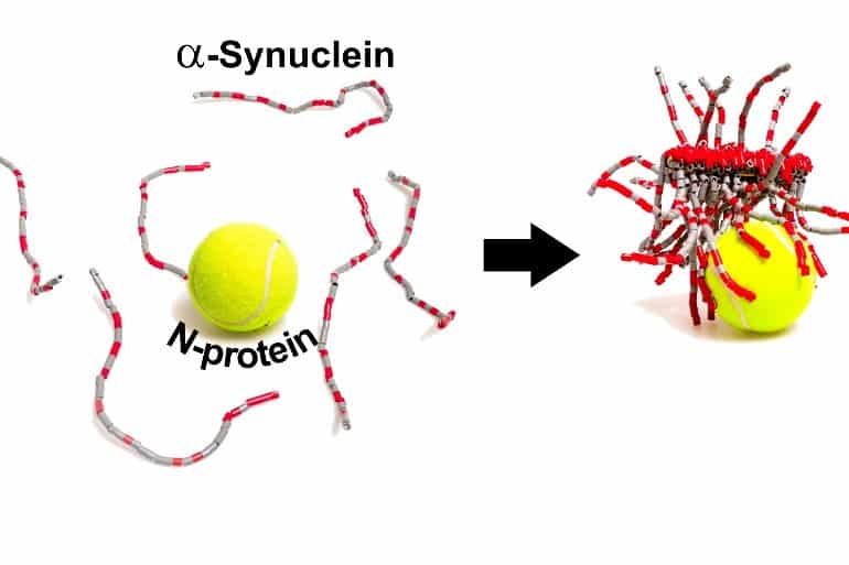
Now, researchers reporting in ACS Chemical Neuroscience have shown that, at least in the test tube, the SARS-CoV-2 N-protein interacts with a neuronal protein called α-synuclein and speeds the formation of amyloid fibrils, pathological protein bundles that have been implicated in Parkinson’s disease.

In addition to respiratory symptoms, SARS-CoV-2 can cause neurological problems, such as loss of smell, headaches and “brain fog.” However, whether these symptoms are caused by the virus entering the brain, or whether the symptoms are instead caused by chemical signals released in the brain by the immune system in response to the virus, is still controversial.

In Parkinson’s disease, a protein called α-synuclein forms abnormal amyloid fibrils, leading to the death of dopamine-producing neurons in the brain. Interestingly, loss of smell is a common premotor symptom in Parkinson’s disease.

This fact, as well as case reports of Parkinson’s in COVID-19 patients, made Christian Blum, Mireille Claessens and colleagues wonder whether protein components of SARS-CoV-2 could trigger the aggregation of α-synuclein into amyloid. They chose to study the two most abundant proteins of the virus: the spike (S-) protein that helps SARS-CoV-2 enter cells, and the nucleocapsid (N-) protein that encapsulates the RNA genome inside the virus.

In test tube experiments, the researchers used a fluorescent probe that binds amyloid fibrils to show that, in the absence of the coronavirus proteins, α-synuclein required more than 240 hours to aggregate into fibrils. Adding the S-protein had no effect, but the N-protein decreased the aggregation time to less than 24 hours.

This shows two tennis balls and strings
The SARS-CoV-2 N-protein can interact with α-synuclein in the test tube and help it form amyloid fibrils, a hallmark of Parkinson’s disease. Credit: The researchers / ACS Chemical Neuroscience

In other experiments, the team showed that the N- and α-synuclein proteins interact directly, in part through their opposite electrostatic charges, with at least 3–4 copies of α-synuclein bound to each N-protein.

Next, the researchers injected N-protein and fluorescently labeled α-synuclein into a cell model of Parkinson’s disease, using a similar concentration of N-protein as would be expected inside a SARS-CoV-2-infected cell. Compared to control cells with only α-synuclein injected, about twice as many cells died upon injection of both proteins.

Also, the distribution of α-synuclein was altered in cells co-injected with both proteins, and elongated structures were observed, although the researchers could not confirm that they were amyloid.

It’s unknown whether these interactions also occur within neurons of the human brain, but if so, they could help explain the possible link between COVID-19 infection and Parkinson’s disease, the researchers say.


Interactions between SARS-CoV-2 N-Protein and α-Synuclein Accelerate Amyloid Formation

First cases that point at a correlation between SARS-CoV-2 infections and the development of Parkinson’s disease (PD) have been reported. Currently, it is unclear if there is also a direct causal link between these diseases.

To obtain first insights into a possible molecular relation between viral infections and the aggregation of α-synuclein protein into amyloid fibrils characteristic for PD, we investigated the effect of the presence of SARS-CoV-2 proteins on α-synuclein aggregation.

We show, in test tube experiments, that SARS-CoV-2 spike protein (S-protein) has no effect on α-synuclein aggregation, while SARS-CoV-2 nucleocapsid protein (N-protein) considerably speeds up the aggregation process. We observe the formation of multiprotein complexes and eventually amyloid fibrils. Microinjection of N-protein in SH-SY5Y cells disturbed the α-synuclein proteostasis and increased cell death.

Our results point toward direct interactions between the N-protein of SARS-CoV-2 and α-synuclein as molecular basis for the observed correlation between SARS-CoV-2 infections and Parkinsonism.

Trace Amyloid Back to Precursor Protein


There is a more extreme view that amyloid itself has almost nothing to do with dementia in the human, unlike the brain of the mouse models which are overloaded with abnormal beta-amyloid protein.

As I’ve expressed in a commentary in the Journal of Alzheimer’s Disease, I do not think it is a stretch to consider that amyloid, senile plaques, neurofibrillary tangles, and neuronal death are just various end-points indicative of a process that is actually causing the dementia, but have no role in causing dementia.

From a molecular neurobiological perspective, it is more reasonable to take amyloid, predominantly an extracellular protein, out of the causal cascade leading to dementia and instead follow its intracellular complement of the gamma-secretase cleavage. This protein, so-called amyloid precursor protein (APP) intracellular domain (AICD), is actually in a better position to affect conditions inside the neuron.

AICD is known to cause hyperphosphorylation of tau, which will continue the cascade leading to dementia: formation of paired helical filaments, which aggregate to form neuropil threads (which are found only in those amyloid plaques associated with dementia and a diagnosis of Alzheimer’s disease), and the neuropil threads amputate axons and dendrites, leading to massive synapse loss — the direct cause of dementia.

The subsequent events, retrograde transport of neuropil threads to neuronal cell bodies, accumulation of neuropil threads to form neurofibrillary tangles, and resulting cell death are not part of the cause of dementia, either — instead, they are just late effects. All of the well-known pathological stigmata of Alzheimer’s disease, plaques, tangles, neuron death, inflammatory response, are all visible but secondary consequences of the underlying process leading to the dementia and should not be mistaken for treatment targets.

The real line to follow for prevention is to understand the APOE genotype and how age leads to the progressive breakdown of the serotonin and norepinephrine neurons, finally ending up damaging the cholinergic neurons. The loss of the basic brainstem projections is associated with an imbalance in the memory mechanisms involved in processing the APP, specifically, failure to activate the alpha-secretase cleavage of the APP, with default excess cleavage by beta-secretase and gamma-secretase, leading to increased production AICD, leading to synapse loss and dementia.

I am hopeful that the field can look to the critical neurobiological processes beyond the amyloid cascade hypothesis so it can quit wasting billions of dollars and realign behind some promising directions.

Studies Cast Doubt on Cancer Drug as Alzheimer’s Treatment.


studies-cast-doubt-cancer-drug-alzheimers_1

Four labs can’t replicate finding that showed large-scale clearance of disease-related plaques. Some hope remains for improving memory

Bexarotene, a cancer drug touted as a potential treatment for Alzheimer’s disease, may not be the blockbuster remedy scientists were hoping for, according to several analyses published in Science on 24 May. Four independent research groups report that they failed to fully replicate striking results published in the journal last year by Gary Landreth, a neuroscientist at Case Western Reserve University School of Medicine in Cleveland, Ohio, and his colleagues.

Landreth’s team reported that the drug bexarotene could lower brain concentrations of the β-amyloid protein that has long been suspected as a key contributor to Alzheimer’s disease, and could even reverse cognitive impairments in diseased mice. But the study garnered particular attention for its claim that the drug could clear 50% of amyloid plaques — sticky clumps of the protein thought to interfere with brain function — in as little as 72 hours.

“That attracted a lot of folks to try to replicate these studies,” says Philip Wong, a neuroscientist at Johns Hopkins University in Baltimore, Maryland. “No drug at the present moment can do things like that.”

None of the follow-up studies published this week replicated the effects of bexarotene on plaques. Two groups did, however, confirm Landreth’s finding that the drug reduced levels of a soluble, free-floating form of β-amyloid, which can aggregate in plaques.

Not all of the papers examined memory in mice, but one group led by Radosveta Koldamova, a neuroscientist at the University of Pittsburgh in Pennsylvania, found that bexarotene treatment led to cognitive improvements.

Sticking points
“It was our expectation other people would be able to repeat this,” says Landreth about the results of the studies. “Turns out that wasn’t the case, and we fundamentally don’t understand that.” He suggests that the other groups might have used different drug preparations that altered the concentration of bexarotene in the brain or even changed its biological activity.

In a response published alongside the comment articles, Landreth emphasizes that some of the studies affirm two key conclusions of the original paper: the lowering of soluble β-amyloid levels and the reversal of cognitive deficits. He says that the interest in plaques may even be irrelevant to Alzheimer’s disease. In the past ten years, some neuroscientists have begun to question whether it is plaques or soluble β-amyloid proteins that are most dangerous to brain health.

As the debate over plaques continues, Koldamova says that the cognitive improvement she and Landreth observed suggests that bexarotene is still very promising. “Patients don’t go to the doctor because they have plaques. They go because they have memory decline,” she says.

Ethical dilemma
Other researchers say the contradictory results suggest that much more basic research is needed before bexarotene is used to treat Alzheimer’s. “The mechanism of action behind bexarotene has not been proven,” says Kevin Felsenstein, a neuroscientist at the University of Florida College of Medicine in Gainesville, and a co-author of one of the dissenting papers. Felsenstein’s group found no evidence that bexarotene lowered levels of soluble or plaque forms of β-amyloid protein.

Felsenstein worries that interest in Landreth’s original results could lead to misuse of the drug because, unlike many experimental treatments, it is already on the market. The US Food and Drug Administration has approved bexarotene to treat skin cancer.

In August 2012, The New England Journal of Medicine published an article anticipating growing demand for unauthorized prescriptions of bexarotene as an Alzheimer’s treatment and urging physicians to wait for evidence from human clinical trials.

Physicians and researchers may get some answers soon: Landreth’s group has begun an early clinical trial to test the drug in healthy participants.

Source: scientificamerican.com


Brain Amyloid Imaging — FDA Approval of Florbetapir F18 Injection.


The Centers for Disease Control and Prevention recently estimated that more than 16 million Americans are living with cognitive impairment.1 Cognitive impairment can be ascribed to a variety of disorders, some of which can be treated (e.g., severe depression or effects of medications) but others of which may signal the development of incurable dementias, such as Alzheimer’s disease. For these reasons, the development and improvement of diagnostic procedures — and neuroimaging procedures, in particular — that aid in characterizing cognitive impairment is a health care priority. Improved diagnostic evaluation of patients with cognitive impairment may also enhance the development of therapies, since reliable diagnoses are critical to the success of clinical trials.

Recently, the Food and Drug Administration (FDA) approved a new radiopharmaceutical agent to assist clinicians in detecting causes of cognitive impairment other than Alzheimer’s disease. Florbetapir F18 injection (Amyvid, Eli Lilly) is indicated for positron-emission tomographic (PET) imaging of the brain in cognitively impaired adults undergoing evaluation for Alzheimer’s disease and other causes of cognitive decline.2 Florbetapir binds to amyloid aggregates in the brain, and the florbetapir PET image is used to estimate the density of β-amyloid neuritic plaque. As a component of a comprehensive diagnostic evaluation, the finding of a “negative” florbetapir scan (as qualified below) should intensify efforts to find a non–Alzheimer’s disease cause of cognitive decline. Florbetapir brain imaging is a new type of nuclear medicine imaging, and the interpretation of the image requires special training. The unique features of the imaging information also require careful consideration when the scan results are integrated into a diagnostic evaluation.

Although the pathophysiological consequences of accumulation of β-amyloid in the brain are uncertain, neuropathological identification of amyloid plaques, typically at autopsy, has long been recognized as essential to confirming the diagnosis of Alzheimer’s disease. Because β-amyloid plaques in the brain have been described as a “hallmark” of Alzheimer’s disease, some clinicians may regard the florbetapir scan as a new test for the disease.3 But the drug was developed exclusively to estimate the density of β-amyloid neuritic plaque in the brain, and these plaques have been detected in patients with a variety of neurologic disorders, as well as in older people with normal cognition (see Florbetapir F18 Scan Usage: Information Summary).

Florbetapir is an 18F-labeled ligand that, in nonclinical studies, was shown to bind to β-amyloid aggregates in postmortem sections of human brains and in brain homogenates.4 In the main clinical studies supporting FDA approval, the accuracy of florbetapir scans was assessed in the brains of terminally ill patients who participated in a brain-donation program. The patients, who had a range of underlying cognitive function, underwent florbetapir scans and were followed until they died. The premortem scan results were subsequently compared with the brain autopsy findings. In all the clinical studies, the florbetapir scans were independently interpreted by multiple readers who had completed training in interpreting florbetapir images.

A binary method of interpretation was developed for relating “positive” or “negative” florbetapir scans to neuropathologically defined categories of density of β-amyloid neuritic plaque. The method designated a positive florbetapir scan as categorically indicative of “moderate to frequent” β-amyloid neuritic plaques, as defined by the consensus criteria for Alzheimer’s disease neuropathology established by the National Institute on Aging. In 59 patients who underwent florbetapir scans and autopsy, scan sensitivity for the detection of moderate to frequent β-amyloid neuritic plaques was 92% (range, 69 to 95), and scan specificity was 95% (range, 90 to 100), on the basis of the median assessment among five readers (ClinicalTrials.gov number, NCT01447719).

One of the challenges of the florbetapir clinical development program was that terminally ill patients are not representative of the population that is likely to undergo florbetapir scanning in medical practice. In addition, β-amyloid content could change between the time of live brain imaging and the time of autopsy. More than 20% of autopsies in the main clinical studies were performed more than a year after the live brain imaging (NCT01447719 and NCT01550549).

To evaluate scan reliability in a wider population, a clinical study had new readers examine images from non–terminally ill patients with Alzheimer’s disease or mild cognitive impairment, as well as persons with normal cognition. The previously obtained images from autopsied patients were also included in the study (NCT01550549). Among five readers who interpreted images from the 151 subjects, the kappa score for interrater reliability was 0.83 (95% confidence interval, 0.78 to 0.88), with the lower bound of the 95% confidence interval exceeding the prespecified reliability success criterion of 0.58. For the autopsy subgroup of 59 subjects, the median scan sensitivity was 82% (range, 69 to 92), and the median scan specificity was 95% (range, 90 to 95) for the five new readers.

Clinical and nonclinical studies verified that florbetapir scans can provide neuropathologically accurate and reliable estimations of the density of β-amyloid neuritic plaque in the brain. Nevertheless, as with other imaging methods, there is potential for clinical interpretive error. In the studies of scan accuracy, such errors were uncommon but when present were due mainly to false negative results, as determined by the density of β-amyloid neuritic plaque at autopsy.

Reader training was an especially important element in the clinical development of florbetapir, because the image-interpretation process differs markedly from that typically used in nuclear medicine. For example, the image reader must be proficient in distinguishing white from gray matter, a distinction that may be particularly challenging in patients with cortical atrophy. Unique “gray–white contrast” characteristics of florbetapir images must be recognized as signals of normal or abnormal isotope distribution (see figureTypical Negative and Positive Florbetapir Scans.). In addition, cognitive status and other clinical or diagnostic information are not considered during the interpretation of florbetapir images. The sole goal of the reader is to determine whether a scan is negative or positive, and this determination should be made only by readers who have completed the sponsoring company’s dedicated training program. The success of the reader-training process will be further evaluated in a postmarketing study of image interpretations performed under the typical conditions of clinical practice.

In approving florbetapir, the FDA did not require clinical data assessing the effect of florbetapir imaging on clinical management or patients’ health. The FDA code of regulations (in 21 CFR 315.5[a]) mandates that the effectiveness of a diagnostic radiopharmaceutical agent should be determined by an evaluation of the ability of the agent to provide useful clinical information related to the proposed indications for use. FDA guidance further recognizes that imaging information may in some instances “speak for itself” with respect to clinical value5 and that diagnostic approval may therefore not require assessment of the effects on clinical management or health outcomes. Two FDA advisory committees endorsed the implicit clinical value of information obtained from brain β-amyloid imaging. Florbetapir approval was based on this endorsement and on clinical data showing sufficient scan reliability and performance characteristics.2

The ultimate clinical value of florbetapir imaging awaits further studies to assess the role, if any, that it plays in providing prognostic and predictive information. For example, the prognostic usefulness of florbetapir imaging in identifying persons with mild cognitive impairment or cognitive symptoms who may be at risk for progression to dementia has not been determined. Nor are data available to determine whether florbetapir imaging could prove useful for predicting responses to medication. These concerns prompted the FDA to require a specific “Limitations of Use” section in the florbetapir label.

The FDA approval of florbetapir F18 injection sets the stage for future studies that increase the value of the technique in addressing the diagnostic challenges associated with cognitive impairment. Further investigation of the drug in the postmarketing context is consistent with the commitment of the FDA to the development of imaging products that aid in the diagnostic evaluation of cognitively impaired patients.

Florbetapir F18 Scan Usage: Information Summary.

A negative florbetapir scan:

• indicates sparse to no neuritic plaques.

• is inconsistent with a neuropathological diagnosis of Alzheimer’s disease at the time of image acquisition.

• reduces the likelihood that a patient’s cognitive impairment is due to Alzheimer’s disease.

A positive florbetapir scan:

• indicates moderate to frequent amyloid neuritic plaques.

• may be observed in older people with normal cognition and in patients with various neurologic conditions, including Alzheimer’s disease.

Important florbetapir scan limitations:

• A positive scan does not establish a diagnosis of Alzheimer’s disease or other cognitive disorder.

• The scan has not been shown to be useful in predicting the development of dementia or any other neurologic condition, nor has usefulness been shown for monitoring responses to therapies.

References

  1. 1

Promoting brain health. Atlanta: Centers for Disease Control and Prevention, 2011 (http://www.cdc.gov/aging/pdf/cognitive_impairment/cogImp_genAud_final.pdf).

  1. 2

Highlights of prescribing information: Amyvid (florbetapir F18 injection). Silver Spring, MD: Food and Drug Administration (http://www.accessdata.fda.gov/drugsatfda_docs/label/2012/202008s000lbl.pdf).

  1. 3

Okie S. Confronting Alzheimer’s disease. N Engl J Med 2011;365:1069-1072
Full Text | Web of Science | Medline

  1. 4

Lister-James J, Pontecorvo MJ, Clark C, et al. Florbetapir F-18: a histopathologically validated beta-amyloid positron emission tomography imaging agent. Semin Nucl Med 2011;41:300-304
CrossRef | Web of Science

  1. 5

Guidance for industry: developing medical imaging drug and biological products. Part 2: clinical indications. Washington, DC: Department of Health and Human Services, Food and Drug Administration, Center for Drug Evaluation and Research, Center for Biologics Evaluation and Research, 2004 (http://www.fda.gov/downloads/Drugs/GuidanceComplianceRegulatoryInformation/Guidances/UCM071603.pdf).


Source: NEJM