Gamma Stimulation Promotes Glymphatic Clearance of Amyloid, Slows Alzheimer’s Progression


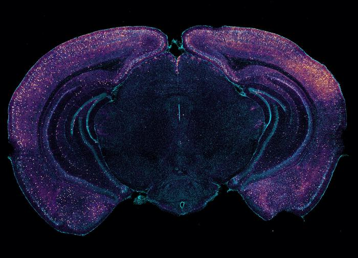
Bright staining highlights VIP-expressing interneurons in this coronal cross-section of a mouse brain. The neurons may help drive glymphatic clearance of amyloid via the release of peptides. [Tsai Laboratory/MIT Picower Institute]

It has been shown that noninvasive optogenetic-driven 40 Hz stimulation promotes neural activity and attenuates pathology in the 5XFAD mouse model of Alzheimer’s disease.

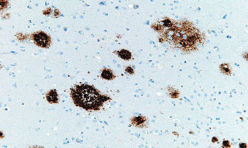
Findings from a new study reveal a key mechanism that may contribute to these beneficial effects: clearance of amyloid proteins, a hallmark of AD pathology, via the brain’s glymphatic system—a recently discovered “plumbing” network parallel to the brain’s blood vessels.

This work is published in Nature in the paper, “Multisensory gamma stimulation promotes glymphatic clearance of amyloid.

“Ever since we published our first results in 2016, people have asked me how does it work? Why 40 Hz? Why not some other frequency?” said Li-Huei Tsai, PhD, professor of neuroscience and director of the Picower Institute and MIT’s Aging Brain Initiative. “These are indeed very important questions we have worked very hard in the lab to address.”

The new paper describes experiments that show that sensory gamma stimulation prompts a particular type of neuron to release peptides that promote increased amyloid clearance via the glymphatic system.

Working with the 5XFAD mouse model of Alzheimer’s, the team first replicated the lab’s prior results that 40 Hz sensory stimulation increases 40 Hz neuronal activity in the brain and reduces amyloid levels. Then they set out to measure whether there was any correlated change in the fluids that flow through the glymphatic system to carry away wastes. Indeed, they measured increases in cerebrospinal fluid in the brain tissue of mice treated with sensory gamma stimulation compared to untreated controls. They also measured an increase in the rate of interstitial fluid leaving the brain. Moreover, in the gamma-treated mice, they measured increased diameter of the lymphatic vessels that drain away the fluids and measured increased accumulation of amyloid in cervical lymph nodes, which is the drainage site for that flow.

To investigate how this increased fluid flow might be happening, the team focused on the aquaporin 4 (AQP4) water channel of astrocyte cells, which enables the cells to facilitate glymphatic fluid exchange. When they blocked APQ4 function with a chemical, that prevented sensory gamma stimulation from reducing amyloid levels and prevented it from improving mouse learning and memory. And when, as an added test they used a genetic technique for disrupting AQP4, that also interfered with gamma-driven amyloid clearance.

More specifically, the authors noted, “Influx of cerebrospinal fluid was associated with increased aquaporin-4 polarization along astrocytic endfeet and dilated meningeal lymphatic vessels. Inhibiting glymphatic clearance abolished the removal of amyloid by multisensory 40 Hz stimulation.”

In addition to the fluid exchange promoted by APQ4 activity in astrocytes, another mechanism by which gamma waves promote glymphatic flow is by increasing the pulsation of neighboring blood vessels. Several measurements showed stronger arterial pulsatility in mice subjected to sensory gamma stimulation compared to untreated controls.

Using single-nucleus RNA sequencing (snRNA-seq), the team saw that gamma sensory stimulation indeed promoted changes consistent with increased astrocyte AQP4 activity. The data also revealed that upon gamma sensory stimulation, interneurons experienced a notable uptick in the production of several peptides. This was not surprising in the sense that peptide release is known to be dependent on brain rhythm frequencies, but it was still notable because one peptide in particular, VIP, is associated with Alzheimer’s-fighting benefits and helps to regulate vascular cells, blood flow and glymphatic clearance.

The team ran tests that revealed increased VIP in the brains of gamma-treated mice. The researchers also used a sensor of peptide release and observed that sensory gamma stimulation resulted in an increase in peptide release from VIP-expressing interneurons.

When the VIP neurons where inhibited, and the mice were exposed to sensory gamma stimulation, there was no longer an increase in arterial pulsatility and there was no more gamma-stimulated amyloid clearance.

While this paper focuses on what is likely an important mechanism—glymphatic clearance of amyloid—by which sensory gamma stimulation helps the brain, it’s probably not the only underlying mechanism that matters. The clearance effects shown in this study occurred rapidly, but in lab experiments and clinical studies weeks or months of chronic sensory gamma stimulation have been needed to have sustained effects on cognition.

Double-Edged


Amyloid beta protein protects brain from herpes infection by entrapping viral particles

amyloid beta plaques

Amyloid beta plaques (brown), a hallmark of Alzheimer’s disease, in the cerebral cortex.

A new study by Harvard Medical School researchers at Massachusetts General Hospital reveals how amyloid beta, the protein deposited into plaques in the brains of patients with Alzheimer’s disease, protects the brain from the effects of herpes viruses.

Along with another study appearing in the same July 11 issue of Neuron, which found elevated levels of three types of herpesvirus in the brains of patients with Alzheimer’s disease, the HMS team’s results support a potential role for viral infection in accelerating amyloid beta deposition and Alzheimer’s progression.


“There have been multiple epidemiological studies suggesting people with herpes infections are at higher risk for Alzheimer’s disease, along with the most recent findings from Icahn School of Medicine at Mt. Sinai that are being published with our study,” said corresponding author Rudolph Tanzi, the HMS Joseph P. and Rose F. Kennedy Professor of Child Neurology and Mental Retardation at Mass General.

“Our findings reveal a simple and direct mechanism by which herpes infections trigger the deposition of brain amyloid as a defense response in the brain,” Tanzi said. “In this way, we have merged the infection hypothesis and amyloid hypothesis into one ‘antimicrobial response hypothesis’ of Alzheimer’s disease.”

Previous studies led by Tanzi and co-corresponding author Robert Moir, HMS assistant professor of neurology at Mass General, found evidence indicating that amyloid beta, which has long been thought to be useless “metabolic garbage,” was an antimicrobial protein of the body’s innate immune system. Amyloid beta appears capable of protecting animal models and cultured human brain cells from dangerous infections.

Brain infection with herpes simplex, the virus that causes cold sores, is known to increase with aging, leading to almost universal presence of that and other herpes strains in the brain by adulthood. The HMS team set out to find whether amyloid beta could protect against herpes infection and, if so, the mechanism by which such protection takes place.

After first finding that transgenic mice engineered to express human amyloid beta survive significantly longer after injections of herpes simplex into their brains than do nontransgenic mice, the researchers found that amyloid beta inhibited infection of cultured human brain cells with herpes simplex and two other herpes strains by binding to proteins on the viral membranes and clumping into fibrils that entrap the virus and prevent it from entering cells.

Amyloid beta plaques

Further experiments with the transgenic mice revealed that introduction of herpes simplex into the brains of 5- to 6-week-old animals induced rapid development of amyloid beta plaques, which usually appear only when the animals are 10 to 12 weeks old.

“Our findings show that amyloid entrapment of herpesviruses provides immediate, effective protection from infection,” Moir said. “But it’s possible that chronic infection with pathogens like herpes that remain present throughout life could lead to sustained and damaging activation of the amyloid-based immune response, triggering the brain inflammation that drives a cascade of pathologies leading to the onset of Alzheimer’s disease.”

“A key insight is that it’s not direct killing of brain cells by herpes that causes Alzheimer’s, rather, it’s the immune response to the virus that leads to brain-damaging neuroinflammation,” Moir said.

“Our data and the Mt. Sinai findings suggest that an antimicrobial protection model utilizing both anti-herpes and anti-amyloid drugs could be effective against early Alzheimer’s disease,” he added. “Later on, when neuroinflammation has begun, greater benefit may come from targeting inflammatory molecules. However, it remains unclear whether infection is the disease’s root cause. After all, Alzheimer’s is a highly heterogeneous disease, so multiple factors may be involved in its development.”

“We are currently conducting what we call the ‘brain microbiome project,’ to characterize the population of microbes normally found in the brain,” said Tanzi, who is director of the Genetics and Aging Research Unit in the MassGeneral Institute for Neurodegenerative Disease. “The brain used to be considered sterile, but it turns out to have a resident population of microbes, some of which may be needed for normal brain health.”

“Our preliminary findings suggest that the brain microbiome is severely disturbed in Alzheimer’s disease and that bad players, including herpes viruses, seem to take advantage of the situation, leading to trouble for the patient,” Tanzi said. “We are exploring whether Alzheimer’s pathogenesis parallels the disrupted microbiome models seen in conditions like inflammatory bowel disease, and the data generated to date are both surprising and fascinating.”