Researchers develop a new way to safely boost immune cells to fight cancer


Researchers develop a new way to safely boost immune cells to fight cancer
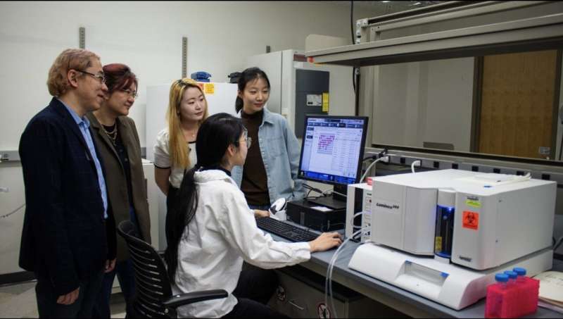
(Standing, from left) Rong Tong, associate professor in chemical engineering; Wenjun “Rebecca” Cai, associate professor in materials science and engineering; Eungyo Jang; and Ziyu Huo gather around Liqian Niu (seated), who is working with a Luminex 200 machine used to analyze tumor cytokine levels. Credit: Hailey Wade for Virginia Tech.

Last year alone, more than 600,000 people in the United States died from cancer, according to the American Cancer Society. The relentless pursuit of understanding this complex disease has shaped medical progress in developing treatment procedures that are less invasive while still highly effective.

Immunotherapy is on the rise as a possible solution. Immunotherapy involves harnessing the power of the body’s immune system to fight against cancer cells. Researchers in the College of Engineering have found a way to revamp a treatment procedure into a groundbreaking practice.

Rong Tong, associate professor in chemical engineering, has teamed up with Wenjun “Rebecca” Cai, associate professor in materials science and engineering, to explore a cancer immunotherapy treatment that has long been of interest to researchers.

In their newly published article in the journal Science Advances, Tong and Cai detailed their approach, which involves activating the immune cells in the body and reprogramming them to attack and destroy the cancer cells. This therapeutic method is frequently implemented with the protein cytokine. Cytokines are small protein molecules that act as intercellular biochemical messengers and are released by the body’s immune cells to coordinate their response.

“Cytokines are potent and highly effective at stimulating the immune cells to eliminate cancer cells,” Tong said. “The problem is they’re so potent that if they roam freely throughout the body, they’ll activate every immune cell they encounter, which can cause an overactive immune response and potentially fatal side effects.”

Tong and Cai, in collaboration with chemical engineering and materials science and engineering graduate students, have developed an innovative approach to employ cytokine proteins as a potential immunotherapy treatment. Unlike previous methods, their technique ensures that the immune cell stimulating cytokines effectively localize within the tumors for weeks while preserving the cytokine’s structure and reactivity levels.

Combining forces to take down cancer cells

Current cancer treatments, such as chemotherapy, cannot distinguish between healthy cells and cancer cells. When someone with cancer is treated with chemotherapy, the treatment attacks all of the cells in their body, which can lead to side effects such as hair loss and fatigue.

Stimulating the body’s immune system to attack tumors is a promising alternative to treat cancer. The delivery of cytokines can jump-start immune cells in the tumor, but overstimulating healthy cells can cause severe side effects.

“Scientists determined a while ago that cytokines can be used to activate and fight against tumors, but they didn’t know how to localize them inside the tumor while not exposing toxicity to the rest of the body,” said Tong. “Chemical engineers can look at this from an engineering approach and use their knowledge to help refine and elevate the effectiveness of the cytokines so they can work inside the body effectively.”

The research team’s goal is to find a balance between killing cancer cells in the body while sparing healthy cells.

To accomplish this goal, Tong and his students used their expertise to create specialized particles with distinctive sizes that help determine where the drug is going. These microparticles are designed to stay within the tumor environment after being injected into the body. Cai and her students worked on measuring these particles’ surface properties.

“In the field of materials science and engineering, we study the surface chemistry and mechanical behavior of materials, such as the specialized particle created for this project,” Cai said. “Surface engineering and characterization, along with particle size, play important roles in controlled drug delivery, ensuring prolonged drug presence and sustained therapeutic effectiveness.”

To ensure successful drug delivery, Tong and his chemical engineering students designed a novel strategy that:

  • Anchors cytokines to these new microparticles, limiting the harm of cytokines to healthy cells
  • Allows the newly particle-anchored cytokines to jump-start immune systems and recruit immune cells to attack cancer cells

“Our strategy not only minimizes cytokine-induced harm to healthy cells, but also prolongs cytokine retention within the tumor,” Tong said. “This helps facilitate the recruitment of immune cells for targeted tumor attack.”

The next step in the process involves combining the new, localized cytokine therapy method with commercially available, Food and Drug Administration (FDA)-approved checkpoint blockade antibodies, which reactivate the tumor immune cells that have been silenced so they can fight back the cancer cells.

“When there is a tumor inside the body, the body’s immune cells are being deactivated by the cancer cells,” Tong explained. “The FDA-approved checkpoint blocking antibody helps ‘take off the brakes’ that tumors put on immune cells, while the cytokine molecules ‘step on the gas’ to jump-start the immune system and get an immune cell army to fight cancer cells. These two approaches work together to activate immune cells.”

Combining the checkpoint antibodies with the particle-anchored cytokine proved to successfully eliminate many tumors in their study.

Engineering an impact on cancer treatment

Team members hope their impact on immunotherapy treatment is part of a greater movement toward cancer treatment approaches that are harmless to healthy cells. The new approach of attaching cytokines to particles also could be used in the future to deliver other types of immunostimulatory drugs, according to the team.

“Researchers are still looking for safer and more effective cancer treatments,” said Tong. “This motivation is what drives us to develop new technologies in the field. The whole class of drugs that are employed to jump-start the immune system to fight cancer cells has largely not yet succeeded.

“Our goal is to create novel solutions that allow researchers to test these drugs with existing FDA-approved therapeutics, ensuring both safety and enhanced efficacy.”

Cai said the nature of cancer treatment research requires expertise across engineering disciplines.

“I view this project as a perfect marriage between chemical engineering and materials science,” Cai said. “The former focuses on the synthesis and drug delivery part, the latter on applying advanced materials characterization. This collaboration not only accelerates immunotherapy research, but also has the ability to transform cancer treatment.”

New compound from blessed thistle may promote functional nerve regeneration


New compound from blessed thistle promotes functional nerve regeneration
Dried blessed thistle (Cnicus benedictus)

Researchers from the University of Cologne have found a new use for cnicin, a substance produced in blessed thistle. Their article “Cnicin promotes functional nerve regeneration” features clinical studies and is published in Phytomedicine.

Blessed thistle (Cnicus benedictus) is a plant in the family Asteraceae and also grows in our climate. For centuries, it has been used as a medicinal herb as an extract or tea, e.g. to aid the digestive system.

Researchers at the Center for Pharmacology of University Hospital Cologne and at the Faculty of Medicine of the University of Cologne have now found a completely novel use for cnicin under the direction of Dr. Philipp Gobrecht and Professor Dr. Dietmar Fischer. Animal models as well as human cells have shown that cnicin significantly accelerates axon (nerve fibers) growth. The study was published in Phytomedicine.

Regeneration pathways of injured nerves in humans and animals with long axons are accordingly long. This often makes the healing process lengthy and even frequently irreversible because the axons cannot reach their destination on time. An accelerated regeneration growth rate can, therefore, make a big difference here, ensuring that the fibers reach their original destination on time before irreparable functional deficits can occur.

The researchers demonstrated axon regeneration in animal models and human cells taken from retinae donated by patients. Administering a daily dose of cnicin to mice or rats helped improve paralysis and neuropathy much more quickly.

Compared to other compounds, cnicin has one crucial advantage: it can be introduced into the bloodstream orally (by mouth). It does not have to be given by injection.

“The correct dose is very important here, as cnicin only works within a specific therapeutic window. Doses that are too low or too high are ineffective. This is why further clinical studies on humans are crucial,” said Fischer.

The University of Cologne researchers are currently planning relevant studies. The Center for Pharmacology is researching and developing drugs to repair the damaged nervous system.

Analyzing the progression in retinal thickness could predict cognitive progression in Parkinson’s patients


Analysing the progression in retinal thickness could predict cognitive progression in Parkinson's patients
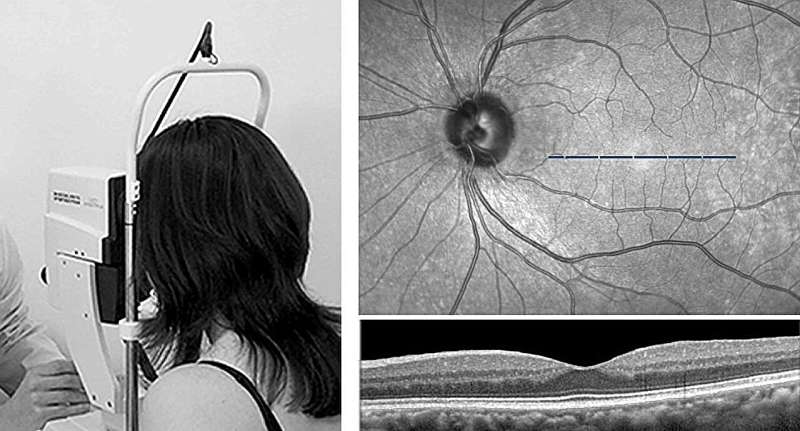
Optical coherence tomography.

Although there are still some aspects pending confirmation for its use in the clinical setting, and its resolution needs to be improved slightly, a study by the UPV/EHU and Biobizkaia has shown that a method routinely used to carry out ophthalmological tests can also be used to monitor the neurodegeneration that occurs in Parkinson’s patients. In the course of the research, it was found that the neurodegeneration of the retina probably precedes cognitive impairment.

When Parkinson’s or another neurodegenerative disease is diagnosed, patients always ask, “And now what? What will happen? What can be expected from the disease?” For neurologists, however, it is not possible to answer these questions precisely, as “the evolution of patients tends to be very varied: some experience no change over the years, while others end up with dementia or in a wheelchair,” explained Ane Murueta-Goyena, researcher in the UPV/EHU’s department of Neurosciences.

Today, identifying Parkinson’s patients at risk of cognitive impairment poses a major challenge, yet this is necessary when it comes to providing more effective clinical treatments and stepping up clinical trials.

In fact, Dr. Ane Murueta-Goyena, in collaboration with Biobizkaia’s research staff, wanted to see “whether the visual system can enable this deterioration to be predicted, in other words, what future the patient can expect within a few years.” The thickness of the retina was used for this purpose.

The retina is a membrane located at the back of the eyeball; it is related to the nervous system and comprises several layers. During the study, a cohort of Parkinson’s patients had the thickness of the innermost layer of their retinas measured using optical coherence tomography.

This type of tomography is a routinely used instrument in ophthalmological tests, as it allows high-resolution, repeatable, and accurate measurements to be made. So the evolution of this retinal layer was analyzed and compared in people with and without Parkinson’s disease over the 2015–2021 period. The results of the analysis of the images of the retinal layers of Parkinson’s patients were also confirmed in a UK hospital.

The results showed that the retinal layer is noticeably thinner in Parkinson’s patients. It was also observed that “during the initial phases of the disease it is in the retina where the greatest neurodegeneration is detected, and, from a given moment onwards, when the layer is already very thin, a kind of stabilizing of the neurodegeneration process takes place.”

“Retinal thinning and cognitive impairment do not occur simultaneously. The initial changes in the retina are more evident and then, over the years, patients are observed to worsen clinically in both cognitive and motor terms,” explained Murueta-Goya. “In other words, the slower retinal layer thickness loss is associated with faster cognitive decline; this slowness is linked to greater severity of the disease.”

The researcher has attached great importance to the results. “We have obtained information on the progression of the disease, and the tool we are proposing is non-invasive and available at all hospitals.”

The results need to be validated internationally, and “by slightly improving the resolution of the technology, we will be closer to validating the method for monitoring the neurodegeneration that takes place in Parkinson’s disease.” The researcher also revealed that they are continuing the research on another cohort of patients and that funding is the key.

Dozens of COVID virus mutations arose in man with longest known case, research finds


Dozens of COVID virus mutations arose in man with longest known case

An immune-compromised man with a year-and-a-half-long COVID infection served as a breeding ground for dozens of coronavirus mutations, a new study discovered.

Worse, several of the mutations were in the COVID spike protein, indicating that the virus had attempted to evolve around current vaccines, researchers report.

“This case underscores the risk of persistent SARS-CoV-2 infections in immunocompromised individuals, as unique SARS-CoV-2 viral variants may emerge,” said the research team led by Magda Vergouwe. She’s a doctoral candidate with Amsterdam University Medical Center in The Netherlands.

The patient in questioned endured the longest known COVID infection to date, fighting with the virus for 613 days before dying from the blood disease that had compromised his immune system, researchers said.

Immune-compromised patients who suffer persistent infections give the COVID virus an opportunity to adapt and evolve, the investigators explained.

For instance, the omicron variant is thought to have emerged in an immune-compromised patient initially infected with an earlier form of COVID, researchers said.

In this latest report, the man was admitted to Amsterdam University Medical Center in February 2022 with a COVID infection at age 72, after he’d already received multiple vaccinations.

He suffered from myelodysplastic and myeloproliferative overlap syndrome, a disease in which the bone marrow makes too many white blood cells, according to the U.S. National Cancer Institute.

Following a stem cell transplant, the man also had developed lymphoma, a cancer of the white blood cells, researchers said.

A drug he took for lymphoma, rituximab, depleted all the immune cells that normally produce antibodies for COVID, they added.

To clear his COVID, the man received a monoclonal antibody cocktail that ultimately proved ineffective.

In fact, gene sequencing showed that the coronavirus started mutating to evade the antibodies he’d received, a step that could have potentially undermined the effectiveness of the treatment in others, researchers said.

Gene sequencing of 27 nasal specimens taken from the man revealed more than 50 mutations in the COVID virus, including variants with changes in the spike protein targeted by vaccines.

“The prolonged infection has led to the emergence of a novel immune-evasive variant due to the extensive within-host evolution,” researchers said.

Such cases pose a “potential public health threat of possibly introducing viral escape variants into the community,” they added.

However, they noted that there had been no documented transmission of any COVID variants from the man into other people.

The researchers will present their findings at the European Society of Clinical Microbiology and Infectious Diseases meeting next week in Barcelona. Findings presented at medical meetings should be considered preliminary until published in a peer-reviewed journal.