Shilajit: Boost Energy, Enhance Brain Function, Mitigate Bone Loss, and More


Oozing from ancient rock is a substance that has been used for centuries in healing.

Shilajit: Boost Energy, Enhance Brain Function, Mitigate Bone Loss, and More
(StockImageFactory.com/Shutterstock)
Sheramy Tsai

By Sheramy Tsai

3/11/2024

Updated:

3/11/2024PrintX 1

0:00

12:40

With a history as rich as its mineral content, shilajit offers a glimpse into nature’s pharmacy, offering benefits from enhanced fertility to increased energy. As science begins to unravel the mysteries of this ancient elixir, questions arise about its efficacy and potential as a natural health solution.

Nature’s Ancient Elixir

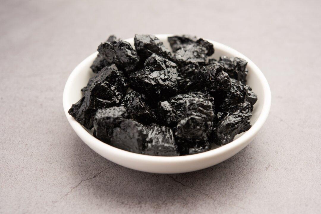
High in the Himalayas, shilajit seeps from the rocks, a tar-like byproduct of centuries-old decomposed plant matter. With a composition rich in minerals, fulvic acid, and humic acids, it occupies a revered spot in Ayurvedic medicine as a “Rasayana,” aimed at promoting longevity and revitalizing the body and mind.

Ayurvedic doctor Sruthi Bhat praised shilajit for its broad spectrum of health benefits, telling The Epoch Times, “The unique composition of shilajit provides a plethora of health benefits.” According to Ms. Bhat, shilajit supports cardiovascular health, aids digestion, and balances hormones, among other benefits. “Shilajit serves as a versatile tonic for overall health and well-being, targeting multiple systems of the body to promote vitality and resilience,” she shared.

Scientific scrutiny supports these claims, revealing shilajit’s composition to be incredibly diverse, with over 85 minerals, vitamins, and phytonutrients. Fulvic acid, a key component of shilajit, is noted for its ability to improve nutrient absorption, elevate energy levels, and facilitate the body’s detoxification. Fulvic acid is critical in transporting nutrients into the cells and expelling toxins, significantly enhancing energy and overall health.

Shilajit, also called salajit, shilajatu, mumie, mineral pitch, and mummiyo, is sourced from diverse locations such as India, Nepal, Russia, and Chile. Research suggests that its health benefits differ based on the geographical origin of the substance.

Dubbed the “destroyer of weakness,” shilajit is prominent in many cultures. According to an ancient Ayurvedic text, the Cha raka Samhita, “There is no curable disease in the universe that is not effectively curable by shilajit, when administered at the appropriate time, adopting the prescribed method.”

Ayurveda’s Golden Elixir for Modern Wellness

In Ayurvedic tradition, shilajit is classified into four types: svarna (gold), rajat (silver), tamra (copper), and loh (iron), each valued for specific therapeutic benefits. Gold shilajit, known for its rejuvenating effects, is especially esteemed. “This form of shilajit is believed to promote vitality, longevity, and overall well-being by balancing the doshas and stimulating cellular regeneration,” Ms. Bhat notes.

According to Ayurvedic principles, the various forms of shilajit target distinct health issues. Silver shilajit is used for its cooling effects, while copper shilajit is favored for its warming properties that boost vitality and metabolism. Iron shilajit is credited with grounding and is often recommended for fighting fatigue and improving endurance.

RELATED STORIES

Environmental Nutrition: What Is Ayurveda?

1/4/2024

Environmental Nutrition: What Is Ayurveda?

Intro to Ayurvedic Herbs – Learn Which Ones Works Best for You

5/12/2023

Intro to Ayurvedic Herbs - Learn Which Ones Works Best for You

Ancient Ayurvedic methodologies emphasize the critical role of purifying shilajit. It undergoes extensive purification to remove contaminants, such as heavy metals, and to enhance its therapeutic effects. Dr. Bhat points out, “Purification of shilajit stands as a pivotal step before its medicinal use.” A common purification process for shilajit includes heating it in an herbal decoction called triphala or in cow’s urine, then filtering and drying the mixture.

“Shilajit can be administered with various anupanas (carriers) to enhance its therapeutic effects,” explains Ms. Bhat. These carriers may include milk, ghee, and or honey.

Six Potential Benefits of Shilajit

Although shilajit has been a staple in traditional medicine for centuries, modern science is just beginning to explore its reported health advantages. Beyond its potential to address cancer, cardiovascular disease, and insomnia, shilajit is also making strides in these six key areas:

1. Enhances Energy and Athletic Performance

Shilajit is making waves for its ability to bolster physical and mental energy. The secret lies in its capacity to enhance mitochondrial efficiency, essentially turbocharging the body’s cellular engines.

Shilajit’s active components optimize mitochondrial function, enabling a more effective conversion of nutrients into energy. When combined with CoQ10, an antioxidant found naturally in the body, shilajit may significantly enhance stamina and endurance.

Sidney Stohs, who holds a doctorate in biochemistry, highlights in his research, “Animal and human data support its use as a ‘revitalizer,’ enhancing physical performance and relieving fatigue with enhanced production of ATP.”

Athletes and individuals enduring fatigue have noticed as evidence mounts in favor of shilajit’s role in enhancing physical performance and tackling conditions such as chronic fatigue syndrome.

A study with 63 participants demonstrated that consuming 500 mg of shilajit daily for eight weeks can significantly help maintain muscle strength after exertion and minimize tissue damage, highlighting its importance as a supplement for energy enhancement and recovery.

2. Improves Brain Health

Research suggests shilajit could play a role in supporting brain health. Studies indicate that fulvic acid in shilajit may help prevent the buildup of tau protein, a hallmark of Alzheimer’s that leads to brain cell degeneration.

Additional research supports the broader cognitive advantages of shilajit, particularly when used alongside other nutritional supplements or dietary measures. Notably, a study showed that when paired with a vitamin B complex, shilajit might benefit individuals with Alzheimer’s disease.

3. Reduces Bone Loss

Shilajit is being explored for its potential benefits in bone health, particularly among postmenopausal women. Recent scientific research suggests that shilajit extract could play a significant role in maintaining bone mineral density, offering hope for those at risk of osteoporosis.

In a detailed study involving sixty women aged 45 to 65, findings indicated that daily doses of 250 milligrams (mg) and 500 mg of shilajit extract significantly preserved bone mineral density over 48 weeks. The study’s authors attribute this effect to shilajit’s rich antioxidant, anti-inflammatory, and collagen-enhancing properties, which may help offset increased bone turnover and oxidative stress associated with estrogen deficiency.

4. Boosts Fertility

Shilajit has the potential to influence hormonal balance and enhance fertility. In a comprehensive study, men aged 45 to 55 who took 250 mg of purified shilajit twice daily for three months saw a remarkable increase in both total and free testosterone, along with dehydroepiandrosterone levels, outperforming those on a placebo.

Further research has underscored shilajit’s effectiveness in improving sperm quality, particularly for those with low sperm counts. Taking 100 mg of shilajit twice a day led to significant enhancements in sperm movement and a 61.4 percent rise in overall sperm count, with a notable 18.9 percent increase in healthy sperm. Moreover, this treatment reduced oxidative stress in semen and boosted blood testosterone levels by about 25 percent.

Shilajit’s fertility-enhancing properties are not limited to men. Neuroscientist Andrew Huberman has noted shilajit’s ability to increase follicle-stimulating hormone, which is essential for egg development, suggesting its broad applicability in fertility treatments. Shilajit is “pro-fertile,” Mr. Huberman stated in a YouTube video titled “Ashwagandha & Shilajit Benefits, Huberman Lab Podcast.”

Beyond aiding in egg growth, shilajit also boosts libido and supports reproductive health, making it a promising natural supplement for those looking to increase their chances of conception.

5. Irons Out Anemia

Thanks to its rich mineral content, shilajit may combat anemia. Animal studies have revealed its capability to raise hemoglobin levels and augment red blood cell counts—essential indicators of blood health. These outcomes are notably relevant given widespread iron deficiency anemia and the quest for natural remedies.

The fulvic acid in shilajit has been found in a study on rats to enhance the body’s ability to absorb and utilize iron, addressing the root cause of anemia by ensuring that iron intake translates into tangible improvements in blood health. Shilajit shows promise as an effective supplement in managing anemia by increasing iron levels and its uptake. Yet, further research is necessary to fully establish its benefits for human anemia treatment.

6. High-Altitude Ally

Known in Sanskrit as the “conqueror of mountains,” Shilajit lives up to its name by aiding those navigating the challenges of high altitudes. This natural substance is celebrated for its ability to address a range of health issues common to mountain explorers.

Research points to shilajit’s effectiveness in fighting altitude sickness, with symptoms such as hypoxia, fatigue, and insomnia being alleviated by its rich fulvic acid and mineral content. These components enhance the transport of nutrients and energy production while bolstering the immune system. Its adaptogenic qualities enable faster acclimatization to high altitudes.

Integrating Shilajit Into Your Health Regimen

In the crowded wellness market, finding authentic shilajit is like searching for a gem among stones. Its promise of natural health benefits makes it a sought-after remedy, yet the risk of counterfeit products means buyers must navigate with caution.

“Authenticity and quality are vital factors to consider,” asserts Ms. Bhat, underscoring the importance of selecting genuine shilajit. Unlike its liquid or pill counterparts, true shilajit is a resin, representing the most potent form of its health-enhancing properties. This natural resin transforms in warmth, becoming pliable and sticky, and dissolves into a golden or reddish hue in warm liquids.

Yet, acquiring shilajit is just the beginning. Integrating it into a daily health regimen requires knowledge and care, especially since it lacks U.S. Food and Drug Administration approval and established dosing guidelines. “Start with small dosages and gradually increase intake,” Ms. Bhat recommends, to allow the body to adjust without adverse reactions. Clinical trials often suggest a daily intake of 200 to 500 mg, divided into two doses, though individual preferences may vary due to its distinctive taste.

Mindfulness in its use is key, particularly for those with pre-existing conditions or those taking other medications. “Consulting a health care professional before commencing any new supplement regimen is paramount,” Ms. Bhat advises, highlighting the need for personalized guidance—especially for pregnant women or individuals with specific health concerns.

As research into shilajit’s efficacy and safety progresses, its potential for improving well-being gains recognition. However, fully integrating this age-old remedy into modern health practices remains a work in progress, calling for further investigation.

How Stress Turns into Fear in the Brain


Our fear response is a survival mechanism that signals us to remain alert and avoid dangerous situations. Those who have suffered episodes of severe or life-threatening stress can later experience intense feelings of fear, even during situations that lack a real threat. Experiencing this generalization of fear is psychologically damaging and can result in conditions such as post-traumatic stress disorder (PTSD). The stress-induced mechanisms that cause our brain to produce feelings of fear in the absence of threats has not been fully understood. Now, neurobiologists at the University of California (UC), San Diego, have identified the changes in brain biochemistry and mapped the neural circuitry that cause such a generalized fear experience.

The findings are published in Science in an article titled, “Generalized fear after acute stress is caused by change in neuronal co-transmitter identity.”

“Overgeneralization of fear to harmless situations is a core feature of anxiety disorders resulting from acute stress, yet the mechanisms by which fear becomes generalized are poorly understood,” the researchers wrote. “In this study, we show that generalized fear in mice results from a transmitter switch from glutamate to γ-aminobutyric acid (GABA) in serotonergic neurons of the lateral wings of the dorsal raphe. A similar change in transmitter identity was found in the postmortem brains of individuals with post-traumatic stress disorder (PTSD). Overriding the transmitter switch in mice prevented the acquisition of generalized fear.”

In their research, former UC San Diego assistant project scientist Hui-quan Li, PhD, (now a senior scientist at Neurocrine Biosciences), Nick Spitzer, PhD, the Atkinson Family Distinguished Professor of the School of Biological Sciences, and their colleagues described the research behind their discovery of the neurotransmitters—the chemical messengers that allow the brain’s neurons to communicate with one another—at the root of stress-induced generalized fear.

The researchers studied the brains of mice in an area known as the dorsal raphe, and discovered that acute stress induced a switch in the chemical signals in the neurons, flipping from excitatory “glutamate” to inhibitory “GABA” neurotransmitters, which led to generalized fear responses.

“Our results provide important insights into the mechanisms involved in fear generalization,” said Spitzer, who is also a member of UC San Diego’s department of neurobiology and the Kavli Institute for Brain and Mind. “The benefit of understanding these processes at this level of molecular detail—what is going on and where it’s going on—allows an intervention that is specific to the mechanism that drives related disorders.”

The researchers then examined the postmortem human brains of individuals who had suffered from PTSD. A similar glutamate-to-GABA neurotransmitter switch was confirmed in their brains as well.

The researchers then found a way to block the production of generalized fear. Prior to the experience of acute stress, they injected the dorsal raphe of the mice with an adeno-associated virus (AAV) to suppress the gene responsible for synthesis of GABA. This method prevented the mice from acquiring generalized fear.

Further, when mice were treated with the antidepressant fluoxetine (branded as Prozac) immediately after a stressful event, the transmitter switch and subsequent onset of generalized fear were prevented.

Not only did the researchers identify the location of the neurons that switched their transmitter, but they demonstrated the connections of these neurons to the central amygdala and lateral hypothalamus, brain regions that were previously linked to the generation of other fear responses.

“Now that we have a handle on the core of the mechanism by which stress-induced fear happens and the circuitry that implements this fear, interventions can be targeted and specific,” said Spitzer.

Gene Therapies Tested in Human Liver System in the Lab


AAV-Adeno-associated-viruses

Scientists from Children’s Medical Research Institute (CMRI) have successfully tested adeno-associated viral vector-based (AAV) gene therapies in whole human livers in the lab. The study, which is published in Nature Communications in a paper titled “Harnessing whole human liver ex situ normothermic perfusion for preclinical AAV vector evaluation,” demonstrates the potential of using a normothermic liver perfusion system—a human liver preserved ex situ in the lab at human body temperature—to test early-stage gene therapies.

This paper is the fruit of a project that began last year when CMRI’s translational vectorology research unit partnered with a team at Royal Prince Alfred Hospital to develop a method of keeping a human liver alive in the lab. The idea was to use the system to test AAV-based vehicles for delivering payloads to treat various inherited diseases. If the system was successful, it would address the challenge of finding effective preclinical models that properly replicate human physiological conditions and systems and reliably predict clinical outcomes. 

The liver is a complex organ with diverse cell types contributing to its structure and function, and that kind of complexity is difficult to replicate using organoids, mice, and nonhuman primates. The results reported in Nature Communications are “very exciting for us because now, for the first time, we can assess the function of gene therapeutics directly in the clinical target organ itself,” said Leszek Lisowski, PhD, unit head of CMRI’s translational vectorology unit and senior author on the publication. “Up to now the gene therapy delivery tools have been tested in animal models, which while invaluable to evaluate safety and targeting of other organs/tissues, do not adequately replicate the functionality of these delivery methods in the patient.”

According to the paper, the researchers used two whole human livers perfused with human blood to evaluate fourteen natural and bioengineered AAV vectors. The liver system “uses an open venous reservoir and incorporates two long-term oxygenators, a gas blender equipped with a pediatric flow regulator for ventilation control, and a flow-adjustable dialysis membrane for water-soluble toxin filtration and perfusate volume control.” Both livers had been deemed unsuitable for transplantation and were consented for research use. The scientists used next-generation sequencing to quantify which vectors had the lowest rates of clearance in the perfusate, and to assess the cell entry performance and transgene expression for each vector, among other studies.

Besides evaluating novel AAV-based therapeutics, Lisowski noted that the liver model could be used to more accurately estimate the effective dose of new therapeutics and identify potential toxic side effects. “The current generation of viral vectors we use to deliver gene therapeutics to the liver are not good enough for the majority of clinical applications. At the moment we often have to use these therapies in high doses to overcome their functional inefficiencies and achieve clinical benefit.”

Metal-Organic Nanoparticles Enable Better Vaccine Delivery, Stronger Immune Response.


An illustration of nanoparticles

Scientists from the Massachusetts Institute of Technology (MIT) and elsewhere have published a paper in Science Advances that describes a type of nanoparticle for delivering vaccines called a metal organic framework (MOF) that can potentially provoke a strong immune response at lower doses. The paper is titled “Zeolitic Imidazolate Frameworks Activate Endosomal Toll-like Receptors and Potentiate Immunogenicity of SARS-CoV-2 Spike Protein Trimer.” 

In the study, which was done in mice, the researchers showed that the MOF successfully encapsulated and delivered part of the SARS-CoV-2 spike protein while simultaneously acting as an adjuvant once it broke down inside cells. More work is needed to ensure that the particles can be used safely in human vaccines, but these early results are promising. 

“Not only are we delivering the protein in a more controlled way through a nanoparticle, but the compositional structure of this particle is also acting as an adjuvant,” according to Ana Jaklenec, PhD, a principal investigator at MIT’s Koch Institute for Integrative Cancer Research and one of the senior authors on the study. “We were able to achieve very specific responses to the COVID-19 protein, and with a dose-sparing effect compared to using the protein by itself to vaccinate.”

The MOF used in this study, called zeolitic imidazolate frameworks 8 or ZIF-8, is a lattice of tetrahedral units made up of a zinc ion attached to four imidazole molecules. Particles typically have diameters that are between 100 and 200 nanometers, making them small enough to get into the lymph nodes directly or through immune cells like macrophages. Prior studies showed that ZIF-8 particles can significantly boost immune responses. What is unclear is exactly how they activate the immune system. 

To answer that question, the researchers devised an experimental vaccine consisting of SARS-CoV-2 receptor-binding protein embedded in ZIF-8 particles. Once the particles entered the cells, the MOFs broke down releasing their viral protein cargo. The imidazole components of the MOFs then activate the toll-like receptors, which help to stimulate the innate immune response. 

RNA sequencing of lymph node cells from the vaccinated mice showed various immune related pathways were activated in response to the vaccine including the TLR-7 pathway, which led to greater production of cytokines and other inflammatory molecules. They also observed that mice vaccinated with the particles had a much stronger response to the viral protein, than those that received just the protein alone. 

Before these particles could be used in vaccines, scientists would have to evaluate not only their safety but also whether they could be scaled up for manufacturing on a larger scale. However, even if ZIF-8 does not work out, the researchers believe that their findings could help guide efforts focused on similar nanoparticles for delivering subunit vaccines, which are usually easier and cheaper to manufacture than mRNA vaccines.

“Designing new vaccines that utilize nanoparticles with specific chemical moieties, which not only aid in antigen delivery but can also activate particular immune pathways, have the potential to enhance vaccine potency,” Jaklenec noted. “Understanding how the drug delivery vehicle can enhance an adjuvant immune response is something that could be very helpful in designing new vaccines.”

Machine Learning Can Spot Tumor-Reactive TCRs, Speed Immunotherapies.


T Cells

The manufacturing process for personalized T-cell therapies hardly begins before it stalls. Why? Right at the start, there is a severe bottleneck: the need to identify patient-derived, tumor-reactive T-cell receptors (TCRs).

To overcome this bottleneck, scientists at the German Cancer Research Center (DKFZ) and the University Medical Center Mannheim have developed predicTCR, a machine learning classifier. According to the scientists, it can identify individual tumor-reactive tumor-infiltrating lymphocyte (TILs) in an antigen-agnostic manner based on single-TIL RNA sequencing.

The scientists also assert that prediTCR can halve the time it takes to get past the bottleneck, helping to reduce the overall time needed to make a personalized T-cell therapy for cancer patients. Since the overall time is at least six months, any reduction in the time needed to complete any manufacturing step is welcome.

Details about predicTCT and its application recently appeared in Nature Biotechnology, in an article titled, “Prediction of tumor-reactive T cell receptors from scRNA-seq data for personalized T cell therapy.”

The paper makes it clear that the scientists’ approach applies to personalized transgenic T-cell therapies, which seek to identify and reinfuse defined tumor-reactive TCRs, either in patient-autologous T cells or in induced pluripotent stem cell–derived, hypoimmunogenic (allogeneic) T cells. These therapies are not produced via the enrichment of tumor-reactive T cells, so they can avoid the problem of T-cell exhaustion. However, they pose a “needle in a haystack” problem, simply because identifying tumor-reactive TCRs is so difficult.

The development of personalized transgenic T-cell therapies is a complicated process. First, doctors isolate TILs from a sample of the patient’s tumor tissue. This cell population is then searched for T-cell receptors that recognize tumor-specific mutations and can thus kill tumor cells. This search is laborious and has so far required knowledge of the tumor-specific mutations that lead to protein changes that are recognized by the patients’ immune system. During this time, the tumor is constantly mutating and spreading, making this step a race against time.

“Finding the right T-cell receptors is costly and time-consuming,” said Michael Platten, MD, head of brain tumor immunology at the DKFZ and director of the department of neurology at the University Medical Center Mannheim. “With a method that allows us to identify tumor-reactive TCRs independently of knowledge of the respective tumor epitopes, the process could be considerably simplified and accelerated.”

To develop such a method, a team led by Platten and co-study head Edward W. Green, PhD, group leader, immunogenomics, began by isolating TILs from a melanoma patient’s brain metastasis and performed single-cell sequencing to characterize each cell. The T-cell receptors expressed by these TILs were then individually tested in the laboratory to identify those that were recognized and killed patient tumor cells. The researchers then combined these data to train a machine learning model to predict tumor-reactive T-cell receptors. The resulting classifier could identify tumor reactive T cells from TILs with 90% accuracy, works in many different types of tumor, and accommodates data from different cell sequencing technologies.

“PredicTCR identifies tumor-reactive TCRs in TILs from diverse cancers better than previous gene set enrichment-based approaches, increasing specificity and sensitivity (geometric mean) from 0.38 to 0.74,” the authors of the Nature Biotechnology article wrote. “By predicting tumor-reactive TCRs in a matter of days, TCR clonotypes can be prioritized to accelerate the manufacture of personalized T-cell therapies.”

“We are now focusing on bringing this technology into clinical practice here in Germany,” Platten added. “To finance further development, we have founded the biotechnology start-up Tcelltech. predicTCR is one of the key technologies of this new DKFZ spin-off.”

Why scientists trust AI too much — and what to do about it


Some researchers see superhuman qualities in artificial intelligence. All scientists need to be alert to the risks this creates.

A robotic arm moves through an automated AI-run laboratory
AI-run labs have arrived — such as this one in Suzhou, China.

Scientists of all stripes are embracing artificial intelligence (AI) — from developing ‘self-driving’ laboratories, in which robots and algorithms work together to devise and conduct experiments, to replacing human participants in social-science experiments with bots1.

Many downsides of AI systems have been discussed. For example, generative AI such as ChatGPT tends to make things up, or ‘hallucinate’ — and the workings of machine-learning systems are opaque.Artificial intelligence and illusions of understanding in scientific research

In a Perspective article2 published in Nature this week, social scientists say that AI systems pose a further risk: that researchers envision such tools as possessed of superhuman abilities when it comes to objectivity, productivity and understanding complex concepts. The authors argue that this put researchers in danger of overlooking the tools’ limitations, such as the potential to narrow the focus of science or to lure users into thinking they understand a concept better than they actually do.

Scientists planning to use AI “must evaluate these risks now, while AI applications are still nascent, because they will be much more difficult to address if AI tools become deeply embedded in the research pipeline”, write co-authors Lisa Messeri, an anthropologist at Yale University in New Haven, Connecticut, and Molly Crockett, a cognitive scientist at Princeton University in New Jersey.

The peer-reviewed article is a timely and disturbing warning about what could be lost if scientists embrace AI systems without thoroughly considering such hazards. It needs to be heeded by researchers and by those who set the direction and scope of research, including funders and journal editors. There are ways to mitigate the risks. But these require that the entire scientific community views AI systems with eyes wide open.ChatGPT is a black box: how AI research can break it open

To inform their article, Messeri and Crockett examined around 100 peer-reviewed papers, preprints, conference proceedings and books, published mainly over the past five years. From these, they put together a picture of the ways in which scientists see AI systems as enhancing human capabilities.

In one ‘vision’, which they call AI as Oracle, researchers see AI tools as able to tirelessly read and digest scientific papers, and so survey the scientific literature more exhaustively than people can. In both Oracle and another vision, called AI as Arbiter, systems are perceived as evaluating scientific findings more objectively than do people, because they are less likely to cherry-pick the literature to support a desired hypothesis or to show favouritism in peer review. In a third vision, AI as Quant, AI tools seem to surpass the limits of the human mind in analysing vast and complex data sets. In the fourth, AI as Surrogate, AI tools simulate data that are too difficult or complex to obtain.

Informed by anthropology and cognitive science, Messeri and Crockett predict risks that arise from these visions. One is the illusion of explanatory depth3, in which people relying on another person — or, in this case, an algorithm — for knowledge have a tendency to mistake that knowledge for their own and think their understanding is deeper than it actually is.How to stop AI deepfakes from sinking society — and science

Another risk is that research becomes skewed towards studying the kinds of thing that AI systems can test — the researchers call this the illusion of exploratory breadth. For example, in social science, the vision of AI as Surrogate could encourage experiments involving human behaviours that can be simulated by an AI — and discourage those on behaviours that cannot, such as anything that requires being embodied physically.

There’s also the illusion of objectivity, in which researchers see AI systems as representing all possible viewpoints or not having a viewpoint. In fact, these tools reflect only the viewpoints found in the data they have been trained on, and are known to adopt the biases found in those data. “There’s a risk that we forget that there are certain questions we just can’t answer about human beings using AI tools,” says Crockett. The illusion of objectivity is particularly worrying given the benefits of including diverse viewpoints in research.

Avoid the traps

If you’re a scientist planning to use AI, you can reduce these dangers through a number of strategies. One is to map your proposed use to one of the visions, and consider which traps you are most likely to fall into. Another approach is to be deliberate about how you use AI. Deploying AI tools to save time on something your team already has expertise in is less risky than using them to provide expertise you just don’t have, says Crockett.

Journal editors receiving submissions in which use of AI systems has been declared need to consider the risks posed by these visions of AI, too. So should funders reviewing grant applications, and institutions that want their researchers to use AI. Journals and funders should also keep tabs on the balance of research they are publishing and paying for — and ensure that, in the face of myriad AI possibilities, their portfolios remain broad in terms of the questions asked, the methods used and the viewpoints encompassed.

All members of the scientific community must view AI use not as inevitable for any particular task, nor as a panacea, but rather as a choice with risks and benefits that must be carefully weighed. For decades, and long before AI was a reality for most people, social scientists have studied AI. Everyone — including researchers of all kinds — must now listen.

Nature