Scientists Are Injecting Alligator Genes Into Catfish


The technique could help prevent infections in the millions of pounds of farmed catfish raised for human consumption


Researcher in white lab coat holds up a catfish
Baofeng Su, a fish genetics researcher at Auburn University, is part of a team of scientists studying the effectiveness of injecting alligator genes into farm-raised catfish.

Americans have a big appetite for catfish: In 2021, fish farms in the United States produced an estimated 307 million pounds of the creatures for human consumption. But aquaculture is complicated by infections and diseases, which kill millions of farmed fish year after year.

Now, researchers say they’ve devised a creative solution to this problem: injecting alligator DNA into farm-raised catfish to make the fish more resistant to disease.

As Greg Garrison writes for AL.com, this innovation “sounds like the start of a Southern gothic horror thriller.” But the scientists spearheading the initiative insist the public has nothing to be afraid of.

In initial tests, the addition of alligator genes did seem to make catfish more impervious to infection. In the future, this could theoretically help minimize the environmental impact of fish farming, decrease waste and make the process overall less resource intensive. And, experts say, diners likely wouldn’t notice a difference when chowing down on genetically modified catfish.

“I would eat it in a heartbeat,” says Rex Dunham, an aquaculture scientist at Auburn University who worked on the project, to MIT Technology Review’s Jessica Hamzelou.

Their work has not yet been peer-reviewed, but the scientists have published a paper describing their findings on the online preprint server bioRxiv.

Alligators have a gene that helps them produce an antimicrobial protein called cathelicidin. Scientists say cathelicidin helps prevent infections from developing in the wounds alligators sustain while fighting with each other.

The catfish researchers wondered whether this same gene might also be able to help catfish ward off disease. To find out, they used the CRISPR gene-editing tool to insert the alligator gene that contains a blueprint for cathelicidin into the genomes of catfish.

The researchers also made a strategic decision about exactly where to inject the gene into the catfish’s genomes. They didn’t want the genetically modified catfish to be able to reproduce, because if they escaped or were released into nature for some reason, they could potentially outcompete wild catfish and set off a chain reaction that might harm the species and its ecosystem.

They decided to inject the alligator gene into the part of the catfish genome that regulates a hormone the fish need to be able to spawn. This sterilized the genetically modified fish.

To test the animals’ resistance to disease, the researchers exposed both gene-edited and unaltered catfish to two types of bacteria that can cause infections. The genetically modified fish survived at rates that were two to five times higher than their unedited counterparts, the findings suggest.

But gene editing may not be a fix-all for farmed catfish. The technique is not straightforward or easy, and scientists would likely need to repeat it for each new round of fish. Beyond that, the Auburn researchers would need to go through the lengthy and arduous process of getting the genetically modified catfish approved for human consumption by the U.S. Food and Drug Administration. They may also have an uphill battle convincing consumers to eat transgenic fish.

“I’m sure you’ll have people that fully expect that catfish to have a big, long mouth with pointy teeth to bite them,” says Greg Lutz, an aquaculture researcher at Louisiana State University who was not involved with the project, to MIT Technology Review.

Still, scientists like Lutz say the idea shows promise. And whether or not genetically modified catfish ever end up on humans’ dinner plates, the findings represent a “breakthrough in aquaculture genetics” research, the scientists write in the paper. Last year, researchers at Auburn mapped the genome of the blue catfish for the first time.

Beyond catfish, scientists have used gene-editing technologies to try to improve strawberriestreat a rare disease called transthyretin amyloidosis, treat cancer and, controversially, alter the genomes of babies.

Postpartum Psychosis: A Life-Threatening Emergency


While rare, this condition elevates risks of suicide and child murder

A photo of a baby monitor with a young mother sitting on the floor, her head in her hands in the background

The recent news cycle has focused on the tragic deaths of three young children in Bostonopens in a new tab or window, allegedly at the hands of their mother. Lindsay Clancy, a labor and delivery nurse, has been charged with homicide in the killings of her 8-month-old, 3-year-old, and 5-year-old children. It is believed that afteropens in a new tab or window she strangled them, she injured herself, then jumped from her home’s second story window. At present, we don’t know what mental health diagnosis she merits. Various sources have suggestedopens in a new tab or window that Clancy was suffering from postpartum psychosis at the time. This harkens back to 2 decades ago and the tragic case of Andrea Yatesopens in a new tab or window, who had been diagnosed with postpartum psychosis long before she murdered her five young children.

The postpartum is romanticized as the perfect time of bonding with a new baby. But the reality is that for many new mothers, it is a difficult time, with a new role with new responsibilities, limited opportunities to sleep, and hormonal shifts. Each of these — along with a family or personal mental health history — can increase the risk of postpartum mental illness.

What Is Postpartum Psychosis?

Postpartum psychosisopens in a new tab or window is one of the most concerning of psychiatric disorders. Fortunately, it is rare, occurring in 1-2 per 1,000 mothers. It can present in an otherwise healthy young woman with no history of mental illness, whose very young infant is completely dependent upon her. It often presents in the first 2 weeks postpartum, after she has left the labour and delivery unit. New mothers develop symptoms not just of psychosis but also of mood disorders and delirium-type symptoms. Their presentation may appear as a fulminant psychosis, with rapid-onset hallucinations (commonly hearing voices or seeing visions) and delusions (tightly held false beliefs), mood swings, and confusion as well. It also develops much more quickly than the delusions of schizophrenia, and changes over time, fluctuating rapidly with periods of lucidity. The absence of postpartum psychosis in the DSM, despite it having been described since the time of Hippocrates, causes difficulties both in making the diagnosis in the emergency room and in forensic casesopens in a new tab or window after infanticides (murders of infants).

Postpartum psychosis is not postpartum depression.

Mothers with postpartum depression need identification and treatment too — but their illness is not the same. Postpartum depression screening is effective and simple using the Edinburgh Postnatal Depression Scaleopens in a new tab or window, freely available online. Postpartum depression occurs in about one in five mothers, who present with signs of depression in the postpartum. Untreated, severe depression can lead to psychotic symptoms, but the progression and treatment is different than in postpartum psychosis.

Postpartum psychosis is not postpartum obsessive-compulsive disorder (OCD). Postpartum OCD, which commonly occurs with postpartum depression, is instead on the spectrum of anxiety disorders. Mothers with postpartum OCD have obsessive distressing thoughts that may focus on their infant’s safety. But they are fears or worries about harming their infant, which they experience as ego-dystonic and distressing. These are fears, not plans.

Furthermore, mothers experiencing symptoms of postpartum psychosis are likely to experience fear of what is happening to them, and internalised stigmatisation about mental health especially in the postpartum, as well as worries about losing custody of their child. They may appear asymptomatic during a brief ER visit. For example, a mother sent from the pediatrics ward to the emergency department due to concerns of risky behaviour upstairs, may act appropriately during a 15-minute interview. This is why collateral information can be so important in making an accurate diagnosis, getting proper treatment, and prevention of risk.

We must be careful not to conflate postpartum psychosis with infanticide. Not all infanticides are due to mental illness — despite common misconceptionsopens in a new tab or window. However, postpartum psychosis is well-known to increase the risk of infanticide. Approximately 4%opens in a new tab or window of cases of untreated postpartum psychosis end in child murder. Similarly, suicide risk is elevated in untreated postpartum psychosis. And mothers who are psychotic have a difficult time focusing on the needs — even hunger — of their vulnerable infants.

Interventions for Postpartum Psychosis

Put simply, postpartum psychosis is a psychiatric emergency, and the vast majority of these mothers require psychiatric hospitalization for safety and treatment. Postpartum psychosis is treatable in the psychiatric hospital. Mood stabilizing and antipsychotic medications may be used. The mother’s support system is engaged in the treatment and safety planning.

In recent decades, huge strides have been made in educating the community about postpartum depression. However, community knowledge lags behind about this rare yet even more dangerous condition. Moms are more likely to go to pediatricians’ offices than to their own healthcare provider after their babies are born, and pediatricians play an important role in identification of maternal mental health concerns too. Educating mothers — and their support system — about postpartum psychosis, and educating ourselves as physicians about what to look for is critical in the diagnosis and treatment of postpartum psychosis.

What Clinicians Need to Know About Loperamide Misuse


While cases have declined, use of loperamide as an opioid substitute still poses serious risks

 A photo of Loperamide products on the self of a Kroger grocery store.

Loperamide, the active ingredient in Imodium, is readily available in over-the-counter (OTC) anti-diarrheal agents that are safe when used as directed. Sources have reported cases of people misusing extreme high doses of loperamide to act as an opioid substitute or to self-manage their opioid withdrawal, which can come with serious health risks.

Through my work as the assistant clinical director at the Upstate New York Poison Center, I was involved in 2012 in the second-known reported caseopens in a new tab or window of loperamide-induced cardiac toxicity and published the first case series of patients with ventricular arrhythmias in connection with loperamide misuse. In the years following, poison control centers and medical examiners saw an increased number of cases with toxicity and death due to high doses of loperamide; however, recent poison center data shows a slight decrease in the number of casesopens in a new tab or window.

It is promising that the number of loperamide cases have declined, however, deaths from opioids remain a public health crisis. The CDC reported a 31% increaseopens in a new tab or window in drug overdose mortality from 2019 through 2020 with these numbers expected to continue to rise. Tragically, in the 12 months ending in April 2021, more than 100,000 people diedopens in a new tab or window from drug overdoses, with more than 70,000 deaths due to synthetic opioids, specifically fentanyl. Despite increased efforts towards harm reduction, resources are still unavailable for many. With that, some may turn to alternative opioid substitutes — and clinicians need to be aware of these agents.

Let’s discuss why healthcare providers need to be aware of cardiac toxicity from loperamide misuse and current initiatives designed to help mitigate misuse and abuse.

Pharmacology of Loperamide

First made available in the U.S. as an OTC product in the late 1980s, loperamide was considered to be free of abuse potentialopens in a new tab or window despite being a mu opioid receptor agonist. At therapeutic doses, loperamide has limited oral bioavailability (<1%) and does not cross the blood-brain barrier. Loperamide is structurally similar to haloperidol (Haldol) and methadone. Although it is an opioid agonist, doses of up to 16 mg do not cause central opioid effects in adults. If taken concurrently with a P-glycoprotein inhibitor or in excessive dosing, opioid effects do occur.

A reviewopens in a new tab or window of the FDA MedWatch system from 1976 through December 2015 described 48 casesopens in a new tab or window of loperamide abuse with serious cardiac events and 10 deaths with a median daily dose of 250 mg (range: 70 mg–1,600 mg). The cardiac toxicity typically occurs with chronic dosing rather than acute overdose.

At high plasma concentrations, loperamide and its metabolite, n-desmethyl-loperamide, interfere with cardiac conduction by blocking both sodium and potassium channels, resulting in widening of the QRS complex and the QT interval and subsequent ventricular arrhythmias. There also have been reports of bradycardiaopens in a new tab or window that suggest a direct myocardial depressant effect.

Efforts to Mitigate Loperamide Misuse

In 2016, the FDA released a warningopens in a new tab or window about the risks of cardiac toxicity and death after supratherapeutic dosing. In addition, in 2018, FDA and the OTC medicine industry announced packaging limits on solid forms of OTC loperamide. The products can only be sold in unit-dose blister packages and in no more than a maximum of 24 tablets (48 mg). Liquid products are still sold in 4- and 8-oz sizes, with no more than 32 mg of loperamide in 8 oz.

The Consumer Healthcare Products Association (CHPA) formed a national advisory board with multiple experts, including myself and other healthcare providers, FDA, pharmaceutical companies, patients, and advocates. Through this work, CHPA developed a national campaign to educateopens in a new tab or window healthcare providers and at-risk patients about loperamide abuse and misuse.

The exact incidence of ongoing loperamide misuse is impossible to quantify. Fortunately, cases of loperamide toxicity in the literature appear to have declined since 2020 and the National Poison Data Center has reported declines in intentional loperamide exposures every year since 2017. Anecdotally, calls to my poison center have decreased since 2020.

The Role of Clinicians and Health Systems

There is no specific antidote for patients with loperamide-induced cardiac toxicity, and the mainstay of care is supportive care. For patients with life-threatening arrhythmias, standard advanced cardiac life support management should be employedopens in a new tab or window. Sodium bicarbonate at doses of 1-2 meq/kg IV bolus may be considered. However, close monitoring of serum potassium is needed because hypokalemia may worsen QT prolongation.

Deaths from drug overdose, the majority of which involve opioids, is a public health crisis. Harm reduction measures, including access to medications for opioid use disorder, need to be readily available and accessible. Clinicians should consider loperamide in the differential diagnosis in patients with opioid use disorder and unexplained syncope or an abnormal electrocardiogram. Clinicians should share the dangers of loperamide misuse and abuse with vulnerable patients, their families, and all members of the healthcare team. For any potential case of loperamide toxicity, call the regional poison center at 1-800-222-1222.

Beyond Memorization: AI Can Revolutionize Medical Education


Tools like ChatGPT could catalyze the trend toward a “flipped classroom”

A photo of the ChatGPT application running on a smartphone over the OpenAI logo.

Since the release of ChatGPTopens in a new tab or window — an artificial intelligence (AI)-driven text generator developed by OpenAI — in November 2022, it has garnered status as the fastest-growingopens in a new tab or window web application in history. On a daily basis, users eagerly share newfound applications of the tool, from creating video gamesopens in a new tab or window to brainstorming holiday gift ideasopens in a new tab or window to writing musicopens in a new tab or window. Healthcare is no exception: physicians have experimented with tasking ChatGPT to summarize medical records, generate treatment plans, and even write letters seeking prior authorizations. On top of that, a studyopens in a new tab or window has reported the ability of ChatGPT to complete the U.S. Medical Licensing Examination (USMLE) with an accuracy near or at the passing threshold.

While ChatGPT and similar tools will not be replacing clinicians anytime soon, the technology does highlight the triviality of the memorization of medical facts. What does it mean for medical education if such an AI tool can, without any special training and in an infinitesimal fraction of the time, perform at a level that takes medical students 4 years and thousands of hours of dedicated study to attain?

The performance of ChatGPT on the USMLEopens in a new tab or window is a wake up call that the medical school curriculum and evaluations systems must change.

For years, leading medical educators like Charles Prober, MD, founding director of the Stanford Center for Health Education, have been advocating for a move away from traditional lectures and a memorization of facts. He advocatedopens in a new tab or window for a “flipped classroom” approach to medical education, where students can gather facts and lectures on their own time, and then come to the classroom to interact with professors and peers to practice problem-solving and data analysis. In this model, medical students learn to solve complex patient cases with their computers and every resource available to them — just like they will in the real world. This approach aims to de-emphasize the memorization of medical facts and focus on interacting with data and resources to develop critical thinking skills.

While this method of teaching is gaining tractionopens in a new tab or window in medical schools across the U.S., ChatGPT can further facilitate this approach. Without AI tools, finding relevant pieces of information can be time-consuming and frustrating, often requiring a search through multiple sources. ChatGPT and similar models provide an order of magnitude reduction in the time it takes to find, synthesize, and present relevant facts.

More than anything, ChatGPT epitomizes the effortlessness of information retrieval in the age of AI and machine learning. The medical education community must take proactive steps to take advantage of these technological advancements. While medical education has traditionally moved at a slow and measured pace — sticking to core concepts and deliberately incorporating new tools and information over time — ChatGPT is not simply another new tool but rather represents a paradigm shift in what it means to learn medicine.

How can AI and machine learning tools like ChatGPT be incorporated into medical training? Just as database searches are a part of medical education today, AI tools can also be introduced to medical trainees. While some may consider using these tools as “cheating,” it’s important for students to engage with and understand the strengths and limitations of this technology. What kinds of medical conditions can we trust ChatGPT to diagnose? What nuances to treatment plans is ChatGPT unable to appreciate? Finally, regardless of how physicians feel about it, patients now have access to ChatGPT; it is therefore the responsibility of clinicians to understand the capabilities of these AI tools so they may play an active role in interpreting and managing the consequences of easily accessible medical — and pseudomedical — information.

The growing adoption of AI tools further underscores the vital role of future physicians as leaders. Given that AI has demonstrated an incredible ability to follow protocols and find correct answers for straightforward medical conditions, we need to spend more effort training physicians to be team leaders. Physicians will likely be responsible for larger patient panels, assisted by interdisciplinary teams of technicians and other clinicians. In this context, physicians must learn better skills around management and communication.

Similarly, AI tools will, in conjunction with telemedicine, triage patients with simpler medical conditions into urgent care clinics or home-based solutions. In-person primary care visits will involve increasingly complex patients approaching inpatient severity, and physicians will need to become comfortable in applying fundamental knowledge in physiology to solve edge cases in medicine. In a similar vein, medical schools ought to acquaint students with exploring the full range of uncertainties and nuances of real-world medical scenarios.

Crucially, students should also focus on learning how to navigate complex social issues that go far beyond the diagnosis and basic treatment guidelines. Medical training should prepare students to tease out the preferences and values of a patient to better inform patient-centered care plans that integrate the medically “right” answer with the patient’s economic and psychosocial environment. This is where doctors of the future will show their worth and skill.

The performance of ChatGPT on the USMLE at the level of a graduating medical student demonstrates the significant potential of AI and machine learning to revolutionize medical education and practice. This revolution will be expedited through several means: competition from other technology such as Google’s Bardopens in a new tab or window and Baidu’s Ernieopens in a new tab or window; OpenAI’s ongoing efforts to improve its language models through GPT-4; and potential future neural networks specialized in medical topics. Although some may bristle at the prospect that these tools undermine the work of physicians, we believe they instead offer an unprecedented opportunity to augment what physicians can do — diagnose diseases with ever greater accuracy and efficiency — and enable what they do best — connect with patients at a human level and ultimately provide the best care possible.

Rheumatoid arthritis drugs may help lower the risk of heart disease


  • More than 1.3 million adults in the United States are affected by rheumatoid arthritis (RA), a chronic autoimmune disease that causes inflammation and swelling in the joints.
  • Research suggests that inflammation can cause atherosclerosis and may contribute to heart disease, which may explain the higher incidence of heart disease among individuals with RA.
  • According to a recent study, medications commonly prescribed to alleviate joint inflammation in RA also appear to decrease the likelihood of developing cardiovascular disease.

A new study, completed by researchers at Columbia University in New York and Brigham and Women’s Hospital in Boston, suggests that some drugs used to treat rheumatoid arthritis (RA) may also help decrease the risk of heart disease.

The research involved 115 participants who had moderate to severe RA and were not responding well to methotrexate treatment.

In RA, the immune system attacks healthy joint tissue, leading to painful and often debilitating symptoms such as joint pain, stiffness, and swelling. Although there is currently no cure, various treatments are available to help manage the symptoms.

When treating moderate to severe rheumatoid arthritis, doctors usually suggest methotrexate as the first treatment. However, most people will also take a tumor necrosis factor inhibitor (TNFi) or a combination of three drugs called triple therapy, which includes methotrexate along with sulfasalazine and hydroxychloroquine, at some point.

Recent research shows that immunomodulatory drugs used to lower inflammation considerably decrease the incidence of heart attacks, strokes, and other cardiovascular events in individuals who have cardiovascular disease.

However, it was uncertain if these medications would have a comparable impact on people who have rheumatoid arthritis, a group that has about a 50% greaterTrusted Source chance of experiencing heart disease than the general population.

What did the research involve?

The participants in the new study were randomly assigned to one of two groups.

At the end of six months, both groups experienced comparable reductions in arterial inflammation, which is an indicator of the risk of heart disease, as well as rheumatoid arthritis disease activity.

Dr. Joan Marie Bathon, a professor of medicine at Columbia University College of Physicians and Surgeons and lead author of the study, explained the background to Medical News Today.

“Individuals who have rheumatoid arthritis (RA) are at significantly increased risk for heart attacks and strokes,” she said. “RA is a very inflammatory disease process and the theory is that the increased inflammation in RA is the main risk factor for ‘extra’ cardiovascular risk. [Other risk factors like high blood pressure, diabetes, obesity, etc still play a role as well.]”

Heart attacks are known to occur when atherosclerotic plaques (the fatty areas in the coronary artery walls) -that have the most inflammation- rupture and a clot in the artery ensues. Statins reduce inflammation in the arteries and reduce heart attacks.

Dr. Joan Marie Bathon

Bathon’s key question in this research was to determine whether the anti-inflammatory medications used to treat RA would also reduce inflammation in the arteries.

“If so, could this reduce the ‘excess’ risk for heart attacks and strokes that RA patients have,” Bathon explained.

An imaging scan known as FDG-PET/CT was used to answer this question. FDG lights up arteries that are inflamed and the PET/CT scan detects the inflammation so it can be measured.

The researchers enrolled RA patients who had inflamed joints and needed to add a medication to their existing treatment (which was methotrexate). They randomized the study participants to one of two treatments:

“We did the FDG-PET/CT scans at the beginning of the study and then again after six months of the added treatment,” Bathon said.

We found that RA medications did indeed reduce inflammation in arteries of RA patients, on the order of 8 to 10% (which is about what a moderate dose of a statin would do). We found further that both RA treatment regimens reduced arterial inflammation equivalently. This is the first time that an RA medication has been shown to improve arterial inflammation.

Dr. Joan Marie Bathon

Bathon noted there were limitations to the research.

“Ideally, we would follow patients with arterial inflammation for many years to determine whether they developed a heart attack or stroke, but this would be a mammoth and hugely expensive study requiring thousands of patients. This was not feasible,” she said.

However, she said the research has implications for RA patients and the public.

The implications of this research

If rheumatologists and patients aggressively manage the RA – i.e., get the joint pain and swelling down to very low levels – there is a good chance that that will also result in a reduction of arterial inflammation and that, in turn, will hopefully reduce their risk for future heart attacks and strokes. Of course, individuals with RA should also aggressively work with their doctors to also keep weight, blood pressure, glucose, and cholesterol levels, under good control at the same time.

Dr. Joan Marie Bathon

Dr. Norman B. Gaylis, a master at the American College of Rheumatology who practices in Florida and was not involved in the study, agreed.

He told Medical News Today that “in my opinion, the paper and the topic are extremely important and timely.”

“The correlation between the presence of inflammation being a risk factor for coronary artery inflammation and increased cardiac morbidity has become more understood. This paper actually measures the value of reducing inflammation objectively resulting in reduced inflammation of coronary artery disease,” Gaylis explained.

Using rheumatoid arthritis (RA) as an example of an inflammatory disease and correlating the reduction of inflammation in RA is extremely valuable both for understanding the importance of being proactive in treating RA and inflammation as aggressively as possible and demonstrating how this helps reduce cardiac morbidity.

Dr. Norman B. Gaylis

Gaylis also highlighted that “the unknown danger of not treating inflammation is not necessarily well established or acknowledged by patients and physicians.”

“Whether it’s RA or inflammation of the gums or gastrointestinal tract, there is more evidence that people with chronic inflammation need to be aggressively treated so the inflammation does not remain and cause an increased risk of cardiovascular disease,” he said.

One of the most important things about this paper is it measures a comparison of immune modulators “biologics” versus non-biologic triple DMARD therapy. Both appear to significantly reduce inflammation of the coronary arteries and thereby reduce the morbidity of cardiovascular disease and RA.

Dr. Norman B. Gaylis

“RA or the presence of inflammation presents a need to search for disease activity and inflammation. Overall, physicians and patients must be proactive in trying to reduce and effectively decrease inflammation as much as possible,” Gaylis said.

“This paper is a novel and very important measurement of how reducing inflammation can lower the risk of cardiovascular disease,” he added.

Gaylis concluded by saying “personally, as a rheumatologist who has treated and lectured on the subject of RA for many years, it illustrates the need to aggressively treat and reduce all signs of disease activity and not allow inflammation to smolder in RA patients.“

Oats May Be the Most Underrated Ingredient in Your Pantry — Here’s Why


You can serve oatmeal sweet or savory; hot or cold (pardon, "overnight"). Stir in cinnamon, nut butter or berries, or put an egg on it.  (HHLtDave5/iStock)

You can serve oatmeal sweet or savory; hot or cold (pardon, “overnight”). Stir in cinnamon, nut butter or berries, or put an egg on it.

One of the healthiest, most underrated superfoods out there? Oats. Ask any dietitian or doctor and they’ll tell you that you should be absolutely be eating more of them. They’re incredibly versatile, affordable and shelf-stable as well as nutritious. You can serve oatmeal sweet or savory; hot or cold (pardon, “overnight”). Stir in cinnamon, nut butter or berries, or put an egg on it. Bake oats into bars or make homemade granola. However you eat them, here are all the healthy benefits you’ll be reaping when you eat oats, according to Cara Harbstreet, M.S., RD, LD, of Street Smart Nutrition.

1. Digestive Health

Oats are a super source of fiber and can help support a healthy digestive system. They contain both soluble and insoluble fiber, and they’re one of the most convenient ways to boost your intake of fiber (a shortfall nutrient most Americans don’t consume enough of).

2. Heart Health

Three grams of soluble fiber from oats, when part of a diet low in saturated fat and cholesterol, can reduce blood cholesterol which may help reduce the risk for heart disease. A serving of good ol’ Quaker Old-Fashioned Oats provides about 2 grams, so add another small daily serving — like a granola bar or yogurt topping — to do your cardiovascular system a serious favor.

3. Satiety

Oats keep you fuller, longer. They’re considered a whole grain — another food group most Americans don’t eat enough of — and can be incorporated into hot, cold, sweet or savory dishes for any time of day. They pair well with other nourishing foods, such as fruits, vegetables, dairy products and plant- and animal-based protein sources, enhancing your overall diet quality. Oats are also super hearty and filling, so you won’t be tempted to stop by the vending machine for candy 20 minutes after you eat them. Finally, thanks to their mild flavor and ease of cooking, all varieties of oats provide a culinary blank canvas for creating nourishing and delicious meals and snacks.

Preventative antibiotic during labour ‘saves lives’


pregnant woman - main

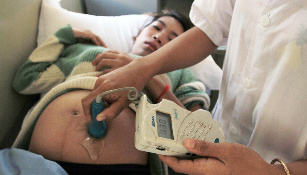
A pregnant woman undergoes a medical exam in Preah Vihear in Cambodia. A new study says preventative administration of azithromycin during labour could prevent maternal sepsis, as well as other infections after childbirth.

Speed read

  • Maternal sepsis is a leading cause of maternal deaths in lower-income countries
  • Trials show preventative dose of azithromycin cuts number of infections
  • Findings comes as UN report warns of setbacks in tackling maternal mortality

A single oral dose of the antibiotic azithromycin given during labour can dramatically reduce the risk of sepsis and death among women following childbirth, according to a large multi-country clinical trial, as a UN report warns of “stagnation” in maternal health.

Sepsis is a life-threatening condition which occurs when the body’s immune system overreacts to infections, damaging tissues and organs. It is a leading cause of maternal and newborn deaths worldwide, especially in low- and middle-income countries.

According to a report released by UN agencies today (23 February), 287,000 women died during pregnancy or childbirth in 2020 – the equivalent of one death every two minutes. It said that progress towards global goals on reducing maternal deaths had, at best, stalled in recent years.

“We are conducting studies on azithromycin resistance to make sure the intervention is also safe in addition to being effective in reducing infections”

Waldemar Carlo, co-director of the neonatology department at the University of Alabama’s Heersink School of Medicine

Researchers behind the azithromycin trials say preventative administration of the antibiotic during labour could not only stop hundreds of thousands of deaths from sepsis, but also ward off other infections after childbirth.

Waldemar Carlo, co-director of the neonatology department at the University of Alabama’s Heersink School of Medicine and one of the lead authors of the study, said the drug was chosen because “it covers many organisms that can be important and common pathogens in maternal infections”.

More than 29,000 pregnant women were enrolled in the trials in seven low- and middle-income countries: Bangladesh, the Democratic Republic of Congo, Guatemala, India, Kenya, Pakistan and Zambia. They were randomly assigned to receive either a two-gram dose of oral azithromycin or a placebo during labour.

Azithromycin, an inexpensive antibiotic effective against a broad range of bacteria, is known to reduce maternal infection when given intravenously during Caesarean delivery. Three smaller trials in the United States and Africa had also shown the potential to reduce maternal infections during vaginal births, says Carlo.

According to the study, published in the New England Journal of Medicine, only 1.6 per cent (227) of the women who received azithromycin developed sepsis or died within six weeks after delivery, compared to 2.4 per cent (344) of those who received the placebo.

Additionally, women who received azithromycin were less likely to develop endometritis, an infection of the lining of the womb, and other infections. They also had fewer hospital readmissions and unscheduled healthcare visits, compared to the placebo group.

“We are conducting studies on azithromycin resistance to make sure the intervention is also safe in addition to being effective in reducing infections,” Carlo added.

Administration of azithromycin did not reduce the risk of stillbirth, newborn sepsis, or newborn death, the research concluded.

The trial, called A-PLUS, was co-funded by the Bill & Melinda Gates Foundation and the Eunice Kennedy ShriverNational Institute of Child Health and Human Development (NICHD), part of the US National Institutes of Health. The NICHD’s Global Network for Women’s and Children’s Health Research conducted the study between September 2020 and August 2022.

According to the researchers, A-PLUS was originally designed to enroll up to 34,000 women. However, based on a recommendation from the study’s independent data and safety monitoring committee, it was stopped early due to the clear maternal benefit of azithromycin.

Steven Simpson, head of the Sepsis Alliance and a professor in the Pulmonary Clinical Care and Sleep Medicine division at the University of Kansas, US, believes the findings could be life-saving.

According to the Global Maternal Sepsis study, 15 women per 1,000 live births in low- and middle-income countries have an infection which results in or contributes to death or life-threatening complications during their hospitalisation, based on data from 2017.

“There are 140 million births per year. More than half occur in Asia,” Simpson tells SciDev.Net. He calculates that the 1.68 million sepsis deaths per year could be reduced to 1.12 million, going by the results of the trial. “Approximately 560,000 women could avoid sepsis or death annually in Asia alone, if every mother received this treatment,” he says.

“Giving children a better chance to have a mother, and especially at relatively low cost, should be an important global initiative,” he adds.

Talimogene Laherparepvec (T-VEC): An Intralesional Cancer Immunotherapy for Advanced Melanoma


Talimogene laherparepvec (T-VEC; IMLYGIC®, Amgen Inc.) is the first oncolytic viral immunotherapy to be approved for the local treatment of unresectable metastatic stage IIIB/C–IVM1a melanoma. Its direct intratumoral injection aim to trigger local and systemic immunologic responses leading to tumor cell lysis, followed by release of tumor-derived antigens and subsequent activation of tumor-specific effector T-cells. Its approval has fueled the interest to study its possible sinergy with other immunotherapeutics in preclinical models as well as in clinical contextes. In fact, it has been shown that intratumoral administration of this immunostimulatory agent successfully synergizes with immune checkpoint inhibitors. The objectives of this review are to resume the current state of the art of T-VEC treatment when used in monotherapy or in combination with immune checkpoint inhibitors, describing the strong rationale of its development, the adverse events of interest and the clinical outcome in selected patient’s populations.

Abstract

Direct intralesional injection of specific or even generic agents, has been proposed over the years as cancer immunotherapy, in order to treat cutaneous or subcutaneous metastasis. Such treatments usually induce an effective control of disease in injected lesions, but only occasionally were able to demonstrate a systemic abscopal effect on distant metastases. The usual availability of tissue for basic and translational research is a plus in utilizing this approach, which has been used in primis for the treatment of locally advanced melanoma. Melanoma is an immunogenic tumor that could often spread superficially causing in-transit metastasis and involving draining lymph nodes, being an interesting model to study new drugs with different modality of administration from normal available routes. Talimogene laherperepvec (T-VEC) is an injectable modified oncolytic herpes virus being developed for intratumoral injection, that produces granulocyte-macrophage colony-stimulating factor (GM-CSF) and enhances local and systemic antitumor immune responses. After infection, selected viral replication happens in tumor cells leading to tumor cell lysis and activating a specific T-cell driven immune response. For this reason, a probable synergistic effect with immune checkpoints inhibition have been described. Pre-clinical studies in melanoma confirmed that T-VEC preferentially infects melanoma cells and exerts its antitumor activity through directly mediating cell death and by augmenting local and even distant immune responses. T-VEC has been assessed in monotherapy in Phase II and III clinical trials demonstrating a tolerable side-effect profile, a promising efficacy in both injected and uninjected lesions, but a mild effect at a systemic level. In fact, despite improved local disease control and a trend toward superior overall survival in respect to the comparator GM-CSF (which was injected subcutaneously daily for two weeks), responses as a single agent therapy have been uncommon in patients with visceral metastases. For this reason, T-VEC is currently being evaluated in combinations with other immune checkpoint inhibitors such as ipilimumab and pembrolizumab, with interesting confirmation of activity even systemically.

Conclusions

The landscape of options for advanced melanoma is rapidly improving and progressing, with drugs and combinations able to significantly expand patients’ window of expectations. However, adverse events occurring during treatment can limit the dose intensity and duration of those treatments, thus consequently affecting their effectiveness. Moreover, despite their tremendous impact on outcomes, most of the patients still relapse and die of their disease, leading to the need of expanding the therapeutic armamentarium with new drugs, sequences or combinations. Furthermore, with the advent of new technologies, therapeutic options are becoming more and more multidisciplinary, being surgery often avoided thanks to the adoption of new approaches requiring radiotherapy or interventional radiology expertise in order, for example, to reach visceral and deep internal lesions for intralesional injection of drugs.

T-VEC is the first genetically modified herpes simplex virus-1-based oncolytic immunotherapy approved by FDA and EMA for the treatment of unresectable, cutaneous, subcutaneous and nodal lesions in patients with melanoma recurrent after initial surgery [72,73]. This intratumoral injectable drug is designed to preferentially replicate in tumors, produce GMCSF, and stimulate antitumor immune responses both locally and systemically. As extensively discussed in this review, it has shown efficacy in monotherapy and in combination with immune checkpoints inhibitors.

Efficacy has been demonstrated mainly on injected lesion, but also an abscopal systemic effect was evident on metastasis in distant organs. Like most of the immunotherapies effectiveness is not immediately translatable is target lesion reduction and, instead, it is worth noting that progressive disease before observing a response is common both in patients treated with T-VEC alone or in combination with checkpoint inhibitors. This pattern of pseudo-progression has been widely described and reinforces the importance of continuing treatment beyond progression in the event of appearance of a new lesion or limited increase in existing ones [55,74].

In advanced melanoma, the combination of oncolytic viruses has been tested in clinical trials using T-VEC together with systemic administration of a checkpoint inhibitor: ipilimumab or pembrolizumab. A Phase 2 trial of T-VEC in combination with ipilimumab met its primary endpoint, resulting in a significantly higher ORR without additional safety signals than ipilimumab (39% vs. 18%, p = 0.002). Another Phase Ib trial tested the association of T-VEC and pembrolizumab with a confirmed ORR of 67% with a CR rate of 43%. In the coupled translational study, this association was able to increase CD8+ T cells, while PD-L1 and IFN-γ upregulation were observed in tumors from responders.

The positive effect of T-VEC, as a monotherapy or in combination with checkpoint inhibitors, which was observed in both injected and uninjected (including visceral) melanoma lesions, indicated that a systemic antitumor immune response was triggered. These results suggest that T-VEC may improve the efficacy of checkpoint inhibitor immunotherapies by changing the tumor microenvironment and support the rationale that combining immunotherapies with complementary mechanisms of action may yield augmented antitumor responses.

Interestingly, adverse events, both in monotherapy and in combination with immune checkpoints inhibitors are mild and easily reversible, leading to a new efficient and well tolerated treatment opportunity in those melanoma patients with injectable lesion and low tumor burden.

Trials are ongoing to confirm clinical results on larger number of patients and in comparison with the best standards of care, in order to confirm this approach is able to achieve high efficacy with low toxicity. Furthermore, new generation clinical trials incorporate regular sampling of both peripheral blood and tumor tissue, allowing basic and translational research, which will give insight on the mechanisms regulating tumor versus T-cells balance in the microenvironment and will characterize the immune response exploring its correlations with clinical outcomes.

Triple-negative breast cancer: Could a cancer-killing virus clear up tumors?


Could a cancer-killing virus aid chemotherapy in eliminating triple-negative breast cancer tumors? Image credit: Drazen_/Getty Images.

  • Although triple-negative breast cancer is challenging to treat, some hope may be on the horizon thanks to a cancer-killing virus called TVEC (talimogene laherparepvec).
  • In a phase 2 clinical trial, 45.9% of patients who received TVEC injections directly to the tumor during chemotherapy had no signs of cancer after surgery.
  • The next step is to conduct a phase 3 clinical trial to confirm the efficacy of TVEC for triple-negative breast cancer in a larger study population.

Triple-negative breast cancer, which accounts for 15%Trusted Source of newly diagnosed breast cancer cases, is characterized by tumors that lack estrogen receptors, progesterone receptors, and human epidermal growth factor receptor 2 (HER2) receptors.

This means that the tumor cells do not respond to hormone therapy or drugs that target HER2.

Currently, the preferred treatment approach for triple-negative breast cancer is neoadjuvant chemotherapy, a type of chemotherapy administered before the main treatment for cancer, such as surgery or radiation therapy.

Doctors administer it to shrink the size of a tumor, making it easier to remove that tumor with surgery, or to make radiation therapy more effective.

Researchers at the Moffitt Cancer Center in Florida are seeking to improve the treatment of triple-negative breast cancer by combining neoadjuvant chemotherapy with the use of a cancer-killing virus — termed an “oncolytic virus” — called TVEC (talimogene laherparepvec).

In 2021, Dr. Hatem Soliman, a medical oncologist specializing in breast cancer, and his coworkers at Moffitt Cancer Center published the findings of a phase 1 study of TVEC combined with neoadjuvant chemotherapy in patients with triple-negative breast cancer. The results demonstrated the safety and feasibility of this approach.

Now, Dr. Soliman and his team have conducted a phase 2 clinical trial to further investigate this treatment. The results of the phase 2 trial have been published in Nature MedicineTrusted Source.

TVEC cooperates with chemotherapy

Some patients with triple-negative breast cancer respond well to chemotherapy and have no signs of cancer in their tissues after treatment. This is known as a “complete pathological response.”

These patients are unlikely to develop cancer again in the next 5 years. Other patients do not achieve a complete response with chemotherapy and are far more likely to develop cancer in the next 2–3 years.

Researchers at Moffitt hoped to find a way to achieve a pathological complete response in patients who may otherwise respond poorly to chemotherapy.

TVEC is already approved for the treatment of advanced melanomaTrusted Source thanks to its ability to kill cancer cells and activate the immune system.

Dr. Soliman and his team predicted that TVEC injections into triple-negative breast cancer tumors during neoadjuvant chemotherapy would result in a higher response rate to chemotherapy, as seen in melanoma.

When asked to explain what TVEC does to the body in patients with triple-negative breast cancer, Dr. Soliman told Medical News Today:

“Talimogene laherparepvec (TVEC) is an oncolytic [cancer-killing] virus that can preferentially infect cancer cells when injected into a tumor. The virus will cause tumor cells to burst open while activating the immune system to attack surrounding cancer cells. Using TVEC in triple-negative breast cancer tumors is a way to wake up the immune system against the tumor while cooperating with chemotherapy to better eradicate the tumors.”

The phase 2 trial

In the trial, 37 patients with stage 2 or 3 triple-negative breast cancer, aged between 27 and 66 years, received five intratumoral TVEC injections with neoadjuvant chemotherapy followed by surgery.

Out of 37 patients, 16 (45.9%) achieved a complete response, that is, they had no signs of cancer after surgery. Another eight patients had a small number of cancer cells left after treatment — known as a “near-complete response.”

Out of 37 patients, 33 (89%) remained cancer-free in the 2 years after treatment, and no recurrences occurred in the patients with a complete or near-complete response.

The side effects of treatment with TVEC and chemotherapy did not differ significantly from those expected of standard chemotherapy, except for higher levels of low-grade fevers, chills, headaches, and injection site pain.

How does it compare to other treatments?

In a recent clinical trial, researchers found that patients with early-stage triple-negative breast cancer who were given a combination of pembrolizumab — a drug that helps the individual’s immune system to fight cancer by preventing the cancer cells from hiding — and neoadjuvant chemotherapy had a higher rate of full recovery compared to those who received a placebo and neoadjuvant chemotherapy.

The results of this trial led to the Food and Drug Administration’s (FDA) approvalTrusted Source of pembrolizumab for high-risk early-stage triple-negative breast cancer.

Although this is a noteworthy development, immunotherapy with pembrolizumab or similar drugs may cause the immune system to attack healthy cells, resulting in several side effects.

Moffitt’s researchers hope that oncolytic viruses could improve the immune response against tumors without causing as many side effects as immunotherapy drugs.

Dr. Howard L. Kaufman, a surgical oncologist at Massachusetts General Hospital and lecturer at Harvard Medical School, told MNT:

“The pembrolizumab/ chemotherapy combination does have significant toxicity and the overall favorable safety profile of TVEC makes it a good agent for combination studies. The findings in the current study may also open the door for other locally delivered immunotherapy approaches as an important new area for clinical investigation into better treatments for patients with high-risk triple-negative breast cancer.”

At the moment, several oncolytic virusesTrusted Source are being studied as a potential treatment for cancer in clinical trials. A phase 1 trial is evaluating the use of a genetically modified smallpox virus, CF33-hNIS or VAXINIA, as a monotherapy or in combination with pembrolizumab to treat metastatic or advanced solid tumors.

Next steps

So far, phase 1 and phase 2 trials with TVEC have shown promising results. The next step is to conduct a phase 3 clinical trial to confirm the efficacy of TVEC for triple-negative breast cancer among hundreds of people.

“While the results of this study are very important for establishing a possible role for TVEC in combination with chemotherapy for neoadjuvant treatment of high-risk triple-negative breast cancer, a larger study is needed with longer follow-up to see the full impact of the therapeutic intervention on survival endpoints,” said Dr. Kaufman.

“Such studies are expensive to perform in terms of both time and money, but the current study provides a strong rationale for supporting such a future clinical trial,” he noted.

Dr. Soliman told MNT it would take 2–3 years for the phase 3 trial to be done and TVEC to become available to patients with triple-negative breast cancer.

Scientists manage to detect pancreatic and prostate cancer with a urine test


Scientists are working on new ways to detect cancers such as pancreatic and prostate cancer. xavierarnau/Getty Images

  • Pancreatic cancer accounted for 3.2%Trusted Source, and prostate cancer accounted for 14.2% of all new cancer cases in 2022 in the United States.
  • Pancreatic cancer, in particular, can be difficult to treat and has a much higher mortality rate than many forms of cancer, what may be due to the fact it is often diagnosed late.
  • Korean researchers recently devised a way to detect both pancreatic and prostate cancer with a urine test.
  • With pancreatic cancer on the rise in younger women, this technology may provide a route to earlier cancer detection and treatment.

Early detectionTrusted Source of cancer is very important to successful treatment and improving a patient’s prognosis. As such, researchers continue looking for ways to improve on detection of various forms of cancer.

A team led by researchers from the Surface & Nano Materials Division of the Korea Institute of Materials Science recently published a study in Biosensors and Bioelectronics showing a new way to detect pancreatic and prostate cancer using a urine test.

They say the test can have up to a 99% detection rate.

Pancreatic and prostate cancer

Pancreatic and prostate cancer affect millions of people in the U.S.. The American Cancer Society estimates that approximately 64,000 people will receive a pancreatic cancer diagnosisTrusted Source in 2023 and that around 288,000 people will receive a prostate cancer diagnosisTrusted Source in 2023.

Some signs and symptoms of pancreatic cancer include:

Unfortunately, pancreatic cancer is generally only detected in later stages, and the death rates with this form of cancer are high. The 5-year survival rate for pancreatic cancer is 12%.

Additionally, pancreatic cancer rates are increasing in the U.S. each year and are increasing at faster rates in younger women, particularly in Black women. A recent study showed that pancreatic cancer rates in Black women under age 55 rose 2.23% compared to Black men of the same age group.

The 5-year survival rate for prostate cancer is 97%. With the exception of skin cancer, prostate cancer is the most common cancer in men, and 1 in 8 men receive a diagnosis throughout their lifetime.

Not all men experience symptoms of prostate cancer, but doctors can detect it through routine screening. However, for men who do experience symptoms, some of those may include frequent urination and painful urination.

Current testing and screening for either pancreatic or prostate cancer can be rather invasive and sometimes expensive.

Some ways doctors test for pancreatic cancer include utilizing CT and MRI scans, and for prostate cancer testing, doctors use blood testing, transrectal ultrasounds, or rectal exams.

Creating a cancer-detecting urine test

Since biofluids can be instrumental in detecting or diagnosing certain conditions, the researchers in this study wanted to utilize these in a more advanced way—by using urine to detect cancer metabolites.

“Urine is a promising biofluid for disease diagnosis because most metabolic components are excreted via the urinary tract,” write the authors.

The researchers note that prior studies show that the metabolite compounds present in urine are different in urine of people without cancer versus the urine of people with cancer, but that previously, the only way to detect those compounds was through a “laborious” process.

By developing an enhanced Surface-enhanced Raman scattering (SERS) sensor, the scientists were able to detect “hot spots” in urine samples. These hot spots picked up and amplified certain molecular compounds that point toward cancer.

After creating a urine test strip and a handheld testing machine, the researchers obtained urine samples from a number of patients with pancreatic or prostate cancer to see how well the test functioned.

In addition to obtaining urine samples from 19 patients with pancreatic cancer and 39 patients with prostate cancer, the researchers collected samples from 40 cancer-free males and 20 cancer-free females.

The test was overall able to detect 99% of the cancerous samples.

Detecting cancer earlier?

“Since early diagnosis is the most important for incurable diseases such as cancer, we expect this technology to provide a new diagnostic method,” says study author and research lead Ho Sang Jung, who is in charge of the research.

Dr. Domenech Asbun, a hepatobiliary, pancreas, and foregut surgeon at Miami Cancer Institute, part of Baptist Health South Florida, spoke with Medical News Today about the study.

“The authors use an interesting combination of technology—including established complex physics and cutting-edge deep learning models—in attempts to solve a long-standing problem: how do we diagnose occult cancers in the general population before it’s too late?” he said.

“The findings in this study may provide an important step forward in finding practical ways to diagnose cancers that oftentimes grow undetected. If the results can be reproduced on a larger scale, these tests may allow for early treatment of cancers that are quite often deadly when undetected for too long.”
— Dr. Domenech Asbun

The future of this technology

Jung spoke with Medical News Today about the future of this research and said they are expanding on it by testing more subjects.

“The number of patients tested using our system is continuously growing. Our team is trying to get as many clinical samples as possible to build more general deep-learning models with better accuracy,” said Jung.

Jung also said they want to be able to test for more than just two types of cancer.

“The next step in this research is broadening applicable cancer types. Now, our team just finished testing clinical samples of pancreatic cancer, prostate cancer, lung cancer, and colorectal cancer simultaneously, and our team is expected to submit an article this year.”

In addition to expanding on cancer types, Jung says they envision at-home testing kits in the future.

At home tests may be within reach

“Because the developed diagnostic method uses urine, it is noninvasive thus it would be more beneficial in terms of usability when it is applied for self-testing or rapid-kit-testing at home, pharmacy, or any places where they can provide some clinical information.”
— Ho Sang Jung, study author