Doxycycline continues to show promise for STI prevention, but not in cisgender women


Studies continue to show that taking doxycycline after having unprotected sex can prevent STIs in transgender women and men who have sex with men. What has been unclear is whether it also works for cisgender women.

On Monday, researchers at the Conference on Retroviruses and Opportunistic Infections presented the first major evidence that it may not.

IDN0223Stewart_Graphic_01
Data derived from Stewart J, et al. Abstract 121. Presented at: Conference on Retroviruses and Opportunistic Infections; Feb. 19-22, 2023; Seattle.

Specifically, a randomized trial conducted in Kenya found that an intervention of doxycycline postexposure prophylaxis — called doxy-PEP — did not significantly reduce STIs among cisgender women compared with testing and treatment alone, according to findings presented by Jenell Stewart, DO, MPH, an infectious diseases physician and researcher at Hennepin Healthcare in Minneapolis.

In a press conference, Stewart suggested three possible reasons that the study failed to show a significant benefit for doxy-PEP in cisgender women: “adherence, anatomy and resistance.”

According to Stewart and colleagues, the women in the study reported taking doxycycline “at least as many days they had sex” 78% of the time.

“They told us in weekly text message surveys that there was a high rate of adherence [to] doxycycline PEP, but they also told us that it was imperfect,” Stewart said.

It is unclear “the extent to which perfect adherence is needed to prevent STIs in the endocervix, or the opening of the uterus,” Stewart explained. She noted that the study did not uncover any drug-resistant chlamydia, but that every gonorrhea sample they tested displayed high levels of resistance to tetracycline, the class of antibiotics doxycycline belongs to.

The researchers enrolled nearly 450 women in the study and randomly assigned them in equal numbers to take either a single 200 mg dose of doxycycline within 72 hours of having unprotected sex or to receive standard of care, which included quarterly screening and treatment for STIs, but not doxy-PEP. More than one-third of the women reported transactional sex.

At follow-up, Stewart and colleagues diagnosed 50 STIs in the doxy-PEP arm and 59 STIs in the standard of care arm (RR = 0.88; 95% CI, 0.60-1.29). The results were similar when they analyzed each STI separately and grouped participants by age, contraceptive use, transactional sex, and whether they had an STI at baseline.

MSM, transgender women still benefit

In another randomized trial presented at the meeting, Jean-Michel Molina, MD, PhD, a professor of infectious diseases at the University of Paris, and colleagues found that MSM were 84% less likely to contract chlamydia or syphilis and about half as likely to contract gonorrhea if they received doxy-PEP within 3 days of having condomless sex, compared with study participants who did not.

The study, which enrolled more than 500 MSM who were already taking PrEP for HIV prevention and had at least one STI in the past year, was stopped early on the advice of a data and safety and monitoring board because of how effective the intervention was.

Molina and colleagues had previously shown that doxy-PEP halved the incidence of STIs among MSM who were being followed as part of another study on HIV PrEP.

In the new trial, Molina and colleagues also gave half of the participants the meningococcal B vaccine Bexsero (GSK) to test its effectiveness at preventing gonorrhea and found that it reduced the incidence of gonorrhea by around 50%.

In 2017, researchers in New Zealand were the first to report that a meningitis B vaccine might prevent gonorrhea, which has grown increasingly resistant to antibiotics in recent years. Bexsero’s potential as a gonorrhea vaccine is being tested in a $10 million NIH study at the University of Alabama at Birmingham.

For now, Molina said, a reduction in gonorrhea would be an “added benefit” of the vaccine. Asked whether the data from his study were enough to begin using the vaccine off-label for gonorrhea, he did not say yes or no.

“It would be nice to reduce the overall burden of gonorrhea infection [and to] confirm these data, but at this point, it would probably be an individual decision to see whether or not you get this vaccine,” Molina said. “What we don’t know is for how long you are going to be protected [and] whether or not you would need a booster injection.”

Modest effect on resistance

Last summer, after Annie F. Luetkemeyer, MD, professor of medicine at the University of California, San Francisco, presented findings at the International AIDS Conference showing that doxy-PEP reduced STIs by more than 60% among transgender women and MSM, the CDC released interim clinical “considerations” for using doxy-PEP as an STI prevention tool for transgender women and MSM — something the agency acknowledged some clinicians and patients were already doing — but it has not published detailed, formal guidance yet, nor has it addressed using doxy-PEP in other populations.

One of the outstanding questions was whether on-demand doxycycline use would promote resistance in STIs — especially gonorrhea — or other common bacteria found on humans, like Staphylococcus aureus.

On Monday, Luetkemeyer had an answer in the form of 12-month findings from the trial she presented last summer: The effect of doxy-PEP on resistance was modest, unlikely to be clinically relevant and must be considered in the context of how effective it was at reducing STIs in the study.

The findings did suggest, however, that doxy-PEP may be less effective at preventing infection with tetracycline-resistant gonorrhea.

“I don’t think this means that doxy-PEP drove tetracycline resistance” in the trial, Luetkemeyer said.

Most efficient strategy

In a fourth study presented Monday at CROI, Michael Traeger, PhD, MS, a research fellow at Harvard Medical School, and colleagues assessed 10 different strategies for prescribing doxy-PEP for STIs and found that the most efficient strategy was to prescribe it based on a patient’s STI history rather than their HIV status or use of PrEP.

The study enrolled more than 10,000 MSM, transgender women and non-binary people assigned male at birth between 2015 and 2020 who had taken at least two STI tests at an LGBTQ health clinic in Boston.

For each of the 10 potential prescribing strategies, Traeger and colleagues estimated the proportion of patients prescribed doxy-PEP, the number of STIs averted and the number of patients who would need to be treated each year to prevent one STI.

The researchers found that prescribing doxy-PEP to all patients prevented 70% of STIs but the number of patients who would need to be treated to prevent one infection was 3.7.

Prescribing it only after an STI diagnosis reduced the proportion of patients receiving the antibiotic from 100% to 41% and prevented 42% of STIs, but the most efficient strategy was prescribing it only to patients with at least two concurrent STIs. Only 1.2 patients would need to be treated to prevent one infection using this strategy, according to Traeger and colleagues.

Providing an outside perspective, CROI vice chair Landon Myer, MD, head of the school of public health and family medicine at the University of Cape Town in South Africa, said doxy-PEP “really has the potential to change public health practice in the immediate future.”

“It’s not for everyone,” Luetkemeyer added, noting that the results of the Kenya study mean there is no intervention for cisgender women yet. “I would say the place to start is people who have really demonstrated an increased risk” — including MSM with a recent STI who are engaging in condomless sex, and people with recurrent STIs.

“That’s the population you really want to focus on. I don’t think this needs to be for all MSM, nor would all MSM want to take it. You really want to focus on past STIs or risk factors for multiple STIs,” she said. “If someone has multiple partners and condomless sex, it would be a discussion I would have with them.”
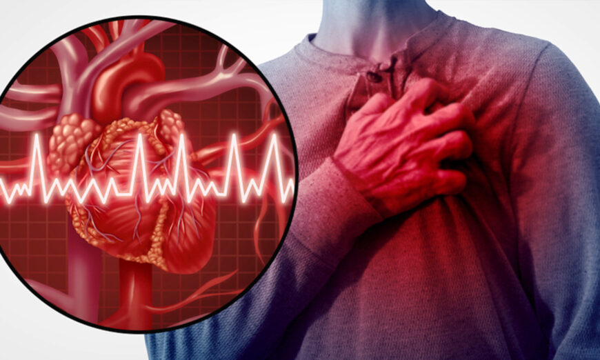
Panic Attack or Heart Attack? How to Know the Difference


Panic Attack or Heart Attack? How to Know the Difference

25% of emergency room patients presenting with chest pain met the criteria for something else entirely, yet attending emergency department cardiologists failed to recognize it 98 times out of 100.

According to the U.S. Centers for Disease Control and Prevention, nearly 805,000 Americans have a heart attack each year, and 605,000 are first heart attacks. Knowing the risk factors, symptoms and how to take early action will increase your chances of survival.

However, what may look and feel like an apparent heart attack may actually be a panic attack, and according to researchers, the cost of misdiagnosing noncardiac chest pain is high. “It is important for physicians to be able to recognize panic attacks and to distinguish them from cardiac disease, thus avoiding unnecessary use of health care resources,” one report states.

An investigation published in 1996 found that 25% of emergency room patients presenting with chest pain met the DSM-III-R criteria for panic disorder, yet attending emergency department cardiologists failed to recognize patients having a panic attack 98% of the time. As noted by the authors:

“Panic disorder is a significantly distressful condition highly prevalent in ED [emergency department] chest pain patients that is rarely recognized by physicians. Nonrecognition may lead to mismanagement of a significant group of distressed patients with or without coronary artery disease.”

So, just how do you tell the two apart? Before we get into those details, let’s take a look at the common signs and symptoms associated with each.

Symptoms of Heart Attack

When a heart attack starts, blood flow to your heart has suddenly become blocked and the muscle can’t get oxygen. If not treated quickly, the muscle fails to pump and begins to die. While often a result of coronary heart disease, a heart attack can also be caused by a blood clot blocking an artery. Some of the most common symptoms of a heart attack include:

  • Chest pain or discomfort
  • Upper body discomfort
  • Shortness of breath
  • Breaking out in a cold sweat
  • Nausea
  • Sudden dizziness
  • Feeling unusually tired
  • Lightheadedness

Symptoms of Panic Attack

A panic attack typically comes on abruptly, producing intense fear and a sense of impending doom or even death that is typically severely disproportionate to the situation at hand. Common symptoms include:

  • Hyperventilation
  • Chest pain
  • Heart palpitations
  • Trembling
  • Sweating; hot or cold flashes
  • Nausea
  • Dizziness or lightheadedness
  • Numbness and/or tingling sensations

Panic attacks tend to peak within 10 minutes, and most subside within 30 minutes. Few last more than one hour. It’s not uncommon for people to seek medical help, thinking they’re having a heart attack or are dying, when panic attacks first set in and they’re unfamiliar with the symptoms.

How to Tell Them Apart

While it can be very difficult to tell a panic attack from a heart attack, some generalizations can be made that can help tell them apart.

  • Pain onset — The chest pain associated with a heart attack will typically start as a feeling of pressure, fullness or aching that escalates, reaching maximum severity after a few minutes, whereas the pain associated with a panic attack tends to be sharp and stabbing in the center of the chest, typically lasting only five to 10 seconds.
  • Pain location — The location of the pain also tends to differ between the two. Whereas panic-associated pain is localized in one small area of the chest, heart attack symptoms typically include pain or discomfort that radiates from the chest into other areas, such as one or both arms, abdomen, back, shoulders, neck, throat or jaw.

Don’t Ignore Your Symptoms

When in doubt, seek immediate medical attention. It’s better to be safe than sorry, as sudden death is the most common symptom of a heart attack. As noted by Dr. Sam Torbati, medical director of the Ruth and Harry Roman Emergency Department in an interview for Cedars-Sinai Medical Center:

“Unfortunately, there is great crossover between the symptoms of panic attack and heart attack, making it very challenging to discern between the two without a physician assessment and testing, such as an EKG.

Common symptoms that may affect patients with either a panic or heart attack include chest pain, shortness of breath, dizziness, sweating, passing out, tingling, or a sensation of impending doom.

These shared symptoms of heart and panic attack may also be caused by other serious conditions such as blood clots, lung infection or collapse, or tear in the large vessels of the chest for patients with certain pre-existing risk factors. So when in doubt, seek immediate medical attention …

The best predictor as to whether symptoms are due to panic versus heart attack is the patient’s age and previous history of panic attacks … Patients should immediately go to the ER if they have new onset chest pain (tightness, squeezing, heaviness), shortness of breath, sweating, lightheadedness, pain that radiates to the jaw or arm, or a ripping sensation in their chest or back.

Heart attacks tend to occur in middle-aged people and older age groups, so the older the person is, the lower a threshold they should have for coming to the ER right away.

Patients with pre-existing coronary artery disease and those with risk factors associated with coronary artery disease should also be evaluated immediately, including those with hypertension, diabetes, obesity, high cholesterol, or a history of smoking.”

Possible Connection Between Panic Disorder and CAD

It’s also worth getting your symptoms checked out even if you’re certain they’re due to a panic attack. Some research suggests there may in fact be a connection between panic disorder and coronary artery disease, although the exact relationship is still unclear. According to a 2008 review in The Primary Care Companion to the Journal of Clinical Psychiatry:

“There are several reasons to consider that a relationship between panic disorder and coronary artery disease (CAD) might exist. First, panic disorder has been linked to other forms of cardiac disease.

Second, the most likely source of the chest pain during panic attacks is ischemia. Finally, there is evidence that panic disorder may be associated with cardiovascular risk factors, such as hypertension, hyperlipidemia, and smoking.

Panic disorder is associated with several cardiac abnormalities. In addition to patients with panic disorder having elevated standing heart rates, 10% have an arrhythmia.

Panic disorder is associated with increased left ventricular mass and diameter, and patients with panic disorder have poorer cardiovascular fitness as demonstrated by lower maximum oxygen consumption and decreased exercise tolerance …

Case reports have linked panic disorder to a descending aortic aneurysm and pulmonary hypertension secondary to an atrial septal defect with pulmonic valve disease. However, the strongest association is between panic disorder and mitral valve prolapse (MVP) … but MVP is not likely to be the source of chest pain.

In addition, the significance of the panic-MVP relationship is unclear … Indirect linkages via autonomic vulnerability or dysfunction have … been proposed. However, the most likely explanation is that the decreased left ventricular volume due to the tachycardia seen in panic disorder produces the MVP.”

The review cites evidence suggesting ischemia is the cause of the chest pain felt during a panic attack, and researchers have found there’s an association between panic attacks and ischemic and nonischemic chest pain alike. According to the authors, “Myocardial ischemia could cause panic attacks via increased catecholamines or cerebral carbon dioxide levels secondary to lactate.”

What’s more, when looking at a large managed care database, researchers found an association between panic disorder and coronary heart disease and this association remained even after controlling for covariates.

Overall, patients with panic disorder were between 80% and 91% more likely to also have coronary heart disease. Patients diagnosed with both panic disorder and depression were, on average, 260% more likely to develop coronary heart disease than patients without those mental health problems.

On the flip side, research published in 2017 also points out that “Anxiety and its associated disorders are common in patients with cardiovascular disease and may significantly influence cardiac health.” According to this paper:

“Both physiologic (autonomic dysfunction, inflammation, endothelial dysfunction, changes in platelet aggregation) and health behavior mechanisms may help to explain the relationships between anxiety disorders and cardiovascular disease.”

Many Women Mistake Heart Attack Symptoms for Anxiety

It’s also important to realize that the symptoms of heart attack can vary from person to person and some may have very few symptoms, especially women. Importantly, research shows women are less likely to report chest pain when having a heart attack.

They’re also less likely to suspect their discomfort is related to a heart problem. Compared to just 11.8% of men, 20.9% of women attributed their chest pain to stress or anxiety. Women also tend to describe their pain differently. They’re more likely to use terms such as “pressure,” “tightness” or “discomfort” in the chest rather than referring to it as “chest pain.”

Doctors are also more likely to dismiss women’s complaints of chest pain as being noncardiac in nature. Overall, 53% of female heart attack patients reported that their doctor did not think their symptoms were heart-related, compared to 37% of male heart attack patients.

Approximately 29.5% of women had actually sought medical help before being hospitalized with a heart attack, compared to just 22.1% of men. What these findings suggest is that women and their doctors tend to misdiagnose or dismiss symptoms of heart attack, placing women at a higher risk of death than men. As noted by the authors:

“The presentation of AMI [acute myocardial infarction] symptoms was similar for young women and men, with chest pain as the predominant symptom for both sexes.

Women presented with a greater number of additional non-chest pain symptoms regardless of the presence of chest pain, and both women and their health care providers were less likely to attribute their prodromal symptoms to heart disease in comparison with men.”

Unfortunately, the absence of chest discomfort is a strong predictor of diagnosis and treatment delays. For this reason, it’s important to remember there are many other symptoms that might indicate a heart attack in progress, including the following:

  • Anxiety attack
  • Back pain
  • Heartburn
  • Hot flashes
  • Extreme fatigue
  • Feeling electric shocks down on the left side of your body
  • Numbness and stiffness in the left arm and neck
  • Feeling like you have a large pill stuck in your throat

Breathe Right to Quell Panic Attacks

When it comes to panic attacks, familiarizing yourself with the function of your fight-or-flight response can be helpful to guide you toward self-help strategies that work for your unique situation.

For example, contrary to popular belief, taking deep breaths can actually worsen a panic attack, as explained by Buteyko Breathing expert Patrick McKeown. A breathing exercise that can help quell anxiety and panic attacks is summarized below.

This sequence helps retain and gently accumulate carbon dioxide (CO2), leading to calmer breathing and reduced anxiety. In other words, the urge to breathe will decline as you go into a more relaxed state.

  • Take a small breath into your nose, a small breath out; hold your nose for five seconds in order to hold your breath, and then release to resume breathing.
  • Breathe normally for 10 seconds.
  • Repeat the sequence several more times: small breath in through your nose, small breath out; hold your breath for five seconds, then let go and breathe normally for 10 seconds.

McKeown has also written a book specifically aimed at the treatment of anxiety through optimal breathing, called “Anxiety Free: Stop Worrying and Quieten Your Mind — Featuring the Buteyko Breathing Method and Mindfulness,” which can be found on Amazon.com.

In addition to the book, ButeykoClinic.com also offers a one-hour online course and an audio version of the book, along with accompanying videos.

EFT — A Long-Term Solution to Anxiety

Energy psychology techniques such as the Emotional Freedom Techniques (EFT) can also be very effective for anxiety and panic attacks. EFT is akin to acupuncture, which is based on the concept that a vital energy flows through your body along invisible pathways known as meridians.

EFT stimulates different energy meridian points in your body by tapping them with your fingertips, while simultaneously using custom-made verbal affirmations. This can be done alone or under the supervision of a qualified therapist. By doing so, you reprogram the way your body responds to emotional stressors.

EFT is particularly powerful for treating stress and anxiety because it specifically targets your amygdala and hippocampus, which are the parts of your brain that help you decide whether or not something is a threat. EFT has also been scientifically shown to lower cortisol levels, which are elevated when you’re stressed or anxious.

In the video above, EFT therapist Julie Schiffman demonstrates how to tap for panic attacks. Please keep in mind that while anyone can learn to do EFT at home, self-treatment for serious issues like persistent anxiety is not recommended. For serious or complex issues, you need someone to guide you through the process. That said, the more you tap, the more skilled you’ll become.

Heart Attack Prevention

As for heart attacks, your best course of action is to take proactive measures to prevent them. According to a 2015 study, more than 70% of heart attacks could be prevented by implementing:

  • A healthy diet
  • Normal body mass index
  • Getting at least 2.5 hours of exercise each week and watching television seven or fewer hours per week
  • Avoiding smoking
  • Limiting alcohol to one drink or less per day

To this I would add maintaining a healthy iron level is important for your heart, as various studies show that both iron deficiency and iron overload can be a significant risk factor for heart attack.

For example, a Scandinavian study found elevated ferritin levels raised men’s risk of heart attack two- to threefold. Another found elevated ferritin raised the risk of a fatal heart attack by 218% in men, while women with high levels were 5.53 times more likely to have a fatal heart attack.

As discussed in “Why Hard Water Decreases Heart Attacks,” magnesium insufficiency has also been implicated in heart attacks, so you want to make sure you’re getting enough magnesium from your diet and/or supplements. In “Could You Have a Heart Attack and Not Know It?” I also review some of the underlying issues that cause heart attacks, and additional steps you can take to lower your risk.

The safety and relative effectiveness of non-psychoactive cannabinoid formulations for the improvement of sleep: a double-blinded, randomized controlled trial


ABSTRACT

The objective of this randomized, double-blinded controlled trial was to evaluate the safety and relative effects of different formulations containing Cannabidiol (CBD) and melatonin, with and without the addition of minor cannabinoids, on sleep. Participants (N=1,793 adults experiencing symptoms of sleep disturbance) were assigned to receive a 4-week supply of 1 of 6 products (all capsules) containing either 15mg CBD or 5mg melatonin, alone or in combination with minor cannabinoids. Sleep disturbance was assessed using Patient-Reported Outcomes Measurement Information System (PROMIS™) Sleep Disturbance SF 8A, administered via weekly online surveys. All formulations exhibited a favorable safety profile (12% of participants reported a side effect and none were severe) and led to significant improvements in sleep disturbance (p<0.001 in within-group comparisons). Most participants (56% to 75%) across all formulations experienced a clinically important improvement in their sleep quality. There were no significant differences in effect, however, between 15mg CBD isolate and formulations containing 15mg CBD and 15mg Cannabinol (CBN), alone or in combination with 5 mg Cannabichromene (CBC). There were also no significant differences in effect between 15mg CBD isolate and formulations containing 5 mg melatonin, alone or in combination with 15mg CBD and 15mg CBN. Our findings suggest that chronic use of a low dose of CBD is safe and could improve sleep quality, though these effects do not exceed that of 5 mg melatonin. Moreover, the addition of low doses of CBN and CBC may not improve the effect of formulations containing CBD or melatonin isolate.

INTRODUCTION

Approximately one third of American adults do not get enough sleep each night.1 Poor sleep can have a profound impact on a person’s quality of life; it can hinder cognitive functioning2 and lead to depression,3 reduced productivity,4 cardiovascular disease,5 and increased healthcare utilization.4 There is strong clinical evidence in support of pharmacologic interventions for the treatment of insomnia (difficulty getting to sleep or staying asleep6) and other sleep disorders, in particular for GABAA receptor agonists, such as benzodiazepines.7 Concerns remain, however, over their many side effects, including ‘hangover effects’, cognitive impairment, abuse, and the considerable risk of dependance.8 Consequently, there is a prevailing need to evaluate safer forms of therapeutic treatment for the improvement of sleep.

Many patients turn to complementary and alternative medicines (CAM) for the treatment of insomnia and other sleep disorders.9 Melatonin is among the most commonly used and well-studied CAM treatments for sleep,10 and clinical evidence supports its efficacy for the improvement of sleep quality, particularly for those experiencing jet lag and delayed sleep-wake phase disorder.1114 Moreover, melatonin exhibits a favorable safety profile and does not demonstrate dependence even when administered at high doses.15

Cannabis preparations have also begun to gain attention for their potential therapeutic effects for the treatment of insomnia and other sleep disorders.16 To date, the preponderance of clinical research on Cannabis and sleep has focused on Δ9-tetrahydrocannabinol (Δ9-THC), the major active constituent of Cannabis sativa.17 Yet use of the non-psychoactive cannabinoid Cannabidiol (CBD) has proliferated in the US, with many new users seeking relief for sleep difficulties.18 Preclinical research has demonstrated that CBD possesses anxiolytic, anti-inflammatory, and analgesic properties,19 which could aid in the improvement of sleep. Evidence from retrospective and prospective observational studies also suggest that the clinical administration of cannabinoids could improve sleep and other related health issues such as pain and anxiety.2022

Clinical research assessing the use of CBD for insomnia and other sleep disorders remains limited, though some small clinical studies have found support for the hypothesis that CBD may improve sleep. In a study of 15 individuals with insomnia, those who received 160 mg CBD reported sleeping longer than those who received placebo.23 Another study of 33 individuals with Parkinson’s Disease revealed that 300 mg of CBD per day led to a transient improvement in sleep quality relative to placebo.24 In small experimental studies, fixed doses of 300 mg, 400 mg and 600 mg of CBD were also found to induce self-reported sedative effects relative to placebo in healthy adults (11 adults, 300 and 600 mg25; 10 males, 400 mg26). Importantly, clinical evidence of CBD also indicates that the cannabinoid has a favorable safety profile,27,28 even when taken at doses as high as 1200 mg daily for up to 4 weeks,29 supporting the exploration of CBD as a potentially safer therapeutic option for the improvement of sleep.

Despite the limited clinical evidence, marketing claims regarding the effectiveness of CBD for sleep abound.30,31 Many manufacturers have also touted the superiority of their CBD products relative to melatonin32,33 though, to date, no clinical study has directly compared the effects of these compounds on sleep. Manufacturers have also combined CBD with melatonin and other minor cannabinoids, claiming that these additions could enhance the effect of CBD or melatonin alone.34 These claims, too, are unfounded. No large scale randomized clinical trial has evaluated whether CBD could impact the effects of melatonin on sleep (or vice versa). No clinical trials have also evaluated whether the addition of minor cannabinoids, such as Cannabichrome (CBC) and Cannabinol (CBN), could contribute to the therapeutic effectiveness of CBD for sleep improvement. CBN, in particular, has gained prominence as a sleep aid additive,35 though the literature is almost entirely devoid of clinical research supporting its effect on sleep quality.36 Multi-arm studies allowing for direct comparisons across cannabinoid and melatonin formulations could provide critical information on the main and interactive effects of these compounds on sleep.

Notably, few clinical trials of CBD have also tested the effect of daily usage of CBD at the lower doses commonly found within commercially available products. Most clinical trials of CBD evaluate doses ranging from 300 to 1500 mg CBD per day,3739 while commercial products’ dosage generally range from 5 to 100 mg CBD per day. Clinical research on CBD has suggested a bell-shaped dose response curve wherein an intermediate dose of CBD exhibits a greater anxiolytic effect than a very low or high dose,40,41 though more clinical studies are needed evaluate the therapeutic benefits of chronic CBD use for sleep at dose ranges reflecting that of commercial products.

This study sought to address these gaps in the literature by investigating the effect of chronic use (daily use over 4 weeks) of low dose CBD and melatonin formulations, alone and in combination with certain minor cannabinoids, on sleep quality. The primary objective of the study was to compare the safety and effects of CBD isolate to CBD combination formulations (i.e., formulations containing CBD and minor cannabinoids, with and without melatonin) to determine if the addition of melatonin and these minor cannabinoids confer any therapeutic benefit to a formulation containing CBD. As a secondary aim, we also sought to determine if the addition of CBD and CBN confer any therapeutic benefit to a formulation containing melatonin.

DISCUSSION

In this randomized, double-blinded controlled trial to evaluate the effects of different orally ingested cannabinoid and melatonin formulations on sleep disturbance, we observed that a 15mg of CBD reduced self-reported sleep disturbance over the course of 4 weeks. The addition of minor cannabinoids (15mg CBN, alone or in combination with 5 mg CBC) did not impact the therapeutic effects of 15 mg CBD. Moreover, we found no evidence of a difference in effect on overall sleep disturbance score between 15mg CBD isolate and formulations containing 5 mg melatonin, alone or in combination with 15mg CBD and 15mg CBN. However, when examining changes in each PROMIS Sleep Disturbance 8a scale question score separately, we observed that those taking the 5mg melatonin in combination with 15mg CBD and 15mg CBN reported greater improvements in the restless and refreshing aspects of their sleep relative to those taking CBD isolate, though changes in self-reported sleep quality, sleep satisfaction, and in difficulties and worries over falling asleep did not vary between any formulation relative to CBD isolate. Secondary analyses also revealed that the addition of 15mg CBD and 15mg CBN did not significantly impact the therapeutic effects of a formulation containing 5mg melatonin on overall sleep disturbance score. Notably, all study arms led to significant improvements in sleep disturbance and exhibited favorable safety profiles.

Few clinical studies have examined the effect of CBD, with or without the addition of minor cannabinoids, for the improvement of sleep. This study is among the first to evaluate the safety and effectiveness of CBD dose ranges and formulations commonly found within commercially available CBD products. Our results demonstrate that the relatively lower doses of CBD found within these orally ingested products may be safe for chronic use and sufficient to produce significant improvements in symptoms of sleep disturbance. These effects, however, do not exceed that of 5 mg melatonin. Moreover, we found no evidence that the low doses of CBN and CBC can improve the effect of formulations containing low doses of CBD or melatonin isolate.

Our findings regarding the lack of additional therapeutic effect from the addition of CBN are noteworthy as cannabis product manufacturers have recently begun to tout the sleep-inducing effects of CBN,44 though preclinical and clinical research in support of these claims is scarce and outdated.36 Indeed, our study represents the first clinical trial to evaluate the use of CBN for sleep using validated sleep measures. Our findings suggest that 15mg of CBN may confer little added benefit to a sleep aid product. We note, however, that our findings reflect a relatively lower dosage of orally ingested CBN and may not be generalizable to higher dosages or other modes of administration of the cannabinoid.

In our secondary analyses, we did not find evidence that the addition of CBC to formulations containing CBD and CBN will impact their therapeutic effect on sleep quality. We did, however, observe substantially higher likelihood of experiencing a MCID among those in the CBD combination arm containing CBC (72%) compared to the arms containing just CBD and CBN (56% and 60% in the Full and Isolate spectrum CBD combination arms, respectively), suggesting CBC might impart some effect on sleep improvement. However, as this was not a planned comparison for the current study, and as the omnibus test between the CBD combination arms did not reach statistical significance, we are unable to fully interpret the meaning of this trend. Thus, further studies are needed to thoroughly characterize the impacts of CBC on sleep.

Preclinical research suggests that certain cannabinoids and other components of the Cannabis plant could work synergistically, stimulating a greater effect than that if CBD or Δ9-THC were examined in isolation – a phenomenon known as the ‘entourage effect’.45 As we observed no difference in effect between the CBD isolate and cannabinoid combination formulations, we found no evidence of an entourage effect with CBD, CBC, and CBN in these trial products. However, these findings may only be generalized to the specific dosages of these cannabinoids in this sample. We cannot exclude, from the present evidence, the possibility that higher concentrations of these minor cannabinoids could modulate the effects of CBD.

We observed that participants assigned to arms without melatonin averaged a higher daily capsule intake compared to participants taking melatonin-containing capsules. Importantly, we did not detect an increase in safety concerns in arms with a higher average daily intake. Although not statistically significant, this trend may suggest that without melatonin, higher doses of cannabinoids may be necessary to induce the desired effects on sleep quality. Moreover, it is possible that melatonin capsule dose was more optimally calibrated relative to the doses of cannabinoids.

Previous clinical research on melatonin for insomnia and other common sleep disorders suggests that its effects are modest and inferior to prescription medications.46 As we did not find any significant differences between CBD isolate and the melatonin or CBD combination formulations, such conclusions regarding modest relative effects could be extended to the formulations in this sample. Nonetheless, as melatonin and CBD both demonstrate a highly favorable safety and tolerability profile, these alternative therapies could still play a role in the treatment of common sleep disorders, especially given the harmful side effects of common pharmacological treatments for these disorders.8

This study has several limitations. First, about 28% of participants did not complete any follow-up surveys and were therefore excluded from the study. While our overall attrition levels still fell below anticipated attrition levels of 45% and the study was adequately powered to detect significant sleep changes, differential loss to follow-up could induce post-randomization confounding.47 As those who were excluded did not fill out any surveys beyond the screener, we were limited in our ability to evaluate characteristics of those excluded and assess potential imbalances across study arms. We were also unable to run a sensitivity analysis including the excluded individuals as they did not provide any PROMIS Sleep Disturbance 8a scores and imputing their data would not be appropriate as their observations were determined to be missing not at random (MNAR; as no study period surveys were completed by these individuals, the missingness is thus dependent on unobserved data). Nonetheless, we did not find any significant differences in the percentage of excluded individuals between study arms, and there were no significant differences in baseline demographic or health characteristics between study arms in the final sample, indicating that balance was maintained across study arms despite changes to the study sample post-randomization.

Additionally, as there was no placebo control within this study, we cannot determine if and how much the observed effects may be due to participant expectations/placebo. In previous clinical research, melatonin has been shown to have modest effects on sleep relative to placebo,48 though clinical evidence of CBD’s effect relative to placebo remains limited, albeit promising.2325 Notably, previous clinical research suggests that placebo response could play a major role in the effect of CBD on stress and anxiety,49 though the impact of this response has yet to be explored for CBD and sleep. Further placebo-controlled studies are needed to determine the therapeutic effects of CBD for sleep.

We see this data as “real world” data, as it was collected from a population that was using the products in a manner and setting like that of real consumers of these products. Without exhaustive eligibility criteria and intensive monitoring, the missingness and heterogeneity of the data may be greater than that of traditional clinical trials. However, many traditional clinical trials have limited external validity because the characteristics and behaviors of participants may not reflect those of real-world users. As such, real world studies have unique value in their ability to provide complementary evidence to support clinical trial designs and help guide regulatory and clinical decisions.50,51

Our study is the first randomized, blinded, controlled trial which compares the effects of CBD and melatonin on sleep, and further investigates therapeutic benefits of combining CBD isolate with melatonin or minor cannabinoids. Our findings represent an essential scientific advancement towards thoroughly characterizing and contrasting the effects of commonly used non-prescription sleep disorder treatments.

How Magnesium Can Help You Sleep


Magnesium is a mineral that is essential for  processes throughout the body Trusted Source Medline Plus MedlinePlus is an online health information resource for patients and their families and friends. View Source , including communication between cells in the nervous system. Sleep is largely controlled by the nervous system, and experts believe that nutrients like magnesium may play a role in sleep health. However, the exact relationship between magnesium and sleep is still being studied.

Despite the unclear relationship between magnesium and sleep, there have been encouraging studies showing that magnesium supplements may improve sleep qualitysleep duration, and benefit people with sleep disorders like insomnia and restless legs syndrome. Future research may clarify the effects of magnesium on sleep as well as the role of magnesium supplements in promoting sleep health.

What is Magnesium?

Magnesium is one of several  electrolytes Trusted Source National Library of Medicine, Biotech Information The National Center for Biotechnology Information advances science and health by providing access to biomedical and genomic information. View Source  in the body. Electrolytes are minerals that the body needs to function properly. Magnesium plays  essential roles Trusted Source National Institutes of Health (NIH) The NIH, a part of the U.S. Department of Health and Human Services, is the nation’s medical research agency — making important discoveries that improve health and save lives. View Source  in the production of proteins, bone, and DNA. Magnesium is also important for maintaining blood sugar, blood pressure, and regulating activity in the muscles, nerves, and cardiovascular system.

Magnesium in the body typically  comes from a person’s diet Trusted Source Merck Manual First published in 1899 as a small reference book for physicians and pharmacists, the Manual grew in size and scope to become one of the most widely used comprehensive medical resources for professionals and consumers. View Source . Additional sources of magnesium include dietary supplements and medicines, including laxatives and over-the-counter remedies for heartburn and indigestion.

Although many people’s diets don’t consistently provide enough magnesium, most people are able to keep the level of magnesium in their body  within normal limits Trusted Source National Library of Medicine, Biotech Information The National Center for Biotechnology Information advances science and health by providing access to biomedical and genomic information. View Source . The kidneys and other tissues normally release excess magnesium into the urine and, in times of low magnesium intake, these tissues can conserve magnesium by reducing the amount that is excreted.

Because much of the body’s magnesium is stored within other tissues, it can be difficult to measure the level of magnesium in a person’s blood. But a blood test for magnesium can help doctors determine if magnesium levels are  dangerously low or high Trusted Source Merck Manual First published in 1899 as a small reference book for physicians and pharmacists, the Manual grew in size and scope to become one of the most widely used comprehensive medical resources for professionals and consumers. View Source .

Having abnormally low or high magnesium levels is uncommon in the general population. Very low magnesium levels can happen in people with low dietary intake of magnesium, those with long-term excessive use of alcohol, and in people taking certain medications. Symptoms of abnormally low levels of magnesium  include :

  • Weakness
  • Muscle spasms
  • Abnormal heart rhythms
  • Nausea and vomiting
  • Sleepiness
  • Personality changes
  • Seizures

People at higher risk for elevated levels of magnesium include those with kidney failure, which prevents the body from properly filtering out and releasing excess magnesium.

Foods with Magnesium

The Recommended Dietary Allowances for magnesium varies depending on age, sex, pregnancy, and whether a person is breastfeeding. For healthy adults over 18 years of age, experts recommend a daily magnesium intake between  310 and 420 milligrams Trusted Source National Institutes of Health (NIH) The NIH, a part of the U.S. Department of Health and Human Services, is the nation’s medical research agency — making important discoveries that improve health and save lives. View Source .

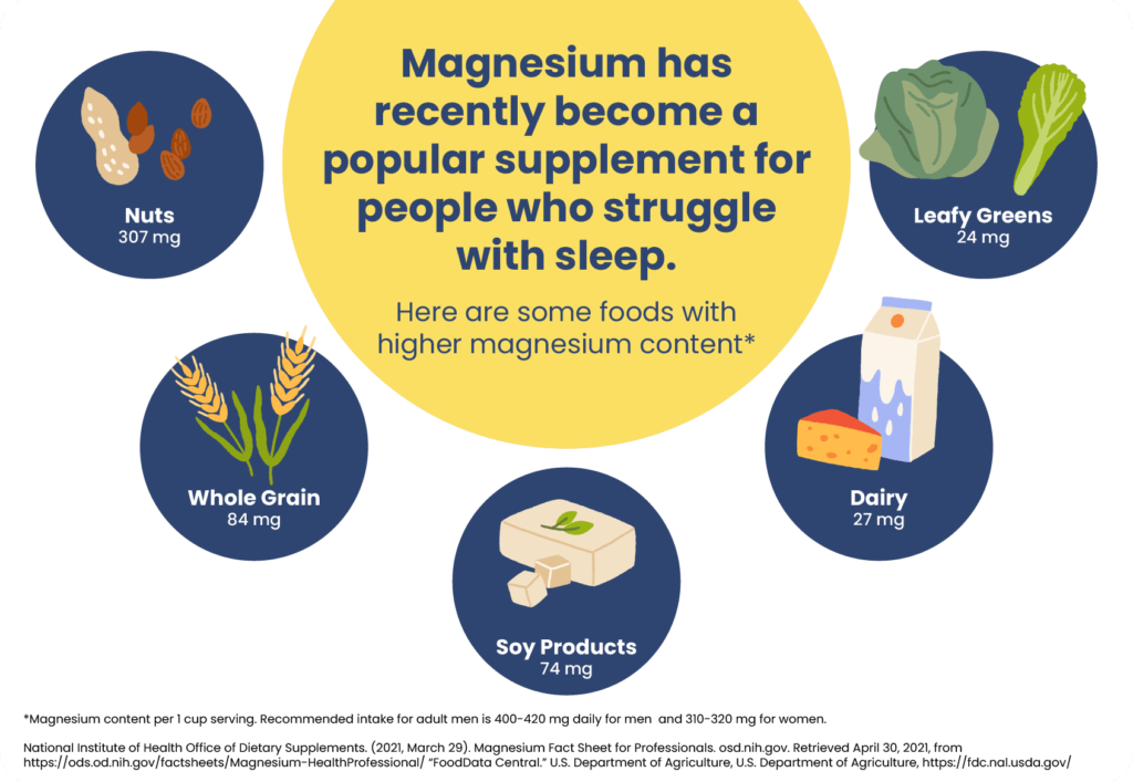
Chart of foods containing higher magnesium content

There are many ways to get adequate magnesium in the diet. Some examples of foods rich in magnesium include:

  • Green leafy vegetables
  • Fish
  • Legumes
  • Whole grains
  • Pumpkin seeds
  • Chia seeds
  • Almonds
  • Cashews
  • Fortified breakfast cereals
  • Soy and soymilk

How Magnesium Impacts Sleep

Magnesium is included in a variety of nutritional supplements and natural sleep aids. Despite claims that magnesium can improve sleep, few studies have investigated this link. Researchers hypothesize that magnesium may relax the central nervous system and cause  chemical reactions Trusted Source National Library of Medicine, Biotech Information The National Center for Biotechnology Information advances science and health by providing access to biomedical and genomic information. View Source  in the body that increase sleepiness.

Studies suggest that magnesium supplements might help to reverse age-related changes in sleep often seen in older adults. Healthy magnesium levels may have an important role in the development of baby and infant sleep cycles. In contrast, low magnesium could be associated with poor sleep quality.

Magnesium and Insomnia

Insomnia is impaired sleep that causes fatigue and daytime sleepiness. Insomnia can also lead to struggles with memorymental health concerns, and a higher risk for accidents. Thus, people with insomnia may turn to supplements like magnesium to help with sleep.

Research is not clear on the role of magnesium supplements in the treatment of insomnia. However, some studies have shown  encouraging findings Trusted Source National Library of Medicine, Biotech Information The National Center for Biotechnology Information advances science and health by providing access to biomedical and genomic information. View Source . One study of older adults with insomnia found that magnesium supplementation at a dose of 500 milligrams daily for eight weeks helped them fall asleep faster, stay asleep longer, reduced nighttime awakenings, and increased their levels of naturally circulating melatonin.

There have also been studies combining magnesium with other supplements that have shown  promising results Trusted Source National Library of Medicine, Biotech Information The National Center for Biotechnology Information advances science and health by providing access to biomedical and genomic information. View Source  for treating insomnia. Yet, because the supplements in these studies contained more than just magnesium, it is difficult to interpret whether the benefits were linked to magnesium, to the other supplements, or to the combination.

Magnesium and Restless Legs Syndrome

Restless legs syndrome is a sleep-related movement disorder that creates an irritating urge to move the legs. The exact cause of this condition remains unclear, but some researchers suggest that magnesium deficiency may play a role in the condition’s development.

Studies have  not demonstrated Trusted Source National Library of Medicine, Biotech Information The National Center for Biotechnology Information advances science and health by providing access to biomedical and genomic information. View Source  a clear relationship between magnesium levels and restless legs syndrome in the general population. However, there is some evidence that magnesium may play a role in restless legs syndrome that develops  during pregnancy Trusted Source National Library of Medicine, Biotech Information The National Center for Biotechnology Information advances science and health by providing access to biomedical and genomic information. View Source  or while  receiving dialysis Trusted Source National Library of Medicine, Biotech Information The National Center for Biotechnology Information advances science and health by providing access to biomedical and genomic information. View Source .

Researchers have also investigated whether magnesium supplementation can reduce the symptoms of restless legs syndrome. Results of these studies are  contradictory Trusted Source National Library of Medicine, Biotech Information The National Center for Biotechnology Information advances science and health by providing access to biomedical and genomic information. View Source . Some have found that magnesium supplementation may be helpful to reduce symptoms in people with restless legs syndrome. But a more recent study of people with many types of cramps, including restless legs syndrome, found  no benefit from Trusted Source National Library of Medicine, Biotech Information The National Center for Biotechnology Information advances science and health by providing access to biomedical and genomic information. View Source  magnesium supplementation.

Other Benefits of Magnesium

Supplemental magnesium may be recommended by a doctor for people with conditions that cause a magnesium deficiency. Magnesium may be given to people who have certain health conditions  such as :

  • Diarrhea
  • Pancreatitis
  • Uncontrolled diabetes

There’s some evidence showing that magnesium supplements may benefit people with certain  chronic conditions Trusted Source National Library of Medicine, Biotech Information The National Center for Biotechnology Information advances science and health by providing access to biomedical and genomic information. View Source  like migraine headaches, high blood pressure, and osteoporosis. But more research is needed to determine how helpful magnesium is in managing these conditions.

Magnesium supplements may also be recommended for people taking medications that can lower magnesium levels, including proton pump inhibitors, diuretics, antibiotics, and some medications used to treat cancer.

Risks of Magnesium

In most cases, magnesium supplements are safe. A dangerously high level of magnesium is rare in otherwise healthy people unless they take a  very high dose  of magnesium.

Symptoms of magnesium toxicity can range from mild to severe, and vary depending on a person’s magnesium level. Symptoms of excessive magnesium include:

  • Nausea
  • Headache
  • Drowsiness
  • Low blood pressure
  • Muscle paralysis
  • Cardiac arrest

Magnesium is also included in over-the-counter products such as Epsom salts, antacids, or laxatives. The risk of magnesium toxicity from using these products is higher in people who have impaired kidney function. Other risk factors for magnesium toxicity include conditions that increase the absorption of nutrients, such as colitis, gastritis or gastric ulcer disease.

Magnesium vs. Melatonin

Melatonin and magnesium supplements are both used to improve sleep, but they work in different ways in the body. Melatonin is a sleep hormone that the pineal gland in the brain produces when the time to sleep is approaching. Experts note that adding supplemental melatonin may help people fall asleep but may be less helpful for staying asleep all night.

Magnesium may help to quiet the nerves in the body that keep people awake. However, while experts recommend melatonin for treating some sleep disorders, magnesium may not be something that a doctor recommends unless a person has another reason to take it, such as evidence of low magnesium levels.

How to Use Magnesium for Sleep

Overall, the evidence for magnesium on insomnia and other sleep disorders remains mixed. However, some evidence shows that otherwise healthy people may benefit from low doses of oral magnesium supplements to help improve symptoms of insomnia. Research suggests doses of up to one gram of magnesium should be taken no more than three times daily.

While magnesium supplements are generally considered safe, there are some people who are at a higher risk for magnesium toxicity or harmful drug interactions. Therefore, before purchasing a magnesium supplement, talk to a health care provider first to find out if taking magnesium is safe and warranted.

The Absolute Worst Cooking Oils for Your Health


By HUIZENG/Shutterstock

Your choice of cooking oil can make a profound difference in your health. I’ve often warned against the use of soybean oil. Not only is partially hydrogenated soybean oil loaded with trans fat, which has been linked to heart disease, it’s also a source of an omega-6 fat called linoleic acid (LA), which is highly susceptible to oxidation and is typically from GMO seeds.

The problem results once you start to digest this fat, as you break it down into harmful sub-components called advanced lipid oxidation end products (ALEs) and oxidized LA metabolites (OXLAMs) that can cause significant damage at the cellular level. For example, an ALE called 4HNE is a mutagen known to cause DNA damage. Studies have shown there’s a definite correlation between elevated levels of 4HNE and heart failure.

Additionally, LA breaks down into 4HNE faster when the oil it is contained in is heated. This is largely why cardiologists recommend avoiding fried foods. ALEs and OXLAMs also play a very significant role in cancer and heart disease.

LA-Rich Soybean Oil Linked to Obesity and Diabetes

In 2015, a UC Riverside research team found soybean oil induced obesity, insulin resistance, diabetes and fatty liver in mice. Two years later, they confirmed this by showing soybean oil modified to be low in LA caused less obesity and insulin resistance than the unmodified soybean oil.

“The dogma is that saturated fat is bad and unsaturated fat is good. Soybean oil is a polyunsaturated fat, but the idea that it’s good for you is just not proven.” ~ Frances Sladek, UC Riverside toxicologist

Then, in 2020, that same team published research showing soybean oil, both the modified and unmodified versions, actually produced genetic changes in the brains of mice, and they were not for the better. However, this time LA was not the primary culprit.

Soybean Oil Linked to Genetic Changes in the Brain

In this surprising study, the researchers compared diets high in three different types of fat.

  1. Soybean oil, which has a high LA content
  2. Soybean oil modified to be low in LA
  3. Coconut oil

It is surprising, because I would have thought that the LA produced the damaging effects, but LA was ruled out. So, they have identified yet another reason to avoid consuming soy products. As reported by the UC Riverside, the researchers:

“… did not find any difference between the modified and unmodified soybean oil’s effects on the brain. Specifically, the scientists found pronounced effects of the oil on the hypothalamus, where a number of critical processes take place.

‘The hypothalamus regulates body weight via your metabolism, maintains body temperature, is critical for reproduction and physical growth as well as your response to stress,’ said Margarita Curras-Collazo, a UC Riverside associate professor of neuroscience and lead author on the study.

The team determined a number of genes in mice fed soybean oil were not functioning correctly. One such gene produces the ‘love’ hormone, oxytocin. In soybean oil-fed mice, levels of oxytocin in the hypothalamus went down.

The research team discovered roughly 100 other genes also affected by the soybean oil diet. They believe this discovery could have ramifications not just for energy metabolism, but also for proper brain function and diseases such as autism or Parkinson’s disease …

[T]he research team has not yet isolated which chemicals in the oil are responsible for the changes they found in the hypothalamus. But they have ruled out two candidates.

It is not linoleic acid, since the modified oil also produced genetic disruptions; nor is it stigmasterol, a cholesterol-like chemical found naturally in soybean oil. Identifying the compounds responsible for the negative effects is an important area for the team’s future research …

‘The dogma is that saturated fat is bad and unsaturated fat is good. Soybean oil is a polyunsaturated fat, but the idea that it’s good for you is just not proven,’ [UC Riverside toxicologist Frances] Sladek said.

Indeed, coconut oil, which contains saturated fats, produced very few changes in the hypothalamic genes. ‘If there’s one message I want people to take away, it’s this: reduce consumption of soybean oil,’ [assistant project scientist Poonamjot] Deol said …”

Seed Oils — A Most Harmful Ingredient in the Modern Diet

While that UC Riverside study claimed the genetic changes in the brain applied only to soybean oil and no other vegetable oils, there are loads of other research showing vegetable oils, also known as seed oils, are some of the most harmful foods you could eat.

In the video above, Dr. Chris Knobbe, an ophthalmologist and founder and president of the Cure AMD Foundation, a nonprofit dedicated to the prevention of age-related macular degeneration (AMD), gives an excellent synopsis of why seed oils are the unifying mechanism behind westernized chronic diseases like heart disease, obesity, cancer and diabetes. Some of the points he makes are:

Heart disease, now the leading cause of death in the U.S., was virtually unknown in the 19th century. The same goes for cancer, which caused 0.5% of deaths in 1811 and 5.8% of deaths in 1900 — spiking to more than 31% of deaths in 2010.

A similar pattern emerged for diabetes, which was rare in the 19th century and had a prevalence of 0.37% in 1935. By 2020, there was a 28-fold increase in 85 years, to a prevalence of 10.5%.

Obesity? Same story. With a prevalence of just 1.2% in the 19th century, obesity increased 33-fold in 115 years, to a prevalence of 39.8% in 2015. By 1990, meanwhile, 24% of U.S. adults were diagnosed with metabolic syndrome, which is a combination of high blood pressure, dyslipidemia, insulin resistance, hyperglycemia and visceral obesity.

By 2015, 88% of U.S. adults failed to meet five criteria for metabolic health, measured by blood glucose, triglycerides, HDL cholesterol, blood pressure and waist circumference.

Macular degeneration and osteoarthritis followed similar striking increases, causing Knobbe to ask, “What was so ubiquitous during this time that could have prompted these changes?” Dietary history provides the answer, with the introduction of four primary processed foods — sugar, industrially processed seed oils, refined flour and trans fats — acting as the culprits.

Seed Oils Are Incredibly Proinflammatory

The problem with seed oils is that they’re incredibly proinflammatory and they increase excessive oxidative damage in your body. This oxidative stress, in turn, triggers mitochondrial damage and dysfunction that then drives the disease process. Several studies have demonstrated the truth of this.

The OXLAMs (LA metabolites) are also cytotoxic, genotoxic, mutagenic, carcinogenic, thrombogenic, atherogenic and obesogenic. Then, there’s the issue of direct toxicity from pesticides and herbicides.

Most of the vegetable oils produced today — especially canola, corn and soy — are made from genetically engineered crops and are therefore a significant source of toxic glyphosate exposure. According to Knobbe, the reason these oils have been able to remain in the food supply, despite their high toxicity, is because they’re not acute biological poisons, but chronic ones.

Seed oils were introduced into the U.S. diet in 1866. By 2010, they made up 32% of Americans’ diet, which amounts to 80 grams per person per day. In contrast, in 1865, most people would have only about 2% to 3% of their caloric intake from omega-6 LA from butter, lard and beef tallow.

Ancestrally raised animals had very low omega-6, but this changed significantly once animals started being raised in concentrated animal feeding operations (CAFOs). CAFO pork, for example, can contain up to 20% omega-6 fats, thanks to them being fed a diet high in omega-6 grains. The results of this dietary change have been significant.

Today, omega-6 fats make up anywhere from 20% to 30% of the average person’s daily calories, with 80% of it being LA. Instead of 80 grams per day, your goal is to get it under 7 grams per day. That would but you into the healthy percentage range of LA that our ancestors from a mere 150 years ago consumed.

According to a report by Jeff Nobbs, 6 in 10 Americans have a chronic disease, and heart disease, asthma, cancer and diabetes have increased 700% since 1935. During this time, Americans have been smoking and drinking less, exercising more and eating “healthier” according to conventional guidelines to lower saturated fat and sodium. Nobbs, too, believes seed oil is the missing link that explains why Americans keep getting sicker.

Processed Seed Oils Harm Your Health in Many Ways

Aside from oxidation, inflammation and mitochondrial dysfunction, processed seed oils and vegetable oils like soybean oil also harm your health in other ways. For example, they’ve been found to:

1.Damage the endothelium (the cells lining your blood vessels) and cause an increase in penetration of LDL and very low-density lipoprotein (VLDL) particles into the subendothelium.

In other words, these oils get integrated in your cell and mitochondrial membranes, and once these membranes are damaged, it sets the stage for all sorts of health problems. With a half-life of 600 to 680 days, it can take years to clear them out of your body. They also get incorporated into tissues such as your heart and brain. One result of this could be memory impairment and increased risk of Alzheimer’s disease. Canola oil, in particular, has been linked to Alzheimer’s.

2. Make your cell membranes more permeable, allowing things to enter that shouldn’t.

3. Make your cell membranes less fluid, which impacts hormone transporters in the cell membrane and slows your metabolic rate.

4. Inhibit cardiolipin, an important fat in the inner membrane of your mitochondria that needs to have non-damaged fat to perform optimally and facilitate optimal function of the electron transport chain and production of ATP.

Cardiolipin also works like a cellular alarm system that triggers apoptosis (cell death) by signaling caspase-3 when something goes wrong with the cell. If the cardiolipin is not saturated with DHA, it cannot signal caspase-3, and hence apoptosis does not occur. As a result, dysfunctional cells are allowed to continue to grow, which can turn into a cancerous cell.

5. Inhibit the removal of senescent cells, i.e., aged, damaged or crippled cells that have lost the ability to reproduce and produce inflammatory cytokines that rapidly accelerate disease and aging.

6. Strip your liver of glutathione (which produces antioxidant enzymes), thereby lowering your antioxidant defenses.

7. Inhibit delta-6 desaturase (delta-6), an enzyme involved in the conversion of short-chained omega-3s to longer chained omega-3s in your liver.

8. Impair your immune function and increase mortality from COVID-19. Saturated fat, on the other hand, may lower your risk of death. The authors of that study noted that unsaturated fats “cause injury [and] organ failure resembling COVID-19.”

More specifically, unsaturated fats are known to trigger lipotoxic acute pancreatitis, and the sepsis and multisystem organ failure seen in severe cases of COVID-19 greatly resembles this condition. In short, linoleic acid contributes to the inflammatory domino effect that eventually kills some people with COVID-19.

How to Avoid These Dangerous Fats

Considering the profoundly serious damage they cause, eliminating seed and vegetable oils from your diet can go a long way toward improving your health. This includes soy, canola, sunflower, grapeseed, corn, safflower, peanut and rice bran oil.

Also, be mindful of olive oil and avocado oil, as both are commonly adulterated with cheaper seed oils. That said, even pure olive and avocado oil are loaded with LA. If, like me, you’re in the habit of eating olive oil, I would strongly encourage you to limit your intake to 1 tablespoon per day or less. In my view, olive oil is not a magic bullet and if you are already consuming 80 grams of LA per day, it will only worsen, not help, your health.

To avoid these oils, don’t cook with them, of course, but also avoid processed foods, condiments, fast foods and restaurant foods. If you eat out, you’re undoubtedly eating unhealthy amounts of seed oils, as most restaurant foods are loaded with it.

Fried foods, dressing and sauces tend to be key culprits. Your best bet is to prepare most of your food at home, so you know what you are eating and, in the case of seed oils, what you’re not.

Conventionally raised chicken and pork are also very high in LA, and therefore best avoided. As mentioned earlier, CAFO animals are routinely fed grains such as corn, and as a result, their meat becomes high in LA, as the corn is loaded with it. You can learn more about this in Joe Rogan’s interview with Dr. Paul Saladino, author of “The Carnivore Code.”

How Much Linoleic Acid Is Too Much?

Many now understand that your omega-6 to omega-3 ratio is very important, and should be about 1-to-1 or possibly up to 4-to-1, but simply increasing your omega-3 intake won’t counteract the damage done by excessive LA. You really need to minimize the omega-6 to prevent damage from taking place.

Ideally, consider cutting LA down to below 7 grams per day, which is close to what our ancestors used to get before all of these chronic health conditions, including obesity, diabetes, heart disease and cancer, became widespread. If olive oil puts you over the limit, consider cooking with tallow or lard instead.

If you’re not sure how much you’re eating, enter your food intake into Cronometer — a free online nutrition tracker — and it will provide you with your total LA intake. The key to accurate entry is to carefully weigh your food with a digital kitchen scale so you can enter the weight of your food to the nearest gram.

Cronometer will tell you how much omega-6 you’re getting from your food down to the 10th of a gram, and you can assume 90% of that is LA. Anything over 10 grams is likely to cause problems.

Are The Risks of Vitamin D Toxicity Overstated?


(Kavun Halyna/Shutterstock)

Vitamin D is a popular yet arguably controversial supplement. One reason for the controversy is that people are concerned about suffering from toxicities if they take over the commonly recommended amount.

Some of this concern is merited: vitamin D is fat-soluble, meaning that compared to water-soluble vitamins such as B-group vitamins and vitamin C, it is stored in the body for a much longer time, which can put people at a higher risk of toxicity.

On the other hand, as vitamin D deficiency has been deemed an epidemic, more people consider supplementing with it. Therefore, compared to other fat-soluble vitamins like vitamins A, K, and E, it will naturally be subject to a higher level of scrutiny.

Yet experts argue that the toxicities of vitamin D have been overstated, with much of the current fear stemming from historical reports and outdated information.

Historical Cases Fuel Current Fears

Much of today’s fear of toxicities comes from decades-old clinical research, reasoned endocrinologist Dr. Michael Holick in his commentary titled, “Vitamin D Is Not as Toxic as Was Once Thought: A Historical and an Up-to-Date Perspective.”

Before the discovery of vitamin D in the late 1920s, rickets, a disease where children developed bowed legs, was a common problem in Europe and the East Coast of the United States.

The implementation of vitamin D fortification started once people realized that vitamin D could prevent rickets in the 1930s. This was met with great success; children who drank fortified milk no longer developed rickets, which led to vitamin D fortification becoming widespread in Western countries.

The first reports of vitamin D toxicities came in the 1940s. Since vitamin D also regulates the immune system and reduces inflammation, it was used to treat rheumatoid arthritis and tuberculosis, often at massive doses of 100,000 to 600,000 international units (IU) per day.

It is unknown why these doses were decided. But while some people reported miraculous effects of improvement, there were also fearful reports of elevated levels of calcium from vitamin D toxicity. Some developed kidney stones, calcification in the kidneys, or, even died from complications of toxicities.

Physicians were alerted about the toxicities of vitamin D and the treatment was stopped, but it took months to years for manifestations of the intoxication to resolve.

Then in the early 1950s, several infants were born with facial abnormalities, heart defects, mental retardation, and hypercalcemia. Two investigations concluded that it was likely caused by excessive intakes of vitamin D through fortified foods including milk (1, 2).

This conclusion was based on literature that reported that pregnant rodents that received intoxicating doses of vitamin D delivered pups with the same abnormalities.

As a result of this, the fortification of any food or produce with vitamin D was forbidden in Great Britain. The concern of toxicity in children led most of the world—except the United States, Canada, Australia, and a few European countries—to also ban vitamin D fortification.

In retrospect, however, Holick speculated that it is likely that these infants suffered from genetic problems that made them averse to vitamin D, including Williams syndrome and other conditions that impair the breakdown of vitamin D.

Nonetheless, the concept that vitamin D is one of the most toxic fat-soluble vitamins “has been instilled in the psyche of health regulators and the medical community,” Holick wrote.

Epoch Times Photo
An x-ray of a person’s legs, showing bowed legs, indicative of rickets.

Vitamin D Toxicity and Tolerance Levels May Be Higher

Surgeon and physician Dr. Joseph Bosiljevac has been in practice for more than 20 years. He observed that most of the guidelines on vitamin D recommendations have not changed over the decades.

This may also be a sign of unchanged notions about the vitamin’s toxicity.

From the 1980s until 2011, the general recommendation for daily vitamin D intake was 400 IU.

In 2011, the Institute of Medicine (IOM) increased the daily intake to 600 IUs for anyone between the ages of 1 and 70. The recommendation persists today and was made under the assumption that people would fulfill most of their vitamin D needs through sun exposure.

Many medical providers and academics alike have criticized the IOM’s daily recommendations for being far too low.

According to the IOM, a person would be considered to meet vitamin D adequacy once their vitamin D serum levels reach 20 ng/ml. Around 100 IUs of vitamin D would increase serum levels by 1 ng/ml. A dose of 600 IUs of dietary vitamin D would translate to 6 ng/ml, therefore the guidelines assume that the rest of vitamin D would be mostly fulfilled through sunlight.

Yet times have vastly changed, says William Grant, who has a doctorate in physics and has published over 200 papers on vitamin D. Grant is also the director of the Sunlight, Nutrition, and Health Research Center. He argues that most people do not spend enough time in the sun to produce adequate vitamin D.

The body produces around 1,000 IU of vitamin D after 10 to 15 minutes of sun exposure. This only happens under broad sunlight, otherwise, it takes even longer.

Most people also wear sunscreen these days, and since the start of the pandemic in 2020, people have spent an increasingly longer time at home. These factors combined drastically reduce a person’s chances of sun production, and reaching adequacy through diet alone is quite difficult.

Grant also argued that most people may be able to tolerate significantly higher levels of vitamin D than the guideline amount.

The guideline shows 50 ng/ml is the upper limit of serum level. However, according to a 2018 review in Frontiers in Endocrinology, symptoms of intoxication start to appear once serum levels for vitamin D reach 150 ng/ml or over, translating to 15,000 IUs of vitamin D daily.

This is over seven times the official recommendation for serum vitamin D levels.

Grant also pointed to a 2011 report that followed two patients who developed hypercalcemia after ingesting more than 900,000 IUs of vitamin D3 every day.

The first patient was the most extreme case. Due to errors in manufacturing and labeling, he ingested over 1.8 million IUs of vitamin D3 daily for two months and developed hypercalcemia, presenting with a vitamin D serum level of 1,220 ng/ml.

Unexpectedly, the two patients were asymptomatic and no longer hypercalcemic once their vitamin D serum levels fell to below 400 ng/ml, which is 20 times the National Institutes of Health’s recommended cut-off.

Both patients recovered without any complications.

Vitamin D Toxicity Report Increased in Recent Years for Various Reasons

Toxicity does pose a problem that cannot be overlooked.

With over 41 percent of the American population vitamin D deficient, vitamin D treatment has seen increased use over the years, and reports of toxicity have also increased.

A study that followed vitamin D exposures reported to the U.S. poison centers from 2000 to 2014 observed a 1,600 percent increase in reports from 2005 to 2011.

The report also found that despite increased reports over the years, the increase in severe outcomes has not been statistically significant.

Literature reports of vitamin D toxicities have also increased since 2010.

A 2018 review article stated that vitamin D toxicities are usually due to prescription errors, accidental ingestion of toxic levels of vitamin D due to product mislabeling, and increasing use of high-dose supplemental products.

Hypercalcemia: The Main Concern of Vitamin D Toxicity

Vitamin D increases the gut’s ability to absorb calcium through the diet; a major consequence of vitamin D toxicity is abnormally high levels of calcium in the blood, also known as hypercalcemia.

According to the Frontiers in Endocrinology review, common symptoms of hypercalcemia include confusion, apathy, recurrent vomiting, abdominal pain, excessive urination and thirst, as well as muscle and bone pain.

In severe cases, it can cause calcification of soft tissue and kidney stones, and deaths have also been reported in very extreme cases.

However, hypercalcemia is rare, and clinical complications of hypercalcemia are even rarer.

Board-certified internist Dr. Ana Mihalcea, who provides vitamin D injections as part of her clinic’s treatment, said that she has yet to see any of her patients develop toxicities from vitamin D injections.

Dr. Patrick McCullough, a board-certified internist who has published several papers on the use of vitamin D in treatment—especially high-dose vitamin D—told The Epoch Times that most of the hypercalcemia he has observed is easily reversible.

Given the large tolerance margin for vitamin D doses, McCullough argued that vitamin D deficiency poses a higher risk.

3 Factors That Increase Risk of Toxicity

1. Mislabeling and Prescription Errors

Mislabeling by manufacturers and prescription errors are the driving forces behind today’s toxicity levels.

Supplements are not subject to regulation by the U.S. Food and Drug Administration (FDA) for their safety, effectiveness, or labeling. Some have dosages that do not match the labeled dosage, often with instructions on dietary intake that are either insufficient or potentially toxic.

In the case report by Holick, the patient consumed vitamin D3 “more than 1,000 times what the manufacturer had led the patient to believe he was ingesting,” the author wrote.

Mistakes in prescription regarding the time between each intake, as well as dosage per intake, have also resulted in vitamin toxicities; therefore, it is always important to verify doses with pharmacists and health care professionals before supplementation.

2. Drug Interactions

Certain drugs or supplements may interact with vitamin D supplementation, leading to toxicities and hypercalcemia.

Taking calcium or ingesting dairy with vitamin D may elevate calcium levels in the blood.

Certain diuretics, which reduce fluid in the body, can concentrate calcium. Taking lithium may also increase the risk of vitamin D toxicity.

Epoch Times Photo
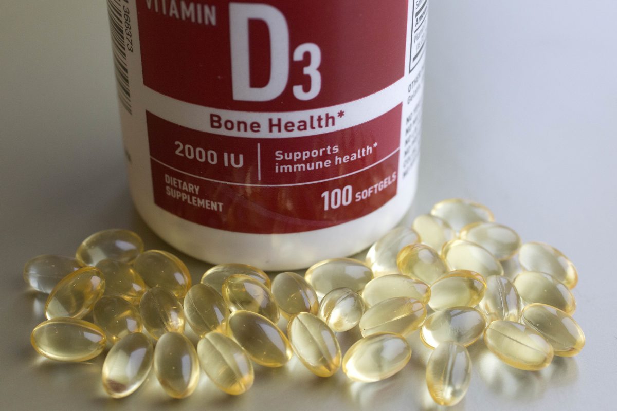
A container of vitamin D capsules.

3. Genetic Factors

Genetic factors can also put certain people more at risk of vitamin D toxicity.

Certain hereditary diseases such as Williams syndrome and familial hypocalciuric hypercalcemia make a person start with a higher calcium level, thus making them susceptible to toxicity.

Sarcoidosis and 24-hydroxylase deficiencies result in elevated vitamin D levels, which in turn increases calcium.

Ways to Reduce Vitamin D Toxicity

Some of the easiest ways to reduce the risk of developing hypercalcemia include drinking water and taking supplements like vitamin K2 and magnesium.

Drinking six to eight glasses of water per day dilutes the calcium concentration in the blood and can reduce the risk of hypercalcemia.

Taking both vitamin K2 and magnesium with vitamin D can reduce calcium levels in the blood by directing it into the bone.

It is also very important for individuals to take vitamins at the dosage most suitable for them.

Some of Mihalcea’s patients would present with a baseline vitamin D level of 30 ng/ml, yet display signs of deficiency, including fatigue and problems with sleeping and concentration.

Some of these patients’ symptoms alleviate once their vitamin D serum levels are increased to 70 ng/ml or higher using supplementation, indicating that their prior vitamin D levels may not have been optimal.

It is also very important to investigate “the different absorption rates in different people,” she added.

While some people experience a dramatic increase in vitamin D levels after supplementation, in others, the increase could be quite subtle.

Mihalcea said that patients who are obese and those with gut problems tend to have a poorer absorption of vitamin D, and for these people, she may need to give them 25,000 IUs a day just so they hit 50 ng/ml.

“There’s a huge variation and I’m always concerned when people just put out this idea that everybody can take the same amount—no, [you can’t],” she said.

What Causes Leg Cramps At Night?


Sharp and painful leg cramps can cause unpleasant sleep disruptions. About 60% of people Trusted Source National Library of Medicine, Biotech Information The National Center for Biotechnology Information advances science and health by providing access to biomedical and genomic information. View Source report experiencing nocturnal leg cramps. Nocturnal leg cramps — also commonly called charley horses — are painful, involuntary contractions of leg muscles Trusted Source National Library of Medicine, Biotech Information The National Center for Biotechnology Information advances science and health by providing access to biomedical and genomic information. View Source during the night. Although nocturnal leg cramps generally pass after minutes, they are unpleasant and can disrupt sleep enough to create problems. Usually, the calves and feet are most affected. Nocturnal leg cramps are more prevalent in women and older adults, but anyone can experience them.

Nocturnal Leg Cramps vs. Restless Legs Syndrome

Nocturnal leg cramps are a distinct condition, but they are often misdiagnosed as restless legs syndrome (RLS) because both disorders involve uncomfortable sensations in the legs. Although symptoms are similar, there are some key differences between these disorders.

You may have nocturnal leg cramps Trusted Source National Library of Medicine, Biotech Information The National Center for Biotechnology Information advances science and health by providing access to biomedical and genomic information. View Source if you are experiencing:

  • Calf or foot cramps during the night
  • Cramping that causes intense pain
  • Sharp pain lasting less than 10 minutes
  • Soreness lingering for hours or days
  • Sleep disruption as a result of cramping
  • Feeling distress around falling asleep

Even though restless legs syndrome also involves leg discomfort at night, its symptoms vary from those found in nocturnal leg cramps. RLS is characterized by the desire or urge to move your legs Trusted Source National Library of Medicine, Biotech Information The National Center for Biotechnology Information advances science and health by providing access to biomedical and genomic information. View Source in the evening. Other symptoms Trusted Source National Library of Medicine, Biotech Information The National Center for Biotechnology Information advances science and health by providing access to biomedical and genomic information. View Source can include:

  • Unpleasant or painful sensations in the legs
  • Discomfort that worsens in the evening or night
  • Relief found by walking or stretching
  • Sensations that worsen with lack of exercise or rest

Symptoms of nocturnal leg cramps and RLS often present in similar ways. Therefore, nocturnal leg cramps might be difficult for your doctor to diagnose. To help your doctor figure out what issue is causing your leg discomfort, track your symptoms and bring detailed notes with you to your appointment.

How Long Do Leg Cramps Last?

Nocturnal leg cramps can occur for as short as a few seconds, and they usually do not last longer than 10 minutes. You may feel soreness or reduced strength in your legs the day after your cramps. Stretching your calf or foot during the cramp may help relieve some of the intense pain.

Causes of Leg Cramps at Night

Leg cramps can be distressing and painful. Experiencing pain during the night can disrupt sleep. The exact causes of nocturnal leg cramps are unknown. However, there are some factors that may increase the chance of experiencing leg cramps during the night.

Medications: Some medications, like diuretics, steroids, and antidepressants, are associated with nocturnal leg cramping. Talk to your doctor about any medications you are taking if you experience leg cramps during the night.

Pregnancy: Leg cramps are a common experience in pregnant women, although some professionals consider pregnancy-related leg cramps different from nocturnal leg cramps. Researchers are unsure if this leg cramping occurs due to pregnancy itself, or because of a lack of blood flow in the veins as a result of pregnancy. Magnesium supplementation has been shown to reduce leg cramping in pregnancy, but you should talk to your doctor before beginning any new supplements.

Dehydration: Not drinking enough water during the day can result in dehydration. Dehydration causes muscle weakness and cramping Trusted Source National Library of Medicine, Biotech Information The National Center for Biotechnology Information advances science and health by providing access to biomedical and genomic information. View Source . Nocturnal leg cramps in particular are not associated with dehydration, but drinking water is still important. The exact amount of water you need to drink each day depends on your body weight, activity level, medications, and local climate.

Standing for Extended Periods: Being on your feet for long periods of time can increase the risk of experiencing nocturnal leg cramps Trusted Source National Library of Medicine, Biotech Information The National Center for Biotechnology Information advances science and health by providing access to biomedical and genomic information. View Source , making leg cramps a problem for people who must stand while they work. Taking breaks from standing and setting aside time to elevate your feet may help.

Exercise: Exercising at the gym or through team sports may lead to muscle cramps. When muscles are fatigued or overworked Trusted Source National Library of Medicine, Biotech Information The National Center for Biotechnology Information advances science and health by providing access to biomedical and genomic information. View Source , they can be more susceptible to cramping. Exercise-associated muscle cramps may be mistaken for nocturnal leg cramps when they occur at night. Stretching during an exercise-induced cramp has been shown to help relieve pain.

Alcohol:  Research of people over age 60 shows that those who drink alcohol are more likely to report Trusted Source National Library of Medicine, Biotech Information The National Center for Biotechnology Information advances science and health by providing access to biomedical and genomic information. View Source nocturnal leg cramping. This cramping could be a result of alcohol’s ability to damage muscle fibers, but more research is needed.

How to Reduce Leg Cramps at Night

Although the exact cause of nocturnal leg cramps is unknown, there are ways to reduce the likelihood that you will experience them. Typically, leg cramps do not indicate a serious health problem. However, if you are experiencing frequent leg cramping, you should reach out to your doctor. You may be able to reduce the likelihood of experiencing leg cramps through:

Hydration: Staying consistently hydrated throughout the day may help reduce the frequency of muscle cramps, since dehydration can cause cramps. Though some research suggests that nocturnal leg cramps are not caused by dehydration, it may help to drink water during long periods of outdoor activity or strenuous activity. There is also research to suggest that drinking pickle juice Trusted Source National Library of Medicine, Biotech Information The National Center for Biotechnology Information advances science and health by providing access to biomedical and genomic information. View Source during a cramping episode helps inhibit the cramp quickly.

Stretching: Doing some stretching or yoga before bed may help you reduce both the frequency and intensity of nocturnal leg cramps. Research suggests that engaging in a stretching routine Trusted Source National Library of Medicine, Biotech Information The National Center for Biotechnology Information advances science and health by providing access to biomedical and genomic information. View Source before bed helps reduce cramps and leg pain after about six weeks.

Baths: Some people claim that taking a bath Trusted Source National Library of Medicine, Biotech Information The National Center for Biotechnology Information advances science and health by providing access to biomedical and genomic information. View Source helps relieve their nighttime cramps, though further research is needed. An epsom salt bath in particular could help reduce muscle pain Trusted Source National Library of Medicine, Biotech Information The National Center for Biotechnology Information advances science and health by providing access to biomedical and genomic information. View Source . Epsom salt contains magnesium sulfate. An epsom salt bath could increase your magnesium levels, which may help relieve leg cramping.

Massage: Massaging your calves or feet before bed may help you reduce cramping during the night by relaxing the muscles in your legs. If you have limited mobility, consider asking a partner to help.

Heel Walking: Some people find that walking on their heels helps reduce nocturnal leg cramping. If you wake up in the middle of the night with a cramp in your calf, try getting up and walking on your heels. Although this type of walking helps release the tight calf muscle, research has found that it is not as effective as stretching during a leg cramp.

When to Talk to Your Doctor

If you are experiencing cramping in your feet and calves frequently during the night that does not go away with changes in lifestyle or diet, consult your doctor. In many cases, the cause of nocturnal leg cramps is unknown. Some leg cramps could indicate a more serious underlying disease, however, including:

  • Cardiovascular disease
  • Cirrhosis
  • Kidney disease
  • Osteoarthritis
  • Narrowing of the spinal canal
  • Nerve damage in legs
  • Circulation disorders

Discussing your symptoms and concerns with your doctor can help you determine the best course of treatment.

Infants Outperform AI in “Commonsense Psychology”


Summary: When it comes to detecting what motivates a person’s actions, infants outperform current artificial intelligence algorithms. The findings highlight fundamental differences between computation and human cognition, pointing to shortcomings in current machine learning and identifying where improvements are needed for AI to fully replicate human behavior.

Source: NYU

Infants outperform artificial intelligence in detecting what motivates other people’s actions, finds a new study by a team of psychology and data science researchers.

Its results, which highlight fundamental differences between cognition and computation, point to shortcomings in today’s technologies and where improvements are needed for AI to more fully replicate human behavior.

“Adults and even infants can easily make reliable inferences about what drives other people’s actions,” explains Moira Dillon, an assistant professor in New York University’s Department of Psychology and the senior author of the paper, which appears in the journal Cognition. “Current AI finds these inferences challenging to make.”

“The novel idea of putting infants and AI head-to-head on the same tasks is allowing researchers to better describe infants’ intuitive knowledge about other people and suggest ways of integrating that knowledge into AI,” she adds.

“If AI aims to build flexible, commonsense thinkers like human adults become, then machines should draw upon the same core abilities infants possess in detecting goals and preferences,” says Brenden Lake, an assistant professor in NYU’s Center for Data Science and Department of Psychology and one of the paper’s authors.

It’s been well-established that infants are fascinated by other people—as evidenced by how long they look at others to observe their actions and to engage with them socially. In addition, previous studies focused on infants’ “commonsense psychology”—their understanding of the intentions, goals, preferences, and rationality underlying others’ actions—have indicated that infants are able to attribute goals to others and expect others to pursue goals rationally and efficiently. The ability to make these predictions is foundational to human social intelligence.

Conversely, “commonsense AI”—driven by machine-learning algorithms—predicts actions directly. This is why, for example, an ad touting San Francisco as a travel destination pops up on your computer screen after you read a news story on a newly elected city official. However, what AI lacks is flexibility in recognizing different contexts and situations that guide human behavior.

To develop a foundational understanding of the differences between humans’ and AI’s abilities, the researchers conducted a series of experiments with 11-month-old infants and compared their responses to those yielded by state-of-the-art learning-driven neural-network models.

To do so, they deployed the previously established “Baby Intuitions Benchmark” (BIB)—six tasks probing commonsense psychology. BIB was designed to allow for testing both infant and machine intelligence, allowing for a comparison of performance between infants and machines and, significantly, providing an empirical foundation for building human-like AI.

Specifically, infants on Zoom watched a series of videos of simple animated shapes moving around the screen—similar to a video game. The shapes’ actions simulated human behavior and decision-making through the retrieval of objects on the screen and other movements.

Similarly, the researchers built and trained learning-driven neural-network models—AI tools that help computers recognize patterns and simulate human intelligence—and tested the models’ responses to the exact same videos.

Their results showed that infants recognize human-like motivations even in the simplified actions of animated shapes. Infants predict that these actions are driven by hidden but consistent goals—for example, the on-screen retrieval of the same object no matter what location it’s in and the movement of that shape efficiently even when the surrounding environment changes.

This shows a drawing of a brain on a computer
The ability to make these predictions is foundational to human social intelligence. Image is in the public domain

Infants demonstrate such predictions through their longer looking to such events that violate their predictions—a common and decades-old measurement for gauging the nature of infants’ knowledge.

Adopting this “surprise paradigm” to study machine intelligence allows for direct comparisons between an algorithm’s quantitative measure of surprise and a well-established human psychological measure of surprise—infants’ looking time.

The models showed no such evidence of understanding the motivations underlying such actions, revealing that they are missing key foundational principles of commonsense psychology that infants possess.

“A human infant’s foundational knowledge is limited, abstract, and reflects our evolutionary inheritance, yet it can accommodate any context or culture in which that infant might live and learn,” observes Dillon.

Study Finds Link Between Chronic Pain and Dementia


Summary: People with chronic pain in multiple parts of the body have a higher risk of developing dementia and accelerated cognitive decline.

Source: Chinese Academy of Science

A research team led by Dr. TU Yiheng from the Institute of Psychology of the Chinese Academy of Sciences has found that people with chronic pain in multiple parts of the body had a higher risk of dementia and experienced broader and faster cognitive decline, including memory, executive function, learning, and attention.

The study was published online in PNAS on Feb. 20.

Multisite chronic pain, where pain is experienced in multiple anatomical locations, affects almost half of chronic pain patients and has been found to place a greater burden on patients’ overall health. However, it has not been clear whether people with multisite chronic pain suffered from aggravated neurocognitive abnormalities.

In this study, after analyzing the records of 354,943 people in the UK Biobank cohort, the researchers found that the risk of neurocognitive abnormality increased with each additional pain site and was mediated by atrophy in the hippocampus, the part of the brain responsible for memory.

Since hippocampal volume decreases with age, the researchers equated the magnitude of the effect of hippocampal atrophy in patients with multisite chronic pain to the effect of aging in healthy people with an average age of 60.

This shows a brain
Since hippocampal volume decreases with age, the researchers equated the magnitude of the effect of hippocampal atrophy in patients with multisite chronic pain to the effect of aging in healthy people with an average age of 60.

“Multisite chronic pain may lead to up to eight years of accelerated hippocampal aging, an effect that may underlie a series of cognitive burdens,” said Dr. TU, corresponding author of the study.

The study provides a quantitative understanding of the impact of chronic pain on cognitive function and the risk of dementia, laying an important foundation for future research into the relationship between chronic pain and cognitive impairment.

It also highlights the excessive burden of multisite chronic pain on patients’ cognition and the brain, and the need to address the overlapping nature of pain conditions in both basic research and clinical studies.

Medication Targeting Brain Stress Response May Help Cardiovascular Symptoms of Alcohol Withdrawal


Summary: Prazosin, an FDA-approved medication for the treatment of hypertension may help to prevent relapse in people with cardiovascular and behavioral symptoms of alcohol withdrawal, a new study reports.

Source: Research Society on Alcoholism

Prazosin, a medication FDA-approved for hypertension and used off-label for alcohol use disorder, may help prevent drinking relapse in people with cardiovascular or behavioral symptoms of alcohol withdrawal, according to a new study involving active-duty soldiers. Alcohol use disorder (AUD) is highly prevalent among soldiers.

Prazosin reduces the noradrenergic signaling that is key to regulating the brain stress response. Overactivity of the noradrenergic system produces irritability, anxiety, “fight or flight” responses, and sleep disturbance.

Noradrenergic activity can increase during abstinence from drinking; consequently, it contributes to mood disturbances, insomnia, and other distressing symptoms of alcohol withdrawal and to the “relief cravings” that can lead to relapse.

Elevated noradrenergic activity produces elevated blood pressure and heart rate symptomatic of alcohol withdrawal. Previous research has pointed to the potential of prazosin for reducing alcohol use and cravings.

For the study in Alcohol: Clinical & Experimental Research, investigators designed a randomized control trial of prazosin among US active-duty soldiers.

Between 2015 and 2018, researchers enrolled 102 soldiers in this placebo-controlled study. The soldiers were undergoing mandated outpatient treatment for hazardous drinking and were expected to achieve abstinence. That program overlapped with the first nine weeks of the study.

Participants were interviewed about their alcohol consumption and were randomized to receive prazosin or placebo for 13 weeks. The soldiers kept diaries recording their number of drinks per day, drinking days per week, and heavy drinking days per week. On weeks nine and 13, they completed questionnaires assessing their alcohol cravings, sleep, and depression symptoms.

Those who met diagnostic criteria for PTSD (48 soldiers) were assessed for symptom severity. Researchers tracked participants’ blood pressure, heart rate, and suicidal thinking. They used statistical analysis to compare outcomes of treatment versus placebo among the soldiers, also considering cardiovascular symptoms or PTSD.

The soldiers’ alcohol use had declined steeply even before they were randomized. Nevertheless, on average, those taking prazosin experienced a modest but significant reduction in drinks per day compared to those taking placebo. The differences were more striking among specific subgroups.

This shows the outline of wine bottles
Prazosin reduces the noradrenergic signaling that is key to regulating the brain stress response.

The 15 soldiers with elevated heart rates were at or near zero drinks by week nine and sustained this through the final four weeks of the study (after the outpatient program ended).

Those taking the placebo, in contrast, experienced an alcohol rebound during the latter period. The 27 soldiers on prazosin who had raised blood pressure reported a reduced number of drinks and drinking days compared to those on placebo, with the contrast more notable in the final four weeks.

Overall, prazosin did not appear to reduce alcohol cravings more than placebo, a finding that may reflect the particular conditions of this study. Still, within the prazosin group, soldiers with PTSD did experience relief from cravings. This is likely a result of the medication’s countering effect on excessive noradrenergic signaling in PTSD. Depression symptoms were marginally lower among the prazosin group than the placebo controls.

Among soldiers with elevated cardiovascular measures, the benefit of prazosin in reducing drinking was notable despite the participants’ low alcohol consumption at the start of the study.

The study findings endorse a “personalized medicine” approach for treating people with AUD at high risk of relapse, including those who experience cardiovascular or behavioral symptoms consistent with alcohol withdrawal. Additional research is needed.