Study points to surprising link between head injury and high blood pressure in retired NFL players
The chance that former professional football players will be diagnosed with high blood pressure — a known risk factor for cardiovascular and cognitive dysfunction — rises in step with the number of concussions the athletes sustained during their careers, according to new research by investigators for the Football Players Health Study at Harvard University.
The results held true even after researchers took into account established risk factors known to drive the risk for high blood pressure, or hypertension, including age, body mass index, race, smoking status, and a diagnosis of diabetes.
The results of the study, published Feb. 7 inCirculation, suggest that high blood pressure may be yet another driver of cognitive decline — a condition strongly linked with professional football play in previous studies and believed to stem primarily from repeated head injury. The findings also point to high blood pressure as a modifiable risk factor that could halt or slow both neurologic and cardiovascular damage in former players.
Given that cardiovascular illness remains a top killer in former athletes and in the general population, the researchers said the results should be an impetus for doctors, former players, and their families to consider a history of prior head injury when screening patients for hypertension — even in the absence of other risk factors for this condition.
“If players, families, and physicians are aware of the cardiovascular effects of head injury, we have a better chance of protecting both their cardiovascular health and long-term cognitive health,” said Rachel Grashow, director of epidemiological research initiatives for the Football Players Health Study. Grashow co-led the new study with Aaron Baggish, professor of medicine at the University of Lausanne, senior faculty member at the Football Players Health Study, and former director of Massachusetts General Hospital’s Cardiovascular Performance Program, which provides comprehensive cardiac care to athletes.
The research — based on a survey of more than 4,000 former National Football League players representing the largest study cohort of former professional football players to date — was conducted as part of the ongoing Football Players Health Study at Harvard University, a research program that encompasses a constellation of studies designed to evaluate various aspects of players’ health across their life span.
Grashow explained that most research on cognitive decline in former professional football players has focused on neurodegeneration caused directly by repeated concussions, a prominent aspect of the game. However, the leading cause of death and disability among former football players — and among Americans in general — is cardiovascular disease, a collection of conditions that affect the heart and blood vessels. Hypertension, the most common cause of these conditions, can also gradually damage blood vessels in the brain and, over time, lead to cognitive decline.
Various aspects of professional football, such as purposeful weight gain during play years and deconditioning after career end, are associated with hypertension. However, Grashow, Baggish, and their colleagues wondered whether concussion might also be independently associated with hypertension.
To answer this question, the researchers collected information from 4,168 former NFL players. The team analyzed known risk factors for hypertension in the general population — diabetes, obesity, age, smoking — as well as players’ number of seasons of play, field position, years since play, and the occurrence of 10 common concussion symptoms. These symptoms were used to calculate a concussion symptom score, or CSS.
The analysis showed that as players’ symptom scores rose, so did their likelihood of being diagnosed with hypertension, even after researchers accounted for known hypertension risk factors. Notably, even using the number of occurrences of just one severe symptom of concussion — loss of consciousness — was enough to accurately predict players’ likelihood of developing hypertension.
Grashow said that while it remains unclear exactly how concussion leads to hypertension, one hypothesis is that repeat concussions could cause a chronic inflammation that prompts blood pressure to rise. Uncovering the precise mechanism underlying concussion-related hypertension will be the subject for future research, she added.
Baggish added that unlike many risk factors for cognitive decline, hypertension is potentially controllable with an array of safe and effective therapies, including routine aerobic exercise, dietary modification, and, in some cases, medication.
“By identifying those at increased risk for hypertension based on their history of head injuries, we could intervene with therapies that not only protect their hearts and blood vessels, but also their brains,” Baggish said.
Authorship, funding, disclosures
Additional authors included Can Ozan Tan, Saef Izzy, Herman Taylor Jr., Marc Weisskopf, Meagan Wasfy, Alicia Whittington, Frank Speizer, and Ross Zafonte.
This work was supported by the Football Players Health Study at Harvard University, which is funded by the National Football League Players Association (NFLPA). The content is solely the responsibility of the authors and does not necessarily represent the official views of Harvard Medical School, Harvard University, and its affiliated academichealthcare centers. The NFLPA had no role in the design and conduct of the study; collection, management, analysis, and interpretation of the data; preparation, review, or approval of the manuscript; and the decision to submit the manuscript for publication.
Zafonte reported receiving royalties from Springer/Demos publishing for serving as co-editor of Brain Injury Medicine; serving on the scientific advisory board of Myomo Inc., and onecare.ai Inc; evaluating patients in the Massachusetts General Hospital Brain and Body–TRUST Program, which is funded by the NFL Players Association; and receiving grants from the NIH. Baggish has received funding from the National Institutes of Health National Heart, Lung, and Blood Institute, the National Football Players Association, and the American Heart Association and receives compensation for his role as team cardiologist from the US Olympic Committee/US Olympic Training Centers, US Soccer, US Rowing, the New England Patriots, the Boston Bruins, the New England Revolution, and Harvard University. Taylor reported receiving grants from the NFL Players Association outside the submitted work and grants from the NIH. Weisskopf reported receiving grants from the NFL Players Association and the NIH during the conduct of the study. Grashow, Whittington, and Wasfy received grant funding from the NFL Players Association.
“It is often said that emotional healing is a solitary journey, however, it helps to remember that people have always faced emotional struggles, and we should not be ashamed to ask for help.”~ Alita Pacio
Emotional healing of past pain can be a long and challenging journey, but it is worth the effort.
Emotional Healing
By taking proactive steps towards healing, you can regain balance in your life and move forward toward living a more fulfilling present. Learn these five powerful steps to help you start your emotional healing journey today!
5 Effective Steps to Emotional Healing You Need to Practice NOW
1. Acknowledge Your Painful Feelings
Acknowledging your painful feelings is the foundation for emotional healing. Identifying and naming your feelings does not require that we immediately try to fix the problem.
We can instead choose to simply be aware of what we are feeling in a constructive way. This takes both self-compassion and self-reflection, but these incredibly powerful tools can be used together to begin laying the path towards emotional healing.
How do we begin acknowledging our painful feelings and memories? The best way to begin is by reaching out for professional help or support from those who are close to us, who can provide us with understanding and compassion when we need it the most.
Once we receive that much-needed validation, and feel comfortable enough to sit with our pain, we can begin taking active steps in working through our feelings.
This will look different for everyone – whether its journaling, meditation, yoga, or any other spiritual practice – it’s important to find something that works best for us.
Personally, journaling gives me comfort. Journaling gives me the opportunity to slow down, understand my feelings and connect with myself. I take time journaling and explore what cause the pain, and why.
Acknowledging these feelings of hurt while learning how to nurture ourselves through difficult times can be an incredibly liberating experience on the path towards emotional healing.
2. Identify the Cause of Your Emotional Pain
To begin your emotional healing journey, you must first gain clarity around what is causing your pain.
If a particular situation or person triggers an intense or overwhelming emotion, it helps to consider both the event and the feeling. When we take the time to understand our emotional triggers, we can start to make sense of our feelings and gather the strength needed to heal.
Similar to what I have mentioned above, if we take time to explore what cause the pain, we nurture ourselves through painful times and this is what makes us stronger to heal.
3. Express Your Emotions in Healthy Ways
It takes courage to be vulnerable and express what we are feeling. But an important part of emotional healing is developing an assertive voice. When we express emotions, it is easier to come to terms with them.
This means finding healthy, constructive revelations for our emotions in order to gain peace and clarity.
Often when we think of emotional healing, we think of being detached from our feelings and burying them away; however, releasing our feelings in a truly healthy way can provide far more empowerment than suppression.
It may feel difficult at first, especially if expressing yourself is not something that comes naturally to you. We all have the capability to process our emotions in healthy ways so that we can heal more deeply and fully. Here are some examples.
Writing, conversations with trusted friends or professionals, art, dance, and physical activities can all be used as tools to help you work through your feelings in a productive way.4.
4. Notice What Is Outside Yourself
It is important to develop an understanding of your emotional experience, but it can be just as useful to notice what is outside yourself.
Learning to take space from our emotions by focusing on the environment around us can be a great tool for emotional healing. Giving yourself permission to observe the natural beauty that surrounds you and become aware of how nature has its own rhythm can bring feelings of acceptance, joy, and contentment.
Exploring the outdoors can help us heal from whatever emotional pain or stress we are experiencing. Nature is our best friend when it comes to healing. Take a walk, see the clouds in the sky, listen to birds singing and spend some time observing our surroundings. These are just a few examples we can do when it involves nature and exploring the outdoors.
It is often said that emotional healing is a solitary journey, however, it helps to remember that people have always faced emotional struggles, and we should not be ashamed to ask for help.
We do not need to carry this burden alone. Many people find that talking to close friends or finding a counsellor can be helpful in working through difficult emotions.
Finding the right support system may require some exploration but can prove invaluable in times of confusion and distress.
OVER TO YOU!
Healing yourself doesn’t have to be a difficult or complicated process. There are simple, natural remedies that you can use to improve your overall wellbeing and bring more balance into your life. I hope these powerful steps can help you heal emotionally.
Man presented with irregular gray-white ulcers on the soft palate, uvula, and tonsils
Why has this asymptomatic man in his 60s with an unremarkable medical history suddenly developed gray-white plaque on his soft palate? That’s the question facing Jianjun Qiao, MD, PhD, of Zhejiang University School of Medicine in Hangzhou, China, and colleagues, as reported in JAMAopens in a new tab or window.
When the man presented to the dermatology clinic for assessment, he explained that his tongue had developed this gray surface over the past week. He had been treated with oral cefuroxime for 3 days, without any improvement.
On questioning, he denied having a sore throat, cough, hoarseness, headache, or swallowing difficulties. He had no symptoms such as nausea, vomiting, fever, or night sweats, nor had he lost weight. He told clinicians that for the past 6 months, he had not had any rashes, ulcers affecting his genitals or rectal area, injuries or sores in his mouth, or a history of oral trauma.
He was a nonsmoker, and did not take any regular daily medications. He reported sexual contact with one male partner over the past 6 months.
On physical examination, clinicians noted irregular gray-white ulcers on the soft palate, uvula, and tonsils. The area around the sores was red and swollen. They also observed bilateral submandibular lymphadenopathy, which was not tender to the touch. His tongue appeared normal, and there was no evidence of skin lesions or mucosal erosions in the anal or genital areas.
Clinicians considered several next steps, including prescribing a trial of oral amoxicillin, starting the patient on topical nystatin, obtaining a biopsy of the gray-white plaque, and ordering blood tests for Treponema pallidum.
They decided against the trial of amoxicillin, since a recent course of cefuroxime had not improved the patient’s oral lesions. Given that the patient’s tongue and inner cheeks had no white patches, they ruled out possible oropharyngeal candidiasis, which would have been treated with topical nystatin. They decided to obtain serologic tests for T. pallidum, and if those returned negative findings, an oral biopsy would be the next and more invasive choice.
Syphilis is typically diagnosed by serologic testing, Qiao and team explained. Initial diagnostic methods may include “a treponemal test such as the T. pallidum particle agglutination assay (TPPA), which tests for specific antibodies against T. pallidum, or a nontreponemal antiphospholipid antibody test such as the rapid plasma reagin (RPR) or VDRL test.”
The patient’s blood test results were positive for TPPA and revealed an RPR titer of 1:256. Findings from polymerase chain reaction testing performed on a sample taken from the patient’s soft palate showed that he did not have human papillomavirus (HPV). Results of oropharyngeal and genital swabs showed that he did not have chlamydia or gonorrhea, and he tested negative for HIV, hepatitis B, and hepatitis C.
Clinicians administered one intramuscular injection of benzathine benzylpenicillin G (2.4 million units). This resolved his oral lesions completely within 2 weeks. They advised him to inform his current sex partner of his syphilis diagnosis, so that he could be tested.
At a follow-up visit 1 year later, the patient had no symptoms and oral examination revealed no abnormalities. His RPR titer had decreased to 1:8.
Discussion
“The key to the correct diagnosis is recognition that oral ulcers may be a manifestation of secondary syphilis,” Qiao and team noted. “Syphilis is an infection caused by T. pallidum, a spirochete that is acquired predominantly through sex but that can be transmitted from mother to fetus during pregnancy and may rarely be acquired hematogenously or through an organ transplant.”
Secondary syphilis occurs when T. pallidum is spread through the bloodstream, usually 4 to 10 weeks after the primary syphilis infection occurs. Clinically, secondary syphilis most often manifests as a maculopapular rash involving the palms and soles.
A secondary syphilis infection may also cause fatigue, muscle pain, fever, joint swelling, headache, sore throat, abdominal pain, hair loss, and alopecia, and may also affect vision and hearing. Up to 50% of cases of secondary syphilis present with oral mucosal manifestations, often affecting the lip, tongue, buccal mucosa, soft palate, and tonsils.
“Findings of oral secondary syphilis include erosive or ulcerative mucosal lesions, mucous patches, gray-white papillary or nodular lesions, and hyperkeratotic plaques,” Qiao and colleagues wrote.
Secondary oral syphilis has a long list of differential diagnoses to consider, including viral, fungal, protozoal, and mycobacterial infections; lichen planus; pemphigoid; pemphigus vulgaris; traumatic ulcerations; and squamous cell carcinoma, they said.
Oral secondary syphilis primarily affects men, they added, citing a study of 206 patients with secondary syphilisopens in a new tab or window, of whom 38 had oral secondary syphilis. Of those patients, 95% were men, and all but 2% of those were men who had sex with men; 39% had no other manifestations of secondary syphilis. “Their ages ranged from 21 to 63 years, 37% had HIV, and 53% had a history of sexually transmitted infections other than HIV (most commonly, hepatitis B, gonorrhea, and condylomas),” Qiao and team noted.
Mean time to diagnosis was 4.5 months, but was significantly longer for patients with isolated oral symptoms (8.8 vs 1.8 months, P=0.02).
In patients whose blood test results are negative, they suggested that diagnosis may be obtained with a tissue biopsy of a secondary syphilitic lesion. Histopathology may not be strongly diagnostic of secondary syphilis, as immunohistochemistry test sensitivity ranges from 49% to 92%.
First-line treatment for all stages of syphilis is benzathine penicillin G. “A single dose of 2.4 million units administered intramuscularly is curative for early, uncomplicated syphilis,” Qiao and colleagues wrote, citing CDC recommendationsopens in a new tab or window.
They stressed the value of monitoring quantitative titers of RPR or VDRL “to assess response to therapy, relapse, and to diagnose reinfection with T. pallidum.”
The CDC also advises that treatment be followed up with clinical assessment and nontreponemal testing, at 6-, 12-, and 24-month intervals for patients without HIV and at 3-, 6-, 9-, 12-, and 24-month intervals for patients with HIV.
Qiao and colleagues advised screening syphilis patients for other sexually transmitted infections, including HIV, and testing sexual partners of those infected, noting that the incidence of syphilis has been increasing steadily worldwide since 2000.
Buprenorphine, rather than a full agonist opioid, should be used for patients taking daily opioids for chronic pain, given its lower risk for overdose or misuse, new guidelines from the Department of Veterans Affairs (VA) and Department of Defense (DoD) recommended.
But “the guideline development group does not recommend use of opioid analgesics in the daily management of chronic pain,” wrote James Sall, PhD, of the VA in New Braunfels, Texas, and co-authors in Annals of Internal Medicineopens in a new tab or window.
“The benefits that opioids can provide are small and are outweighed by the risks to the patient,” they continued. “If the decision is made to use long-term opioid therapy for a patient, then buprenorphine should be considered because of its lower risk profile.”
The updated guideline also calls for behavioral health assessments for all chronic pain patients and preoperative opioid and pain management education.
The field of pain medicine is likely to embrace this new buprenorphine recommendation, noted Chinazo Cunningham, MD, MS, of the New York State Office of Addiction Services and Supports in New York City, and Joanna Starrels, MD, MS, of the Albert Einstein College of Medicine in New York City, in an accompanying editorialopens in a new tab or window.
“The updated VA/DoD guideline is both conservative and radical,” Cunningham and Starrels observed — conservative because much is consistent with the CDC’s guidanceopens in a new tab or window but “potentially transformative” by recommending buprenorphine instead of full agonist opioids.
“Although the VA/DoD guideline recommends that buprenorphine be prescribed for chronic pain if daily opioids are prescribed, the recommendation itself is likely to change decision-making about whether opioids should be prescribed,” Cunningham and Starrels pointed out.
While the guideline is specific to the VA/DoD, “its influence is likely to expand into the greater U.S. healthcare system,” they noted.
“Because buprenorphine is an opioid, with long-term risks like physical dependence, it will be important to take precautions to clearly and carefully message to patients and clinicians, closely monitor buprenorphine prescribing patterns by indication and formulation, evaluate public health benefits and harms, and identify unintended consequences,” the editorialists continued.
Importantly, the quality of evidence for the buprenorphine recommendation is low and the recommendation is not clear about formulation, dosing, and the target patient population, they added.
VA and DoD leadership approved the joint clinical practice guidelineopens in a new tab or window in May 2022. The guideline development group used data from a systematic evidence review and graded recommendations and evidence as strong or weak. Besides using buprenorphine, the group recommended:
Screening for additional mental health conditions that potentially increase risk in chronic pain patients
Assessing for behavioral health conditions, history of traumatic brain injury, and psychological factors associated with higher risk for harm
Screening for pain catastrophizing and co-occurring behavioral health conditions to identify those at higher risk for negative outcomes when opioids are being considered in acute pain patients
Providing patients with pre-operative opioid and pain management education to reduce the risk for prolonged opioid use after surgery
The guidance is intended for clinicians who may be considering opioid therapy to manage patients with chronic pain, Sall and colleagues noted. It includes three one-page algorithms to help guide clinical decision-making.
“The guideline development group identified that more studies are needed examining the comparative effectiveness of different analgesic agents, the effectiveness of different tapering strategies, and the effectiveness of different risk mitigation strategies on the management of patients receiving long-term opioid therapy,” they added.
It’s an eerie feeling: You walk into a place you know you’ve never been before but are overwhelmed by a sense of familiarity—a memory you can’t quite reach. Has this all happened before?
Most people experience this sensation, known as déjà vu, at some point in their lives. It’s a hard feeling to study, though, because it tends to arise spontaneously and be shaken off easily, scientists say. Re-creating it on command in a laboratory is tricky business.
Nevertheless, scientists think that déjà vu actually provides a peek into how the memory system works when it goes a little off-kilter. The feeling may arise when parts of your brain that recognize familiar situations get activated inappropriately, says Akira Robert O’Connor, a cognitive psychologist at the University of St. Andrews in Scotland, who researches déjà vu. When this happens, another region of the brain then checks this feeling of familiarity against your recall of past experiences. When no actual matches are found, the result is a discomfiting sense of having seen it all before, accompanied by the knowledge that you haven’t.
“You get this: ‘Huh, weird, all of these experiences I’m having don’t quite match up.’ So it’s at that stage that you realize that you’ve made an error,” O’Connor says, “which is why it feels like an error, even though it’s probably actually the avoidance of an error.”
In some people with dementia, this feeling of familiarity occurs without the recognition of an error, he says. In those cases, people may go about their business as if they actually have seen it all before, complaining that every show on television is a rerun or refusing to visit the doctor because they’re sure they already have.
Déjà vu means “already seen” in French, a term possibly coined by French philosopher Émile Boirac in a letter to the editor of Revue Philosophique de la France et de l’Étranger in 1876. Boirac speculated that perhaps residues of long-forgotten perceptions triggered the feeling. There is now some laboratory evidence that vague similarities between one scene and another can indeed lead to déjà vu. Cognitive psychologist Anne Cleary of Colorado State University and her colleagues have developed a way to spark it in the lab by showing participants virtual scenes that have some subtle similarities to one another, such as the placement of the furniture relative to a painting on the wall. In a 2009 study, the researchers found that viewing these sneakily similar scenes was more likely to cause feelings of déjà vu than viewing dissimilar scenes—suggesting that perhaps there is some environmental trigger for the brain to call out, “Hey, I recognize that!” even when it’s never seen the scene before.
While Cleary’s research shows that a slight familiarity can result in déjà vu, it’s not clear that true familiarity is necessary to kick off the sensation. “Those sorts of ideas make a fair amount of sense,” O’Connor says, “but we’re actually really good at telling apart very similar things.”
In spontaneous déjà vu cases, he says, it’s quite possible that the familiarity feeling is random. At times, the part of the brain responsible for detecting familiarity—the medial temporal lobe, which is located just behind your temple and plays a large role in encoding and retrieving memories—may fire off overenthusiastically for no particular reason, O’Connor says. Supporting this random-misfire hypothesis is the fact that young people actually experience more déjà vu than older people. Younger brains are a little more excitable, prone to fire more quickly rather than holding back, O’Connor says.
Older people may also be less adept fact-checkers when false feelings of familiarity arise, says Chris Moulin, a cognitive neuropsychologist at the Grenoble Alpes University in France, who studies déjà vu. The fact-checker of the brain sits in the frontal cortex, behind the forehead. In older adults, this region may be less likely to put the brakes on a false sense of familiarity.
Older adults still recognize such false familiarity. “It’s not perhaps that older adults are not generating false familiarity,” Moulin says. ”It’s just that they don’t have, anymore, that certainty that what they’re experiencing is false.”
This is a normal part of aging, not the conflation of déjà vu with reality that people with dementia may experience. So enjoy the feeling of having felt it all before while it lasts, Generation Z. “As I age, it’s disappointing to me,” Moulin says, “because I used to have much more déjà vu than I have now.”
Commuting creates a liminal space that allows people to transition between home and work, which remote work doesn’t provide
The following essay is reprinted with permission from The Conversation, an online publication covering the latest research.
For most American workers who commute, the trip to and from the office takes nearly one full hour a day – 26 minutes each way on average, with 7.7% of workers spending two hours or more on the road.
Many people think of commuting as a chore and a waste of time. However, during the remote work surge resulting from the COVID-19 pandemic, several journalists curiously noted that people were – could it be? – missing their commutes. One woman told The Washington Post that even though she was working from home, she regularly sat in her car in the driveway at the end of the workday in an attempt to carve out some personal time and mark the transition from work to nonwork roles.
As managementscholars who study the interface between peoples’ work and personal lives, we sought to understand what it was that people missed when their commutes suddenly disappeared.
In our recently published conceptual study, we argue that commutes are a source of “liminal space” – a time free of both home and work roles that provides an opportunity to recover from work and mentally switch gears to home.
Based on our review, we developed a model which shows that the liminal space created in the commute created opportunities for detachment and recovery.
However, we also found that day-to-day variations may affect whether this liminal space is accessible for detachment and recovery. For instance, train commuters must devote attention to selecting their route, monitoring arrivals or departures and ensuring they get off at the right stop, whereas car commuters must devote consistent attention to driving.
We found that, on the one hand, more attention to the act of commuting means less attention that could otherwise be put toward relaxing recovery activities like listening to music and podcasts. On the other hand, longer commutes might give people more time to detach and recover.
In an unpublished follow-up study we conducted ourselves, we examined a week of commutes of 80 university employees to test our conceptual model. The employees completed morning and evening surveys asking about the characteristics of their commutes, whether they “shut off” from work and relaxed during the commute and whether they felt emotionally exhausted when they got home.
Most of the workers in this study reported using the commute’s liminal space to both mentally transition from work to home roles and to start psychologically recovering from the demands of the workday. Our study also confirms that day-to-day variations in commutes predict the ability to do so.
We found that on days with longer-than-average commutes, people reported higher levels of psychological detachment from work and were more relaxed during the commute. However, on days when commutes were more stressful than usual, they reported less psychological detachment from work and less relaxation during the commute.
Creating liminal space
Our findings suggest that remote workers may benefit from creating their own form of commute to provide liminal space for recovery and transition – such as a 15-minute walk to mark the beginning and end of the workday.
Our preliminary findings align with related research suggesting that those who have returned to the workplace might benefit from seeking to use their commute to relax as much as possible.
To help enhance work detachment and relaxation during the commute, commuters could try to avoid ruminating about the workday and instead focus on personally fulfilling uses of the commute time, such as listening to music or podcasts, or calling a friend. Other forms of commuting such as public transit or carpooling may also provide opportunities to socialize.
Our data shows that commute stress detracts from detachment and relaxation during the commute more than a shorter or longer commute. So some people may find it worth their time to take the “scenic route” home in order to avoid tense driving situations.
Ongoing research into long COVID overlooks the potential of vaccine injury, but many clinicians are seeing distinctions
What’s the difference between long COVID and vaccine injury?
The COVID-19 pandemic is almost over—at least, officially.
Yet many long-haulers and the vaccine-injured see no end in sight as they wake up every day to debilitating symptoms.
Critical care pulmonary specialist Dr. Pierre Kory, who shares a private practice treating both long COVID and COVID-19 vaccine injuries, told The Epoch Times that his clinic has treated more than 200 of these patients since February 2022.
He has only gotten “five or six off of medicines completely.” For most of them, it’s a chronic illness that needs chronic medication.
While long COVID has received plenty of media coverage and research, long-lasting post-vaccine symptoms rarely have been mentioned. Some may wonder if post-vaccine symptoms even exist.
Kory and the many doctors who treat these patients say yes: Vaccine injuries do exist, and the illness looks quite similar to long COVID.
Long COVID Versus Vaccine Injuries
Long COVID is defined by persistent symptoms after a COVID-19 infection, while vaccine injuries are symptoms that manifest due to vaccination.
As early as 2020, a preprint study reported on more than 200 post-COVID symptoms that had persisted for several months, with the most common symptoms reported after six months being fatigue, post-exertional malaise, and cognitive dysfunction. The preprint paper was later published in The Lancet in August 2021.
It’s not unusual for a viral disease to take a person out for a few weeks or months. The Epoch Times previously interviewed several long-haulers who hadn’t seen improvements for months or even years.
Across the United States, long-COVID clinics have popped up one after another, yet many long-haulers feel that their problems are not being addressed.
Compared to long COVID, COVID-19 vaccine injuries receive significantly less media coverage and research.
In the literature, the term “vaccine injury” has long been used with previous vaccines, including flu vaccines, polio vaccines, MMR (measles, mumps, and rubella) vaccines, and many more. Such injuries have mostly been documented as vaccine adverse events, meaning untoward health events associated with the vaccine, but they may not actually be vaccine-related. It’s up to the doctors during diagnosis to decide whether a patient’s symptoms are related to a vaccine.
There Are No Diagnostic Tests for Long COVID and Vaccine Injuries
There has been controversy over whether some of the current long-COVID cases are actually vaccine injury events.
Currently, there is no approved diagnostic test for either long COVID or vaccine injury. Existing clinical tests often yield normal results, even though many patients report discomfort and sickness.
These two conditions also have similar clinical presentations, making it even harder to tell them apart.
Doctors, therefore, need to look at a patient’s medical history and determine the events that led up to the symptom onset to come to a diagnosis.
The criteria for diagnosis mostly pertain to whether the chronic symptoms were preceded by a COVID-19 infection, in which case the illness is likely long COVID; if symptoms were preceded by vaccination, the patient may have a vaccine injury.
Some doctors have developed their own diagnostic methods.
Dr. Sabine Hazan, a California-based gastroenterologist and CEO of Progenabiome, uses gut bacterial composition as supplementary information in her diagnosis and treatment.
She told The Epoch Times that she sees differences in microbiome composition between these two groups of people, which helps her make a diagnosis, though more research is still needed to confirm.
Long COVID and Vaccine Injury Likely Have Same Cause
Both long COVID and vaccine injury have been theorized to be caused by spike proteins, though by very different mechanisms.
In long-haulers, the COVID virus and its spike proteins likely entered through the lungs as part of the infection. If the infection is not cleared, some of the virus—especially its smaller spike proteins—may enter the blood vessels and cause systemic damage to the body.
With the vaccines, the individual gets a dose of mRNA or DNA shot into the deltoid, bypassing the lungs. The mRNA or DNA enters the cells in the deltoid and induces the cells to start producing spike protein, which may enter the blood vessels and cause systemic damage, according to current studies.
Since the spike protein can traverse and damage multiple organs, doctors theorize that spike protein-induced injuries are a multisystem syndrome rather than a disease.
The distinction of spike protein-injury syndromes is important, as it highlights that the condition is systemic and can be related to many organs and body systems.
There are also spike protein injuries that involve only a single organ, such as myocarditis and pericarditis. While these adverse events may also be caused by vaccine spike protein, treatment is more straightforward, since only the heart is affected.
Systemic spike injuries may lead to inflammation, impairments in the gut microbiome, mitochondrial dysfunction, allergic reactions, activation of latent viruses, blood clotting, and injuries to other organs.
The impairment of these mechanisms can therefore cause a collection of symptoms including cognitive problems, migraines, fatigue, malaise, breathing problems, rapid heart rates, neuropathic pain, and seizures.
Some doctors hypothesize that, compared to long-haulers, people who experience vaccine adverse events have a higher amount of spike protein in their bodies. A hypothesis paper reasoned that in infection, the virus tends to be restricted to the lungs, while the vaccine introduces its content straight into the muscles..
Differences in Symptoms Onset
Though spike protein invasion routes all have similarities, their differences may lead to different symptom progressions.
There tend to be two major groups of long-COVID patients. The minority progresses from acute COVID-19 into long COVID without a period of symptom alleviation in between.
Kory said that most of the long-haulers he sees experienced a period of recovery or symptom alleviation for a few days to weeks before progressing into long COVID. This has also been observed in research.
Board-certified internist Dr. Keith Berkowitz, also the founder of the Center for Balanced Health in New York, told The Epoch Times that some of his patients developed long COVID after a mild or asymptomatic infection. This has also been reported in peer-reviewed studies, and other doctors have seen this trend.
Since COVID-19 infects the lungs, many long-COVID patients develop persistent shortness of breath caused by systemic pneumonia. Because the vaccine bypasses the lungs, pneumonia is seen less in vaccine-injured individuals.
There are also two major groups of symptom onset of vaccine injuries.
One group of people experiences symptoms within the first few minutes to hours of receiving the vaccine. These responses are likely due to sensitivities to the vaccine contents, which include lipid nanoparticles in the mRNA vaccines and polyethylene glycol (PEG).
The second group of people develops symptoms later, within days or weeks of vaccination. Since it takes time for cells to make spike protein, it may take a longer time for the vaccines to start causing damage. A preprint study on mRNA lipid nanoparticles in cell culture found that cells need around three to seven hours to start to make spike protein carried in the lipid nanoparticles, though the duration may vary when applied to the human body. Another study detected spike proteins in the plasma one day after vaccination.
Clinicians noticed other minor differences between patients who were diagnosed as long-haulers and those diagnosed as vaccine-injured.
Kory’s vaccine-injured patients tend to have more neurological symptoms, including neuropathies, seizures, tremors, and tinnitus, while Berkowitz said that he observes more cardiac problems among his vaccine-injured patients.
Board-certified internist Dr. Syed Haider, founder of MyGoToDoc.com, an online platform that connects over 50,000 long-COVID patients with health care professionals, said that with his patients, those who developed symptoms after the vaccine usually have one or two particularly prominent symptoms, while the long-COVID patients tend to have more of an even mix.
Hazan, on the other hand, notices subtle differences.
“The differences in the presentations are all in the history taking,” she said.
From early 2022 to now, Kory and Berkowitz have seen a shift in the patients presenting in their clinics. A year ago, the majority of patients they treated had long COVID; now, people who first developed symptoms after vaccines make up the majority.
For Haider, the majority of his patients are still long-haulers, while Hazan sees around a 50-50 split.
While Berkowitz and Kory have continued to see long-COVID symptoms in people infected with Omicron, both say that long COVID after Omicron tends to be less prevalent.
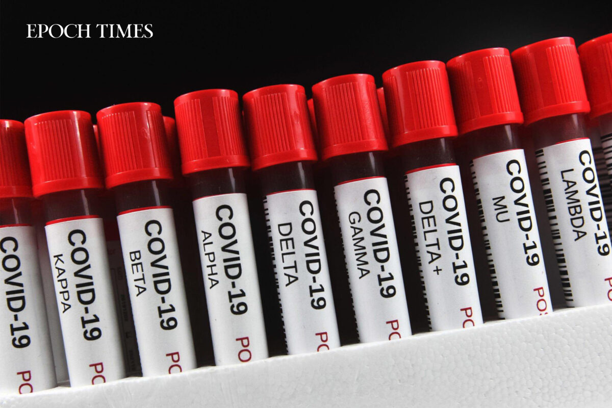
SARS-CoV-2 variants. (Getty Images)
The Majority Now: Vaccinated and Infected
The unfortunate situation now is that most people have both been infected with COVID-19 and vaccinated, complicating diagnosis and treatment.
Regardless of doctors’ own diagnostic methods to determine whether symptoms are caused by infection or vaccination, the consensus among clinicians interviewed by The Epoch Times is that people who have been harmed by the spike protein, whether through long COVID or vaccinations, should avoid getting reinfected, infected for the first time, and getting a booster.
A 2022 survey conducted by React19 on 98 long-COVID or post-vaccine sufferers found that around one-third of the people reported worsening symptoms after a COVID-19 reinfection. (pdf)
Subsequent vaccinations are also ill-advised. “I’ve had patients who took the first shot, really got kind of sick in the weeks afterwards, and actually got a second shot,” Kory said.
People who fell ill after the first shot are advised to speak to their physicians about potential health risks to decide whether they should continue vaccinating.
While there are long-COVID cases in which patients feel better after vaccination, these cases tend to be rare, with most patients experiencing symptom aggravations after a subsequent infection or vaccination.
An interesting thing Berkowitz noticed was that while infections with previous COVID-19 variants such as Alpha and Delta may worsen a post-vaccine patient’s symptoms, he sees less of this with post-vaccine patients infected with Omicron. This suggests that these patients’ immune systems may be better able to control Omicron infections.
Similar Treatment Protocols
Treatment-wise, there is not a clear difference between treatment protocols for these two conditions.
“My approach to treating both syndromes is essentially the same,” said Haider, explaining that clinicians currently do not know how to remove the lipid nanoparticles, polyethylene glycol, intact mRNA, or fragmented mRNA from people who have been vaccinated with the mRNA shots, so there’s no specific way to address the differences.
A biodistribution study (pdf) released upon a Freedom of Information Act request on mRNA vaccines found that when lipid nanoparticles were injected into mice, most of the particles would stay in the injection site while some would sequester in the liver, adrenal glands, spleen, and ovaries. Another study in rats found that those injected with vaccine lipid nanoparticles tended to have a lower immune response, though it’s unknown if this relationship is causal.
Since treatment protocols are similar for the two conditions, vaccine-injured patients may benefit from treatments available at long-COVID clinics, provided that they are receiving proper treatment.
Kory said that a majority of his patients tried going to primary care doctors and long-haul clinics, received little treatment or help, then came to him in despair.
“The other plight of the long-hauler and the vaccine-injured is that the majority have normal tests,” said Kory. “You might find some abnormalities [but] there’s no smoking gun to point to what the problem is, in testing.”
Therefore, a large portion of long-COVID and vaccine-injury treatments aim to target the underlying mechanisms that may be causing the symptoms, hoping that the mechanism that is targeted is the right one.
Many of these treatments aim at clearing the remnant spike proteins and relieving the inflammation they cause, while also boosting overall health to help with self-recovery.
Sometimes these patients simply need time to recover. Kory observed that his long-haul patients tend to see improvement in their symptoms over time, while he found that there seems to be less of a time benefit for people who have post-vaccine symptoms.
“We use this phrase: a tincture of time,” he said.
Long-COVID patients also tend to have more pulmonary problems from their prior infection; therefore, they may be prescribed steroids such as prednisone to control pneumonia.
Berkowitz, on the other hand, sees patients of various presentations and has to adjust his treatments to suit each patient. Vaccine-injured patients especially tend to have more symptoms, and this inevitably increases their recovery time.
Hazan treats patients by replenishing their lost gut microbiome. The type of bacteria lost tends to vary among patients, so everyone is treated differently.
Research
While research on spike protein injury has exclusively focused on long-COVID patients, Kory suspects that some of these cohort studies have also included people who were harmed by the vaccines rather than the disease.
“I don’t think [the studies are] purely about long-haul, unless it’s [published in] 2020 before the vaccines came out,” Kory said.
Therefore, data on long COVID may be confounded and impure.
Focused attention on long COVID while denying vaccine injury syndromes promotes a vaccination agenda, as people may be led to think the vaccines are safe and without harm.
“It will continue to propagate this non-recognition of the scope and scale of vaccine injuries,” Kory said. “If anything, it could make people want to get vaccinated because they don’t want to get long COVID.”
Hazan, who has clinical trial experience of more than 30 years, told The Epoch Times that there has been a lot of resistance against published research that went against the mainstream narrative on vaccine safety and early treatment.
Though she has yet to publish any studies on vaccine injury, her previous papers that speculate on the possible benefits of ivermectin have faced scrutiny, and there have been multiple external attempts to remove them.
A pill that could change the way postpartum depression is treated has been submitted to the U.S. Food and Drug Administration for priority review, pharmaceutical companies Sage Therapeutics and Biogen announced.
The drug, zuranolone, would be taken once-a-day for two weeks and could replace other postpartum depression treatments that take much longer to have any effect. Because the FDA is giving the application priority review, the drug might be approved this calendar year.
“Most current approved therapies may take weeks or months to work. We are committed to advancing treatments that could help physicians and patients by addressing depression symptoms quickly,” Laura Gault, MD, chief medical officer at Sage, said in a news release. “We believe zuranolone, if approved, could offer a new way for physicians to support patients.”
About one in eight U.S. women giving birth experience postpartum depression, or about 500,000 annually, the drug companies said. The pill also could be used to treat major depressive disorder, which is diagnosed in about 14 million people yearly.
A clinical study of 200 women with PPD found depression decreased for about 26.5% of them in three days when they took zuranolone, compared to 12.5% who got a placebo. In two weeks, those figures went up to 57% for those getting zuranolone and 38.9% who got the placebo.
In 2019 the FDA approved a Sage Therapeutics drug that was the first anti-depressant specifically designed for PPD, The Boston Globe reported. That drug, Zulresso, was given intravenously and was a commercial failure because of its high price, about $34,000 per patient, The Globe said.
But the effectiveness of Zulresso motivated pharmaceutical companies to start new research into postpartum depression, which has been often under-studied for years, The Globe reported.
Cognitive decline naturally occurs as we age, with a common belief that dementia is the cause in most cases.
Researchers at the Ohio State University say there are more factors causing cognitive decline than we once thought.
Socioeconomic factors, physical health measures, and behaviors, including exercise and smoking, accounted for 38% of the variation between participants in their level of cognitive function at age 54, researchers reported.
Cognitive or mental declineTrusted Source — a deterioration in functions such as the ability to think, remember, understand, and learn — naturally occurs as we age.
Generally speaking, most people associate cognitive decline with dementia diseases such as Alzheimer’s disease.
However, only about 41% of this decline can be accounted for by dementia. A number of risk factors that may also contribute to cognitive decline, including socioeconomic status, education, race, physical health measures, and behaviors such as exercise and smoking, have previously been identified. But researchers at The Ohio State University and the University of Michigan have now found that these factors only account for a small amount of the variation in mental abilities among older people highlighting gaps in the knowledge needed to reduce cognitive decline.
For this study, the research team applied a statistical approach to examine associations between various lifestyle factors and cognitive decline in older Americans.
“Prior studies have produced some mixed findings regarding some determinants of cognitive functioning, which could come from different datasets, research designs, time periods, and countries,” Dr. Hui Zheng, professor in the Department of Sociology at The Ohio State University, and lead author of this study explained to Medical News Today.
“They may also stem from the fact that each individual study only assessed a limited number of predictors without accounting for intercorrelations with unobserved confounders,” he continued. “They also tend to have a narrow-age cohort or short follow-up. Moreover, we want to effectively estimate both the statistical significance and the substantive contribution of the predictors of cognitive trajectory.”
Dr. Zheng and his team analyzed data from over 7,000 American adults born between 1931 and 1941 who had participated in a larger study called the Health and Retirement StudyTrusted Source where their cognitive function had been regularly measured from 1996 to 2016.
The Health and Retirement Study also collected information on participants’ lifestyle behaviors, including exercise, smoking, physical health, and socioeconomic factors such as education, income, and occupation.
Upon analysis, the research team found the many factors included in the study statistically accounted for only 38% of the variation between participants in their level of cognitive function at age 54. They found the factors with the biggest effect included personal education, household wealth and income, race, occupation, level of depression, and parental education.
“These could be linked through material, psychosocial, and neuropathologic mechanisms,” Dr. Zheng said when explaining how these factors could contribute to cognitive decline. “For example, education can improve cognitive reserve and delay the manifestation of neuropathologyTrusted Source that occurs as a result of aging-related or pathologic process.”
The factors with the least effect on cognitive decline included early life conditions and adult behaviors and diseases. In addition, interestingly, these factors accounted for only 5.6% of the variation in how cognitive function declined in people aged between 54 and 85.
Dr. Zheng said they were a bit surprised at the factors statistically accounting for 38% as they had included a wide range of factors.
“One thing I want to emphasize is that individual socioeconomic status (e.g., education, income, wealth, occupation, race, parental education) matters a lot,” he added. “These findings point to the predominant importance of socioeconomic conditions in shaping the level of cognitive functioning.”
MNT spoke with Dr. David A. Merrill, a psychiatrist, and director of the Pacific Neuroscience Institute’s Pacific Brain Health Center at Providence Saint John’s Health Center in Santa Monica, CA, about this study. He said the presumption has been that individual choices and behaviors matter “the most” in terms of maintaining intact cognitive function with aging.
“However, this study raises the possibility that social determinants of health may play as important, if not more important role, in determining the odds of achieving successful aging outcomes, including maintaining normal memory into and throughout older adulthood,” Dr. Merrill said.
Dr. Karen D. Sullivan, a board certified neuropsychologist and creator of the I CARE FOR YOUR BRAIN education program, said what brain scientists now understand about cognitive aging is that it is multiple decades-long processes that come together to determine one’s chances of having cognitive decline.
“Socioeconomic factors, vascular health, and health behaviors all come together in the individual and interact with genetic predispositions to establish our brain health as an older adult,” Dr. Sullivan explained to MNT.
“Socioeconomic factors, in particular the quality and quantity of one’s early education, exerts an influence on future cognitive health through the contribution to cognitive reserve,” she added. “Cognitively complex activities contribute to our ‘brain bank’ by building layers and layers of neural networks that can better withstand future neurodegeneration.”
MNT asked Dr. Sullivan how these findings might help doctors slow the rate of cognitive decline in people.
“Brain health is whole-person health,” she answered. “The idea that focusing on only one aspect of health, like diet or exercise, will result in a healthy older brain is naïve. Science, as reported in this study, makes it clear we need to make sustained efforts across all aspects of wellness to remain cognitively vibrant into old age.”
“Brain health should be important to us across the lifespan — not just when we hit 65,” she continued. “Brain health is cumulative and what we do in earlier phases of development matters in terms (of) who experiences cognitive decline and who does not. We can either decide to have a cumulative advantage or disadvantage — we get to decide with our behaviors.”
Dr. Merrill said these findings draw attention to the important and urgent need of our society at large to structure itself in ways that promote healthy behaviors as the default for not just the lucky few born into wealth, but for all of us as members of a larger interdependent community.
“We need to make common sense health behaviors like resources for getting regular physical exercise and having access to nutritious foods the expected norm for all, not the few,” he added.
In a study in voles, the so-called love hormone was not necessary to promote pair bonding, but why? Image credit: Anastasia Mihaylova (Shpara)/Stocksy.
Previous studies on the role of oxytocin in social bonding used drugs to block the oxytocin receptor, but researchers have now used genetic technology to generate prairie voles that lack functional oxytocin receptors.
Surprisingly, these mutant prairie voles still displayed the behavioral traits thought to be dependent on oxytocin, namely pair bonding and parental care.
A better understanding of the role of oxytocin in social attachments is required, but the researchers believe that their new genetic model will facilitate future research.
Oxytocin is a hormone produced by the brain. For more than 30 years, research has indicated that oxytocin is involved in the formation of social and emotional bondsTrusted Source in humans, prairie voles, and other species. This is why oxytocin is also known as “the love hormone.”
Animal model studies have shown that the binding of oxytocin molecules to specific receptors in the brain, called “oxytocin receptor signaling,” drives pair-bonding behavior — the formation of strong relationships between monogamous partners.
Now, new research from the University of California and Stanford University is challenging these long-accepted beliefs.
Dr. Devanand Manoli, senior author of the study and assistant professor of psychiatry at the University of California San Francisco, notes that he and his team “were all shocked that no matter how many different ways we tried to test this, the voles demonstrated a very robust social attachment with their sexual partner, as strong as their normal counterparts.”
Researchers studying the role of oxytocin in social bonding have often used prairie voles in their studies because these rodents are known to be monogamous.
Partner voles spend time nestled closely together and show a distinct preference for each other over other potential mates. This pair-bonding behavior is uncommon in the animal world. Prairie voles also exhibit bi-parental care of vole pups.
Yet prairie voles are not commonly used in genetic studies in the same way as mice. This meant that Dr. Manoli and coworkers had to develop new tools and protocols to generate prairie voles that lack functional oxytocin receptors.
They used a revolutionary gene editing technology called CRISPR — which stands for “Clustered Regularly Interspaced Short Palindromic Repeats” — that allows scientists to precisely target and manipulate specific genes within an organism’s DNA.
The researchers sought to determine if the mutant voles exhibit impairments in pair bonding by housing sexually inexperienced males and females together for 7 days, which has previously been demonstrated to be sufficient for establishing pair bonds. They also observed the prairie voles’ ability to provide parental care for their offspring.
When male and female prairie voles without oxytocin receptors were exposed to unfamiliar, non-mutant control voles they spent more time huddling with their partners.
They also displayed aggression toward the unfamiliar members of the opposite sex, indicating rejection of other potential mates.
Like normal prairie vole parents, mutant parents spent the majority of their time in the nest, in direct contact with their litters, and, in the case of mothers, nursing pups.
However, the pups born to mutant mothers weighed significantly less at weaning than pups born to normal mothers, indicating a potential issue with milk ejection or a slight deficiency in nursing behavior.
In the study report, the authors point out that their findings were consistent across three laboratories.
Dr. Larry J. Young, William P. Timmie professor of psychiatry and director of the Silvio O. Conte Center for Oxytocin and Social Cognition at Emory University, told MNT:
“These results were surprising to me because previous research in my lab and others ha[s] clearly shown that oxytocin receptor signaling [is] important for pair bonding in normal adult prairie voles. […] I believe their results because we have also created […] voles [without oxytocin receptors] and obtained similar results which are not yet published.”
Dr. Young believes that prairie voles have “hard-wired circuits for pair bonding.” Normal prairie voles experience oxytocin release during social situations, and their circuits become dependent on oxytocin.
On the other hand, prairie voles without oxytocin receptors never experience oxytocin release, and although their pair-bonding circuits still exist, they are not regulated by oxytocin.
“If you cover someone’s eyes, they cannot read,” said Dr. Young. “However, if someone is blind from birth, there are changes in neural circuits so that they develop very sensitive somatosensory systems and they can read braille.”
“So perhaps if an animal never experiences oxytocin, i[t] can use some other processes to facilitate social interactions and bonding, but in a complex situation it may not be as strong and efficient as in a wildtype,” he hypothesized.
Although the behavior of the mutant voles appears normal in a lab setting, Dr. Young expects that “in the wild, there would be nuanced differences that may make the [mutant voles] socially awkward and possibly unable to bond.”
Prof. Donatella Marazziti, a professor of psychiatry at the University of Pisa in Italy, not involved in the current study, hypothesized that the phenomenon may have something to do with the relationship between oxytocin and vasopressinTrusted Source, which also contributes to social bonding and parental care, according to existing research.
“We do […] know that oxytocin interacts with vasopressin receptors, and maybe some subtypes of these might replace the oxytocin ones,” Prof. Marazziti suggested.
Based on observations in prairie voles and other mammals including humans, clinical trials have used oxytocin to improve social attachment behavior in individuals with certain psychiatric conditions.
Researchers have also administered oxytocin to some children on the autism spectrum, but these studies have produced mixed results.
While Dr. Manoli and coauthors hypothesize that their findings may explain these inconsistent results, Dr. Young disagrees.
“The reason for the [oxytocin clinical trial] failures has to do with poor penetration of the oxytocin into the brain after sniffing, and the fact that oxytocin needs to be paired with behavioral therapyTrusted Source, which most studies do not do,” he told MNT.
Prof. Marazziti also strongly expressed caution in interpreting the study’s findings. “My opinion [is] that it might be difficult and dangerous, albeit sensational, to cloud and delete decades of converging data on the role of oxytocin in mating and bonding,” she told us.
Still, she acknowledged that the study’s findings may warrant further investigation: “Although I would recommend caution before drawing any conclusions, nevertheless, it also true that a paper not aligned to common paradigms may be a challenge and cannot be neglected.”
Overall, the study reveals the need for a deeper understanding of the molecular processes behind social attachment behaviors. The authors speculate that oxytocin-blocking substances used in earlier research might have impacted yet-undiscovered pathways that play a role in pair bonding.
Dr. Manoli anticipates that innovative genetic models, such as the oxytocin receptor-lacking prairie voles generated in this study, will enable a more thorough examination of the molecular and circuit mechanisms that regulate attachment behavior and its impairment in patients with neuropsychiatric disorders.