Summary: Even modest consumption of alcohol can accelerate brain atrophy and increase amyloid plaque formation. The findings reveal alcohol consumption can accelerate Alzheimer’s disease pathologies.
Source: Wake Forest University
Alzheimer’s disease is the most common form of dementia, accounting for 60% to 80% of dementia cases, according to the Alzheimer’s Association. While current research suggests alcohol use disorder is a risk factor in Alzheimer’s disease, the impact alcohol use disorder has on Alzheimer’s disease pathology is an area of continued research.
In a new preclinical study, scientists at Wake Forest University School of Medicine showed that even modest amounts of alcohol can accelerate brain atrophy, which is the loss of brain cells, and increase the number of amyloid plaques, which are the accumulation of toxic proteins in Alzheimer’s disease.
The study appears in Neurobiology of Disease.
“These findings suggest alcohol might accelerate the pathological cascade of Alzheimer’s disease in its early stages,” said Shannon Macauley, Ph.D., associate professor of physiology and pharmacology at Wake Forest University School of Medicine.
The study was a collaboration led by Macauley and Jeffrey Weiner, Ph.D., professor of physiology and pharmacology at Wake Forest University School of Medicine, through the medical school’s Alzheimer’s Disease Research Center and Translational Alcohol Research Center.
Using mouse models of Alzheimer’s disease-related pathology, researchers used a 10-week chronic drinking approach where mice were given the choice to drink water or alcohol, mimicking human behavior regarding alcohol consumption. They then explored how voluntary, moderate consumption of alcohol altered healthy brain function and behavior and whether it altered the pathology associated with the early stages of Alzheimer’s disease.
The researchers found that alcohol increased brain atrophy and caused an increased number of amyloid plaques including a greater number of smaller plaques, potentially setting the stage for increased plaque proliferation in later life.
even modest amounts of alcohol can accelerate brain atrophy, which is the loss of brain cells, and increase the number of amyloid plaques, which are the accumulation of toxic proteins in Alzheimer’s disease.
Interestingly, researchers also noted that acute withdrawal of alcohol increased the levels of amyloid-beta, which is a key component of amyloid plaques that accumulate in Alzheimer’s disease.
Further analysis showed that chronic alcohol exposure poorly regulated brain and peripheral metabolism—another way to accelerate Alzheimer’s disease pathology. Macauley previously showed that elevated blood sugar increases amyloid-beta and amyloid plaques.
In the current study, researchers found that even moderate drinking caused elevations in blood sugar and markers of insulin resistance, which increases the risk not only for Alzheimer’s disease but also for other diseases such as type 2 diabetes and cardiovascular disease.
The study also found that moderate alcohol use altered anxiety and dementia-related behaviors.
“These preclinical findings suggest that even moderate consumption of alcohol can result in brain injury,” Macauley said. “Alcohol consumption may be a modifiable risk factor for Alzheimer’s disease and dementia.”
Ethanol exposure alters Alzheimer’s-related pathology, behavior, and metabolism in APP/PS1 mice
Epidemiological studies identified alcohol use disorder (AUD) as a risk factor for Alzheimer’s disease (AD), yet there is conflicting evidence on how alcohol use promotes AD pathology. In this study, a 10-week moderate two-bottle choice drinking paradigm was used to identify how chronic ethanol exposure alters amyloid-β (Aβ)-related pathology, metabolism, and behavior.
Ethanol-exposed APPswe/PSEN1dE9 (APP/PS1) mice showed increased brain atrophy and an increased number of amyloid plaques. Further analysis revealed that ethanol exposure led to a shift in the distribution of plaque size in the cortex and hippocampus.
Ethanol-exposed mice developed a greater number of smaller plaques, potentially setting the stage for increased plaque proliferation in later life. Ethanol drinking APP/PS1 mice also exhibited deficits in nest building, a metric of self-care, as well as increased locomotor activity and central zone exploration in an open field test. Ethanol exposure also led to a diurnal shift in feeding behavior which was associated with changes in glucose homeostasis and glucose intolerance.
Complementary in vivo microdialysis experiments were used to measure how acute ethanol directly modulates Aβ in the hippocampal interstitial fluid (ISF). Acute ethanol transiently increased hippocampal ISF glucose levels, suggesting that ethanol directly affects cerebral metabolism. Acute ethanol also selectively increased ISF Aβ40, but not ISF Aβ42, levels during withdrawal.
Lastly, chronic ethanol drinking increased N-methyl-d-aspartate receptor (NMDAR) and decreased γ-aminobutyric acid type-A receptor (GABAAR) mRNA levels, indicating a potential hyperexcitable shift in the brain’s excitatory/inhibitory (E/I) balance. Collectively, these experiments suggest that ethanol may increase Aβ deposition by disrupting metabolism and the brain’s E/I balance.
Furthermore, this study provides evidence that a moderate drinking paradigm culminates in an interaction between alcohol use and AD-related phenotypes with a potentiation of AD-related pathology, behavioral dysfunction, and metabolic impairment.
The destruction of our gut microbiome may be wreaking havoc on our nervous systemr
Parkinson’s disease affects movement as it takes its toll on the nervous system.
Overuse of antibiotics can lead to an increased risk of Parkinson’s disease according to a study published recently in the journal Movement Disorders.
Parkinson’s disease is a progressive nervous system disorder associated with build-up of alpha-synuclein, a neuronal protein that affects movement. Symptoms progress gradually over many years and can begin with simple hand tremors and can culminate in complete disability and even death. It afflicts up to 10 million people worldwide with 1 million cases in the United States alone. The prevalence of the disease increases with age, particularly after the age of 50.
The gut microbiome is the community of microbes including bacteria, viruses, protozoa, fungi, and their collective genetic material, which is present in the gastrointestinal tract. This diverse community of microbes weighs around 4 1/2 pounds (two kilograms) in an adult and helps maintain the immune system, manage inflammation, detoxify the body, and much more. Damage to the microbiome can have long-ranging effects as it disrupts these important functions. The vagus nerve is the primary parasympathetic nerve controlling the organs and it is one of the primary communication routes between the gut and the brain.
A well-known side effect of antibiotics is the destruction of the gut microbiome’s valuable diversity. This effect is compounded by the use of modern, more powerful, broad-spectrum antibiotics.
As early as the 1970s, scientists had theorized that there could be a connection between Parkinson’s disease and the gut. More recently, Swedish researchers had finished a 40-year study that followed more than 9,000 patients and compared the prevalence of Parkinson’s disease in patients who had their vagal nerves severed for medical purposes compared to patients who hadn’t. Researchers had come to the conclusion that there was a protective effect against Parkinson’s disease in patients with severed vagal nerves. If Parkinson’s disease begins in the gut and is potentially microbial in origin, the vagal nerve could be the path by which it travels to the brain.
In the past decade, there has been a flood of research regarding Parkinson’s disease, the gut, and any link between the two. It was known that the bacterial composition of the intestine in Parkinson’s patients was abnormal, but the cause was unknown. Many Parkinson’s patients report chronic gut problems and these pathological changes have been observed up to 20 years before diagnosis. Constipation, irritable bowel syndrome, and inflammatory bowel disease have all been associated with a higher risk of developing Parkinson’s disease.
Results in the recently published study suggest that some commonly used antibiotics, which are known to strongly influence the gut microbiota, could be a predisposing factor to Parkinson’s disease. The strongest associations were found for broad-spectrum antibiotics and those that act against fungi and anaerobic bacteria. A delay of 1 to 15 years between excessive antibiotic use and the onset of Parkinson’s disease was observed in the study with the strongest associations seen at 10 to 15 years from the date of antibiotic use. The study compared antibiotic exposure between 1998 and 2014 in more than 10,000 Parkinson’s disease patients and compared it to more than 40,000 non-affected persons matched for age, sex, and residence.
Strongest associations were found with macrolide antibiotics, such as azithromycin, and lincosamide antibiotics, such as clindamycin. There were also associations found with tetracyclines, sulfonamides and trimethoprim, and antifungal medications.
This study further supports the larger concept of the gut-brain axis. The gut-brain axis consists of two-way communication between the central nervous system and the nervous system of the gut, linking the cognitive and emotional centers of the brain with gut functions. The gut microbiome plays a hugely important role in this interaction. The signaling from the gut microbiome to the brain and from the brain to gut microbiome is by means of neural, endocrine, immune, and humoral links.
While we have known about the actions of the brain to gut for hundreds of years, we are only recently discovering the actions of the gut to brain. The vagus nerve is a bidirectional pathway and the simple act of understanding this concept opens the door to new and exciting viewpoints in the diagnosis and treatment of many diseases.
This discovery will hopefully have implications for antibiotic prescribing practices in the future. With our new understanding of the effects the microbiome has on the gut, brain, and the downstream implications on a multitude of diseases, we should always consider the long-lasting effects of antibiotics on the gut microbiome as well as the long-standing problem of antibiotic resistance.
Armen Nikogosian, MD, practices functional and integrative medicine atSouthwest Functional Medicine in Henderson, Nev. He is board-certified in internal medicine and a member of the Institute for Functional Medicine and the Medical Academy of Pediatric Special Needs. His practice focuses on the treatment of complex medical conditions with a special emphasis on autism spectrum disorder in children as well as chronic gut issues and autoimmune conditions in adults.
Is it merely a coincidence that COVID-19 and aging are both accompanied by cognitive decline? Could COVID-19 alone cause aging? Do aging and COVID-19 combine to make cognitive function worse, or do they impair cognitive function separately? What can we do to prevent cognitive decline? This paper will examine these questions.
Summary of Key Facts
COVID-19 accelerates brain aging and increases the risk of dementia even after two years in severe cases.
Researchers found common ground at the genetic level for both COVID-19 and aging.
Meditation can help preserve brain function and volume at the genetic level.
COVID-19 Accelerates Brain Aging and Increases Risks of Dementia
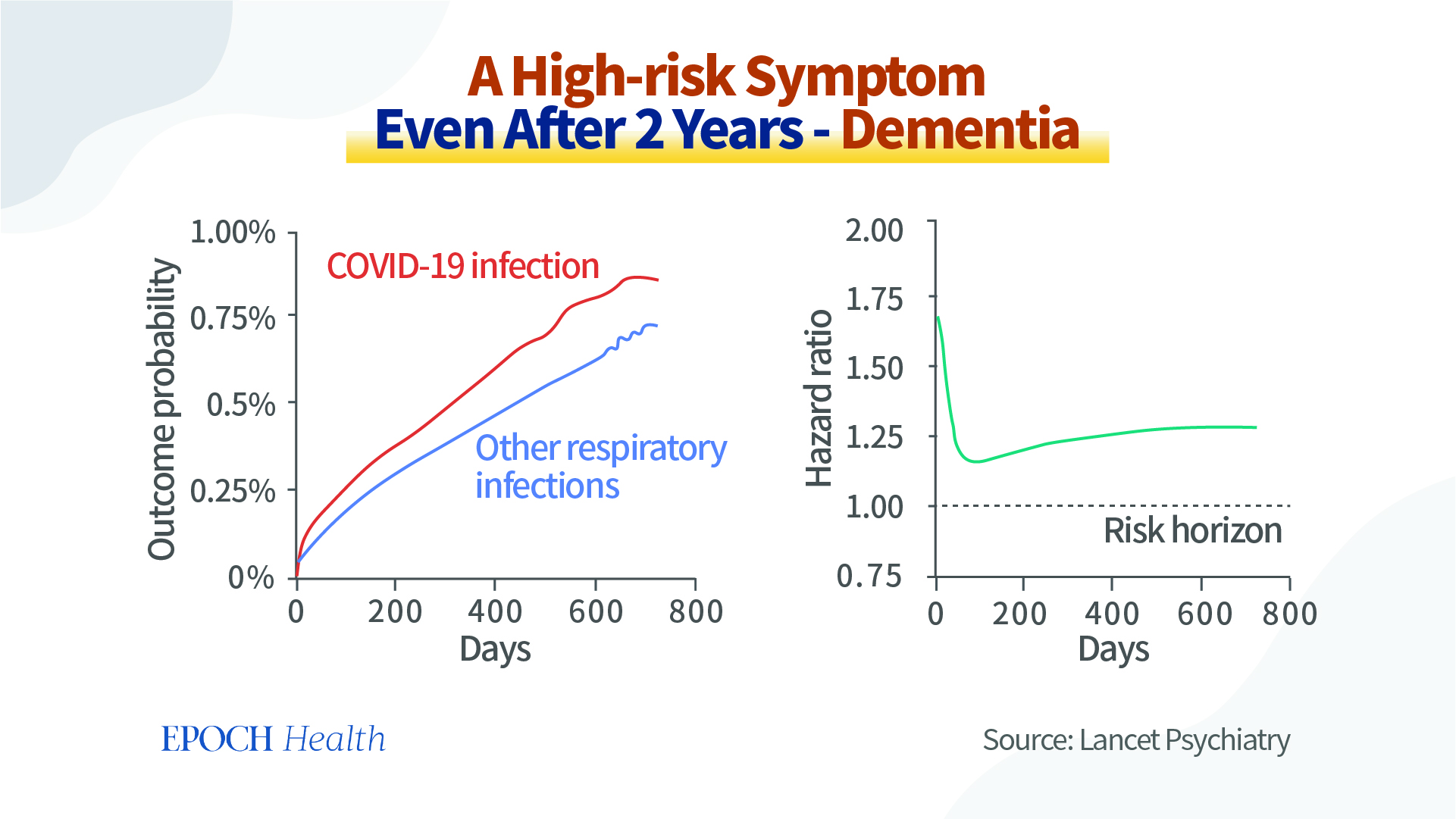
A large-scale UK study published in Lancet Psychiatry analyzed a two-year retrospective cohort including 1,284,437 patients. It revealed that the risk of dementia was significantly higher in the COVID-19 group than in the control group. Moreover, this risk remained higher even two years after recovery.
Risk of dementia increases even after two years in COVID-19 patients. (Lancet Psychiatry)
Another UK study evaluated the cognitive function of 46 patients with severe COVID-19 infection.
These patients had a significant decline in attention, complex problem-solving skills, and memory. Their cognitive deficits were equivalent to aging by two decades and losing 10 IQ points.
Profile of cognitive deficits after severe COVID-19 similar to more than two decades of age-related decline. (eClinical Medicine)
How COVID-19 Accelerates Aging
Research in Europe investigated the correlation between COVID-19 infection and aging. The authors compared the brain images in seven hospitalized patients in the acute phase, one month later, and six months after COVID-19 onset.
During the acute phase, all seven patients presented severe cognitive dysfunction and prominent low metabolism condition in the frontal cortex.
After one and six months of recovery, though symptoms improved in seven patients, they still reported abnormal cognitive function. Their brain images displayed lower metabolism status in the frontal cortex area.
The long-term changes in brain structure and function suggested that COVID-19 infection might be responsible for persistent cognitive and behavioral symptoms.
Molecular-level Explanation of Aging
Scientists have discovered that our internal epigenetic clock controls human experiences of birth, aging, illness, and death. It is similar to the observation that everything in our universe has its cycle of formation, stasis, degeneration, and destruction.
Cells become senescent as we age. That means they stop dividing and enter a stasis. Instead of dying off as they usually would, they persist but change shape and size and secrete inflammatory molecules that cause other nearby cells to become senescent.
In an article published in Nature Reviews Genetics, Steve Horvath, a professor of human genetics and biostatistician at the University of California–Los Angeles, concluded that as people age and have more senescent cells, there are characteristic changes in the methylation status of human DNA. DNA methylation could regulate the gene expression level.
Genes are like seeds lying dormant in the soil. Some will grow, but some will not. The “switches,” including methylation switches, determine whether these seeds will succeed.
Aging Genes Regulated in COVID-19 Patients
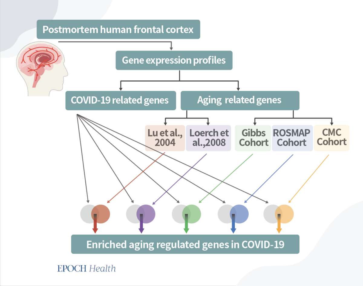
Recent research by Maria Mavrikaki at Harvard Medical School has found convincing evidence that COVID-19 is causing brain aging at the gene level.
Researchers performed a complete gene sequencing analysis in 54 postmortem brain samples. Results showed striking similarities in gene expression patterns between COVID-19 samples and naturally aged controls.
It demonstrated that those genes up-regulated in aging were also up-regulated in severe COVID-19 infection; likewise, other genes down-regulated in aging were also down-regulated in severe COVID-19.
To further validate the results, the researchers collated gene-wide datasets from five independent aging cohort studies and confirmed this association.
Mavrikaki’s research showed that the cognitive deficits reported in COVID-19 patients might result from aging-associated changes in brain structure and gene expression.
Genes expression in five independent aging cohorts was associated with that in COVID-19 cases.
Meditation Can Preserve Brain Function and Volume
The work by Mavrikaki is preliminary but informative. It could guide treatment for people who have lingering cognitive difficulties after COVID-19.
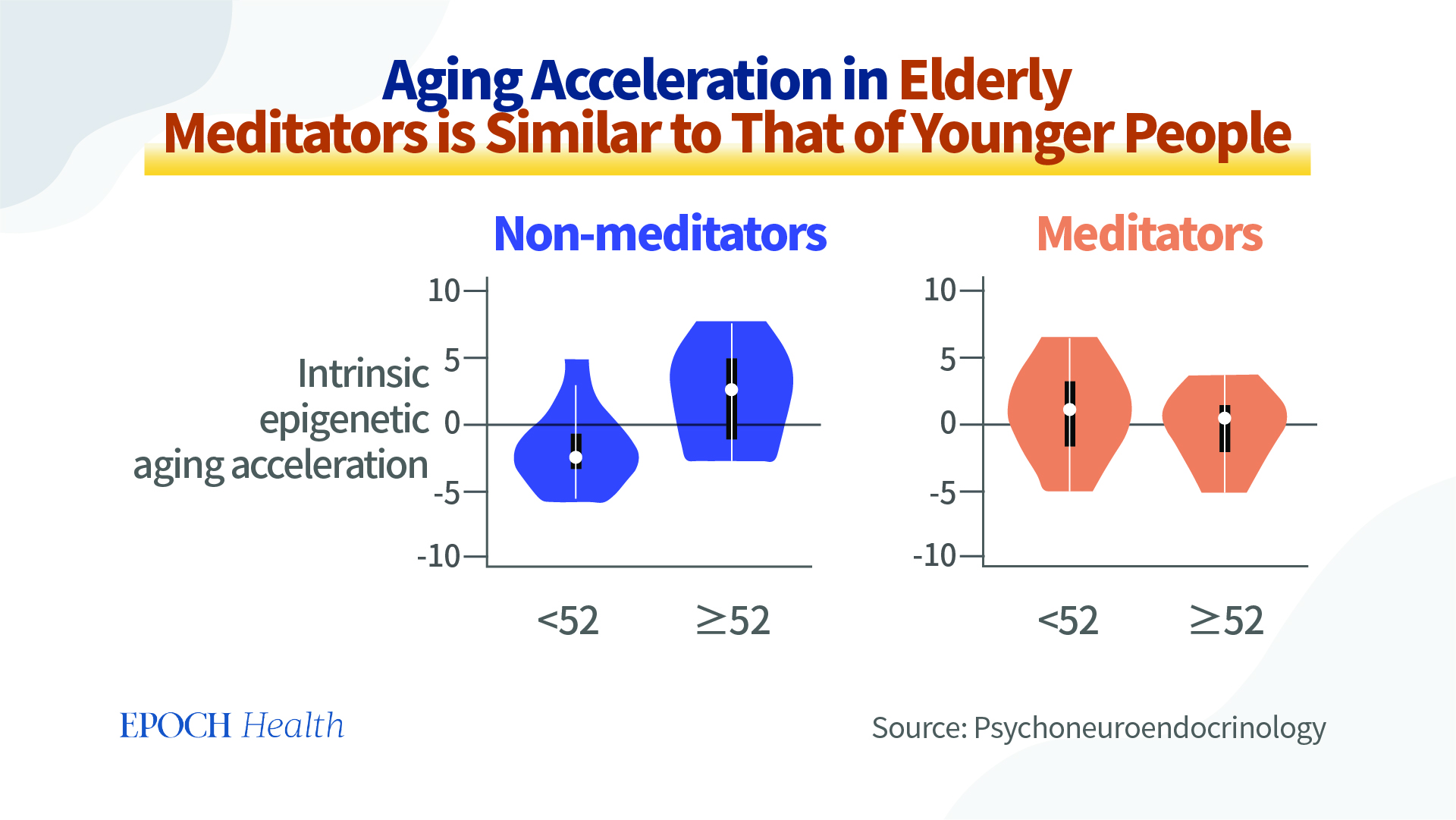
Research in 2014 analyzing a large sample (n = 100) of long-term meditators and control subjects aged between 24 and 77 years revealed that meditation could preserve brain volume.
A 2017 study suggests long-term meditation could reduce age-associated structural and functional brain changes. For example, researchers found increased gray matter volume and brain glucose metabolism markers in elderly expert meditators compared to controls.
A 2020 review summarized 25 published studies on mindfulness meditation and confirmed the effects of mindfulness meditation on increasing brain size, based on brain imaging studies and cognitive function assessment.
Meditation Can Slow the Aging Process at the Gene Expression Level
Furthermore, meditation could influence the expression level of genes.
One key feature of epigenetic information is its potential reversibility.
In the studies above, the aging rate in meditators significantly decreased. Meditation every day has the potential benefits to slow down the aging process and reverse COVID-19 disease at genetic levels.
Conclusion
Aging and severe COVID-19 have epigenetic-level connections.
Meditation practice can target the gene expression involved in both aging and COVID-19. Accordingly, meditation can prevent or reverse COVID-19-induced dementia, cognitive decline, and COVID-19-induced aging. Even for people who have not experienced COVID-19 infection, meditation has many benefits, including slowing the process of cognitive decline.
Summary: Children constantly assist others every time they need help if there is no personal cost to helping, a new study reports.
Source: University of Queensland
University of Queensland researchers have found children will help people in distress unless there is a personal cost.
Dr. James Kirby from UQ’s School of Psychology and his team worked with 285 children aged 4 and 5 to investigate what drives them to be compassionate.
“We tested if their compassion response changed depending on who they were interacting with,” Dr. Kirby said.
“Adults are an authority figure and children sometimes do what is required just because an adult is asking, which is why we also used puppets who are more on a child’s level.
“Our research found children will assist every time if there is no personal cost to helping and this didn’t change if it was an adult or puppet.”
To understand if the compassion response changed if there was a cost involved, stickers were given to the children when they completed tasks.
“We then saw that if they had to give up their reward stickers it was awfully hard for the children to help, even if the adult or puppet showed distress,” Dr. Kirby said.
To understand if the compassion response changed if there was a cost involved, stickers were given to the children when they completed tasks.
“It didn’t mean the children were deliberately selfish because adults also really struggle giving up rewards and resources. Just because they valued the reward it didn’t mean they were uncompassionate, as many of them offered passive compassion such as condolences like ‘that’s okay,’ or ‘maybe next time.’
“Most importantly, this study highlighted that if there were no personal costs or the children didn’t have to give up rewards, they were deeply compassionate and helpful.
“Understanding what drives children to be compassionate is important for setting up positive learning and family environments.”
Abstract
Testing the bounds of compassion in young children
Extensive research shows that, under the right circumstances, children are highly prosocial. Extending an already published paradigm, we aimed here to determine what factors might facilitate and inhibit compassionate behaviour. Across five experiments (N = 285), we provide new insight into the bounds of 4- to 5-year-old children’s compassionate behaviour.
In the first three experiments, we varied cost of compassion by changing the reward (Study 1), using explicit instructions (Study 2) and ownership (Study 3). In the final two experiments, we varied the target of the compassionate behaviour, examining adults compared with puppet targets (Study 4), and whether the target was an in-group member (Study 5). We found strong evidence that cost reduces compassionate responding.
By contrast, the recipient of compassion did not appear to influence responding: children were equally likely to help a human adult and a puppet, and an in-group member and neutral agent.
These findings demonstrate that for young children, personal cost appears to be a greater inhibitor to compassionate responding than who compassion is directed toward.
Summary: Study reveals functional connectivity abnormalities in brain areas associated with reward processing, habit formation, and decision-making in those with substance use disorders and addiction.
Source: University of Montreal
Some 10% to 15% of people will have a substance abuse problem at some point in their life, making it one of the most common psychiatric disorders.
Risks associated with substance abuse include dangerous driving, missed work, problems with depression, anxiety, health and money problems. Yet people with addictions seem to experience a sort of myopia, getting in deeper and deeper to cope with unpleasant emotions and to feel good, regardless of the cost.
Research has clearly established that psychosocial and environmental factors play a role in the development of substance abuse disorders. But there is more to the story, and now functional magnetic resonance imaging (MRI) studies are highlighting the importance of biological factors, in particular how the brain functions.
“The influence of such factors can be as much as 50%—this astonishingly high figure is why we’re so interested in what’s going on in the brains of substance abusers,” explained Stéphane Potvin, a professor in Université de Montréal’s Department of Psychiatry and Addiction whose research focuses primarily on the harmful effects of cannabis and alcohol on the brain structures of people with schizophrenia.
Zeroing in on markers
Potvin, who coordinates the Neurobiology and Mental Health division of the Centre de recherche de l’Institut universitaire en santé mentale de Montréal, wanted to zero in on the neurobiological markers associated with addiction. With his student Jules R. Dugré, a Ph.D. candidate in biomedical sciences, he conducted a meta-analysis of 96 studies involving a total of 5,757 subjects with some form of substance dependence—alcohol, nicotine, cannabis, psychostimulants or another drug.
Rather than focusing on brain activity as a whole, in his analysis Potvin and Dugré looked at what is known as “functional connectivity,” in other words, how well different parts of the brain communicate with each other. This is the first time an analysis of this type has been done.
Published in November in Addiction Biology, the study reveals a number of anomalies in the brain’s reward, decision-making and habit formation systems:
1. Reward
“The brains of substance abusers tend to show hyperconnectivity between the ventromedial prefrontal cortex and ventral striatum, two key areas of the reward system,” said Potvin. “This could explain the tendency to choose immediate gratification and why the substance’s motivational value increases over time despite the negative impact on other spheres of life.”
2. Decision-making
“We also see reduced connectivity in certain brain areas involved in decision-making, such as the prefrontal cortex and amygdala,” added Potvin. “This is consistent with the fact that substance abusers can seem indifferent to the harmful consequences of their choices.”
3. Habit formation
The most original finding is the presence of anomalies in the brain regions associated with habit formation, including the dorsal striatum and premotor cortex. “This hyperconnectivity could explain the compulsive nature of substance abuse,” Potvin noted.
No charges in impulse control
The meta-analysis did not, however, show changes in the brain regions and networks associated with impulse control. This contrasts with previous studies, which showed abnormalities in these regions using other neuroimaging approaches.
Research has clearly established that psychosocial and environmental factors play a role in the development of substance abuse disorders.
Potvin is quick to point out the limitations of his study. “In our analysis, we included all relevant studies of substance abusers, whatever the substance. But are brain systems equally affected by different substances? We really don’t know.”
Ultimately, these findings will help guide the development of neuromodulation-based interventions in the field of addiction.
“First we need a better understanding of the brain systems involved in substance abuse,” said Potvin. “This will allow us to pinpoint the areas to stimulate or inhibit in order to rebalance the brain and change behavior. The more solid evidence we have, the more treatment centers will be motivated to invest in the equipment needed to carry out this type of intervention.”
Abstract
Disrupted functional connectivity of the brain reward system in substance use problems: A meta‐analysis of functional neuroimaging studies
Extensive literature suggests that the brain reward system is crucial in understanding the neurobiology of substance use disorders. However, evidence of reliable deficits in functional connectivity across studies on substance use problems remains limited.
Therefore, a voxel-wise seed-based meta-analysis using brain regions of the reward system as seeds of interest was conducted on 96 studies representing 5757 subjects with substance use problems. The ventromedial prefrontal cortex exhibited hyperconnectivity with the ventral striatum and hypoconnectivity with the amygdala and hippocampus.
The executive striatum showed hyperconnectivity with the motor thalamus and dorsolateral prefrontal cortex and hypoconnectivity with the anterior cingulate cortex and anterior insula.
Finally, the limbic striatum was found to be hyperconnected to the orbitofrontal cortex and hypoconnected to the precuneus compared with healthy subjects.
The current study provided meta-analytical evidence of deficient functional connectivity between brain regions of the reward system and cortico-striato-thalamocortical loops in addiction.
These results are consistent with deficits in motivation and habit formation occurring in addiction, and they highlight alterations in brain regions involved in socio-emotional processing and attention salience.
Summary: Researchers report a greater concentration of alpha-synuclein aggregates was found in stool samples from Parkinson’s patients who suffered isolated REM-sleep behavior disorder.
Source: Heinrich-Heine University Duesseldorf
Isolated REM-sleep behavior disorder is a condition that can provide an indication of Parkinson’s disease well in advance. A research team headed by Professor Dr. Erdem Gültekin Tamgüney from Heinrich Heine University Düsseldorf (HHU) has shown that a greater concentration of α-synuclein aggregates can be detected in the stool samples of sufferers.
In the journal npj Parkinson’s Disease, they now present a method for detecting these aggregates, which they have developed in collaboration with the University Hospital Cologne, Jülich Research Center (FZJ) and the company attyloid GmbH.
There are two forms of Parkinson’s disease (for short: PD). In 70% of cases, it originates in the central nervous system. However, in around 30% of cases it originates in the nervous system of the intestine (“enteric nervous system”).
The latter form is referred to as “body-first Parkinson’s disease” (for short: body-first PD) and the characteristic deposits of aggregates of the body’s own α-synuclein protein are formed in the neurons in the intestine.
A preliminary form of body-first PD is isolated REM-sleep behavior disorder (for short: iBRD). It causes in part complex movements during a specific phase of sleep—REM-sleep—insofar as the patient experiences vivid and disturbing dreams. These movements can endanger the sufferer themselves or others.
A research team headed by Professor Erdem Gültekin Tamgüney from the Institute of Physical Biology at HHU now reports that it is possible to detect an elevated level of α-synuclein aggregates in the stool samples of patients. To achieve this, the team used a new surface-based fluorescence intensity distribution analysis (sFIDA) to detect and quantify individual particles of α-synuclein aggregates.
Professor Tamgüney says, “We are the first to prove the presence of α-synuclein aggregates in stool samples. Our results show a significantly higher level of α-synuclein aggregates in iRBD patients compared with healthy individuals or patients with Parkinson’s.
“These findings could lead to a non-invasive diagnostic tool for prodromal synucleinopathies—including Parkinson’s—which could in turn enable therapies to be initiated at an early stage before symptoms occur.”
However, more research is required before the process can find its way into clinical practice, for example investigation into why the level is lower in Parkinson’s patients.
The study was conducted in collaboration with the Institute of Biological Information Processing—Structural Biochemistry (IBI-7) at Jülich Research Center (FZJ), the Department of Neurology at the University Hospital Cologne and the HHU/FZJ spin-off attyloid GmbH.
To achieve this, the team used a new surface-based fluorescence intensity distribution analysis (sFIDA) to detect and quantify individual particles of α-synuclein aggregates.
HHU worked with the University Hospital Cologne to establish a biobank with stool samples from patients and control subjects, and with FZJ to develop the test procedure and conduct the tests on the samples. attyloid GmbH is a cooperation partner and is working towards the commercial exploitation of the results. It is necessary to verify that the test procedure is safe and can be used in normal operations in order to gain a license.
In body-first PD, the deposits of fibrils of the body’s own α-synuclein protein, which are characteristic of Parkinson’s, are first formed in the neurons of the enteric nervous system, which serves the gastrointestinal tract. The aggregates then spread to the central nervous system in a way similar to prions, i.e. an existing aggregate combines individual α-synuclein proteins in its vicinity into further aggregates in a nucleation process; these aggregates then spread further through the body.
The influence of what happens in the gastrointestinal tract on the brain is referred to as the “gut-brain axis.” The gastrointestinal tract is exposed to the environment and it is possible that harmful substances such as chemicals, bacteria or viruses ingested directly with food or via interaction with the microbiome of the gastrointestinal tract may trigger the pathological formation of α-synuclein aggregates.
Semaglutide injections are a prescription medication used to treat obesity. They are given as an injection under the skin, usually once a week.
Struggling to lose weight despite diet and exercise? Semaglutide injection has been proven to be successful in helping people shed those extra pounds. But if you’re looking for an alternative that doesn’t involve needles, this article is for you! Read on to learn about the top 5 over-the-counter supplements that can help with your weight loss journey .
We’ll discuss the potential benefits and side effects of each of these supplements, as well as how they can help you attain your weight loss goals. Then, we’ll take a look at what type of diet and exercise plan should be implemented with the supplement to maximize its effectiveness. Finally, we’ll provide an overview of semaglutide injection for those who are considering it for their weight loss. With this information in hand, you can make an informed decision about whether or not this injectable drug is right for you.
What are Semaglutide injections?
Semaglutide injections are a prescription medication used to treat obesity. They are given as an injection under the skin, usually once a week.
Semaglutide works by increasing the release of the hormone glucagon-like peptide-1 (GLP-1) from the intestine. GLP-1 is a satiety hormone that signals the brain to reduce hunger and slow down the emptying of the stomach. This leads to reduced food intake and weight loss.
If you are considering using semaglutide for weight loss, talk to your doctor about whether it is right for you.
How does Semaglutide work?
The appetite-suppressing and satiety-increasing effects of the drug semaglutide make it an effective aid in the treatment of obesity. It does this by simulating the actions of a hormone known as GLP-1, which is produced when we consume food. GLP-1 sends a signal to the brain that tells it we are no longer hungry, and it also lowers the quantity of food that we have the desire to consume. Semaglutide is an appetite suppressant that helps individuals lose weight by limiting the quantity of food they desire to consume while also helping them feel fuller after eating.
Quick Heads Up
Semaglutide is one of several drugs for weight reduction that is now available, but it is a prescription drug administered through injection. However, in our testing and when looking at other Semaglutide reviews online and comparing then to PhenQ – there is a clear winner –and it is PhenQ
The top 5 over the counter alternatives to Semaglutide injections
If you are looking for alternatives to Semaglutide injections for weight loss, there are a few over-the-counter options to consider. Here are the top 5:
PhenQ
PhenGold
Trimtone
LeanBean
PrimeShred
Each of these possibilities has components that are intended to assist you in shedding excess fat, controlling your appetite, and boosting your levels of energy. In addition to that, there is a possibility that they will boost your mood, make it easier to concentrate, and speed up your metabolism. Although there is no assurance that any of these items will work for everyone, people seeking an over-the-counter alternative to Semaglutide injections may find that these medicines provide a suitable replacement for the injections.
Before beginning any new weight reduction program or using any supplements, it is imperative that you consult with your primary care physician first. This is one of the essential things to keep in mind.
This all-natural weight loss supplement contains components that have been shown in clinical trials to help in the process of shedding excess pounds. The substance known as capsaicin, which gives chili peppers their distinctively hot taste, is the principal source of energy in PhenQ. Capsaicin is also found in chili peppers. Capsaicin, which is found in chili peppers, has been demonstrated to accelerate the body’s metabolic process and assist in the breakdown of fat. PhenQ is made up of a number of different components, some of which include caffeine, chromium picolinate, nopal, and L-carnitine furmarate.
Thermogenesis, which is the process by which heat is created in the body, and lipolysis are both boosted while taking PhenQ. Thermogenesis is the process by which heat is produced in the body (the breakdown of fats). Even if you don’t perform any exercise, this will cause an increase in the total amount of calories that you burn during the day. In addition, PhenQ suppresses hunger, which leads to a reduction in overall food intake and, thus, a reduction in overall body fat. Last but not least, the components of PhenQ operate to prevent the absorption of new fat from the food that you consume. This makes it possible to lose weight more rapidly than it would be otherwise feasible.
PhenQ offers a number of benefits, some of which include the quickening of weight loss, the squelching of hunger, the quickening of the metabolism, the increase of energy levels, and the improvement of mental clarity. In addition, after commencing therapy with the supplement, some people who have used PhenQ have reported that they have seen a drop in their desire for unhealthy meals.
Taking PhenQ is not known to cause any major adverse effects in most people who use it. Insomnia, jitteriness, nausea, headaches, and dizziness are some of the moderate adverse effects that may be experienced by some persons who use this medication. If you encounter any of these unwanted effects while using PhenQ, it is strongly suggested that you immediately discontinue the use of the product.
In general, PhenQ is an excellent weight reduction product that may assist you in losing weight in a timely manner without putting your health at risk. However, it is essential to keep in mind that no supplement can take the place of a nutritious diet and regular physical activity. If you want to see the greatest effects, combine PhenQ with a healthy lifestyle, and you’ll be well on your way to achieving the results you want from your weight reduction efforts.
2. PhenGold
PhenGold is a weight loss supplement that has become popular in recent years . It is claimed to help people lose weight by increasing their metabolism and suppressing their appetite. PhenGold is also said to have other benefits, such as reducing stress and improving sleep quality. However, there is no scientific evidence to support these claims. In this article, we will take a closer look at PhenGold, its claimed benefits, and whether or not it is effective.
PhenGold contains a powerful blend of ingredients that have been clinically proven to help with weight loss. The main ingredient in PhenGold is green coffee bean extract, which has been shown to boost metabolism and help the body burn fat more efficiently. Other ingredients include garcinia cambogia extract, which helps to suppress appetite, and green tea extract, which is a powerful antioxidant that helps to boost energy levels. PhenGold also contains chromium, which helps to regulate blood sugar levels and prevent cravings.
PhenGold is a popular weight loss supplement that has been shown to be effective in helping people lose weight. The main benefit of PhenGold is that it can help you lose weight by increasing your metabolism and burning more calories. Additionally, PhenGold can also help to suppress your appetite and reduce cravings, making it easier for you to stick to your diet. Furthermore, PhenGold is also free of any harmful side effects and is safe for most people to take.
PhenGold is a powerful thermogenic weight loss supplement that has been shown to be effective in clinical trials. However, like all supplements, it comes with some potential side effects that users should be aware of. The most common side effects of PhenGold include:
Increased heart rate
Increased blood pressure
Jitteriness
Insomnia
While these side effects are generally mild and temporary, they can be more severe in some people. If you experience any of these side effects, it is important to discontinue use of the supplement and consult with a healthcare professional.
This supplement is not a “miracle pill” and it will not help you lose weight overnight. In order to see results, you need to be consistent with your use of the product and make sure that you are following a healthy diet and exercise plan.
If you are committed to making a change in your life and want to improve your overall health and wellness, then PhenGold may be a good option for you. Just remember to be patient and consistent with your use of the product, and don’t expect miracles overnight.
PhenGold is a great supplement for those looking to lose weight and boost their energy. It offers a number of benefits, such as increased metabolism and suppressed appetite, which can help you reach your goals faster. However, it’s important to remember that PhenGold has some potential side effects, so make sure to consult with your doctor before starting any new dietary supplements. With the right diet and exercise program combined with taking PhenGold regularly, you should see results in no time at all!
The side effects of TrimTone are rare, but may include:
Nausea
Vomiting
Diarrhea
If you experience any of these side effects, stop using TrimTone and consult a doctor. TrimTone should be used as directed on the label for best results.
There are many benefits of taking Leanbean, including weight loss, improved energy levels, and reduced cravings. Additionally, there are no known side effects of taking this supplement. However, it is important to follow the directions on the label carefully in order to avoid taking too much and experiencing negative side effects.
When used as directed, Leanbean can be an effective weight loss tool. For best results, take it before meals or snacks in order to help control your hunger.
GLP-1 is responsible for regulating hunger and satiety signals in the brain. By increasing levels of GLP-1, PrimeShred helps to reduce hunger cravings and promote feelings of fullness after meals. PrimeShred also contains thermogenic ingredients that help to boost your metabolism and promote fat burning.
The benefits of PrimeShred include weight loss, reduced hunger cravings, increased energy levels, and improved fat burning. PrimeShred is generally well tolerated, with few side effects reported. The most common side effects include nausea, vomiting, diarrhea, constipation, headache, and dizziness. PrimeShred should be taken twice daily with meals for best results.
Factors to be considered before choosing the best over the counter alternative to Semaglutide injections
Over-the-counter (OTC) options forSemaglutide injections are limited, but there are a few things to consider before choosing the best one for you.
1. The main factor to consider is whether or not the OTC option contains the same active ingredient as Semaglutide. Some OTC options may contain a similar active ingredient, but at a lower dosage, which may not be as effective.
2. Another factor to consider is the price of the OTC option. Semaglutide injections can be expensive, so it’s important to find an OTC alternative that is affordable.
3. Finally, consider the side effects of the OTC option. Some OTC alternatives may have fewer side effects than Semaglutide, so it’s important to weigh the pros and cons before making a decision.
What are the benefits of Semaglutide Injections?
The benefits of Semaglutide Injections for weight loss have been well-documented in clinical studies. When used as directed, Semaglutide Injections have been shown to help people lose weight and keep it off long-term.
Some of the specific benefits of Semaglutide Injections for weight loss include:
• Helps people lose weight quickly – In clinical studies, people who used Semaglutide Injections lost an average of 9% of their body weight after just 16 weeks. This is a significant amount of weight loss that can make a real difference in someone’s health and quality of life.
• Safe and effective – Semaglutide Injections are approved and have been proven to be safe and effective for long-term use. There are no serious side effects associated with the use of Semaglutide Injections.
• Easy to use – Semaglutide Injections come in pre-filled syringes that are easy to use. There is no need for mixing or measuring, so you can be sure you’re getting the correct dose every time.
Precautions
Before using Semaglutide injections, it is important to take the following precautions:
Talk to your doctor about whether this medication is right for you. This is especially important if you have a history of diabetes or other medical conditions.
Read the instructions carefully before use. Make sure you understand how to properly prepare and administer the injection.
Do not inject more or less Semaglutide than prescribed by your doctor. The recommended dose is 0.25mg once daily injected under the skin of the abdomen, thigh, or upper arm.
If you miss a dose, inject it as soon as possible. If it is almost time for your next dose, skip the missed dose and continue with your regular schedule. Do not inject two doses at once.
Store Semaglutide in a cool, dry place away from light.
Conclusion: Semaglutide Weight Loss
Semaglutide injection is a powerful prescription medication used to help people lose weight . While it can be effective, it’s not always the right decision for everyone. For those who don’t want to use this option, there are many over-the-counter alternatives that you can consider. From diet pills and supplements to meal replacement shakes and lifestyle changes, there are plenty of ways to kickstart your journey towards healthier living without taking such a drastic approach as semaglutide injections. The best part? These alternative options are more affordable and come with fewer risks than taking a prescription drug like semaglutide injection.
Gastroesophageal reflux disease (GERD) is a commonly diagnosed digestive disorder characterised by the regurgitation of gastric contents into the oesophagus. Histamine (H2) receptor antagonists (H2RAs) and proton pump inhibitors (PPIs) are the primary acid-suppressive medicines used in this condition.[1] Both Omeprazole and Pantoprazole are PPIs approved by the Food and Drug Administration Agency (FDA) for the treatment of GERD.
Clinical studies have revealed that the efficacy of Pantoprazole (20 mg) and Omeprazole (20 mg) were comparable in maintaining endoscopic and symptomatic remission in patients with healed erosive oesophagitis. The efficacy and tolerability of Pantoprazole (40 mg) and Omeprazole multiple unit pellet system (MUPS) (40 mg) were explored in patients with moderate to severe GERD. It was observed that both these drugs were equivalent in healing after 4 and 8 weeks of treatment in patients with reflux oesophagitis grade II/III. The tolerability and safety of the drugs were also comparable. Another study also reported that the efficacy of Omeprazole MUPS 20 mg and Pantoprazole 40 mg were similar in the management of reflux oesophagitis. The patient satisfaction for these two drugs was also comparable.
While treating GERD in the geriatric population, other comorbid conditions and concomitant medications should be taken into consideration. Therefore, the drug interaction profile of these drugs needs to be screened to ensure safer therapeutics for the patients. It has been reported that Omeprazole has a higher risk for drug interactions as compared to Pantoprazole. Therefore, Pantoprazole is considered to be safe for elderly patients receiving multiple medications.