A Novel, Powerful Tool to Unveil the Communication Between Gut Microbes and the Brain


Summary: Researchers develop a novel tool that allows for the study of the communication of microbes in the gastrointestinal tract and the brain.

Source: Baylor College of Medicine

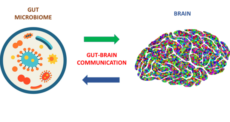
In the past decade, researchers have begun to appreciate the importance of a two-way communication that occurs between microbes in the gastrointestinal tract and the brain, known as the gut–brain axis.

These “conversations” can modify how these organs work and involve a complex network of microbe- and brain-derived chemical signals that are challenging for scientists to decouple in order to gain an understanding.

“Currently, it is difficult to determine which microbial species drive specific brain alterations in a living organism,” said first author, Dr. Thomas D. Horvath, instructor of pathology and immunology at Baylor College of Medicine and Texas Children’s Hospital.

“Here we present a valuable tool that enables investigations into connections between gut microbes and the brain. Our laboratory protocol allows for the identification and comprehensive evaluation of metabolites – compounds microbes produce – at the cellular and whole-animal levels.”

The gastrointestinal tract harbors a rich, diverse community of beneficial microorganisms collectively known as the gut microbiota. In addition to their roles in maintaining the intestinal environment, gut microbes are increasingly being recognized for their influence on other distant organs, including the brain.

“Gut microbes can communicate with the brain through several routes, for example by producing metabolites, such as short-chain fatty acids and peptidoglycans, neurotransmitters, such as gamma-aminobutyric acid and histamine, and compounds that modulate the immune system as well as others,” said co-first author Dr. Melinda A. Engevik, assistant professor of regenerative and cellular medicine at the Medical University of South Carolina.

The role microbes play in the health of the central nervous system is highlighted by the links between the gut microbiome and anxiety, obesity, autism, schizophrenia, Parkinson’s disease and Alzheimer’s disease.

“Animal models have been paramount in linking microbes to these fundamental neural processes,” said co-author Dr. Jennifer K. Spinler, assistant professor of pathology and immunology at Baylor and the Texas Children’s Hospital Microbiome Center.

 “The protocol in the current study enables researchers to take steps toward unraveling the specific involvement of the gut-brain axis in these conditions, as well as its role in health.”

A road map to understand the complex traffic system in the gut-brain axis

One strategy the researchers used to gain insight into how a single type of microbe can influence the gut and the brain consisted of growing the microbes in the lab first, collecting the metabolites they produced and analyzing them using mass spectrometry and metabolomics.

Mass spectrometry is a laboratory technique that can be used to identify unknown compounds by determining their molecular weight and to quantify known compounds. Metabolomics is a technique for the large-scale study of metabolites.

This shows a diagram from the study
This protocol gives researchers a road map to understand the complex traffic system between the gut and the brain and its effects in health and disease.

“The effect of metabolites was then studied in mini-guts, a laboratory model of human intestinal cells that retains properties of the small intestine and is physiologically active,” Engevik said. “In addition, the microbe’s metabolites can be studied in live animals.”

“We can expand our study to a community of microbes,” Spinler said.

“In this way we investigate how microbial communities work together, synergize and influence the host. This protocol gives researchers a road map to understand the complex traffic system between the gut and the brain and its effects.”

“We were able to create this protocol thanks to large interdisciplinary collaborations involving clinicians, behavioral scientists, microbiologists, molecular biology scientists and metabolomics experts,” Horvath said.

“We hope that our approach will help to create designer communities of beneficial microbes that may contribute to the maintenance of a healthy body. Our protocol also offers a way to identify potential solutions when miscommunication between the gut and the brain leads to disease.”

Interrogation of the mammalian gut–brain axis using LC–MS/MS-based targeted metabolomics with in vitro bacterial and organoid cultures and in vivo gnotobiotic mouse models

Interest in the communication between the gastrointestinal tract and central nervous system, known as the gut–brain axis, has prompted the development of quantitative analytical platforms to analyze microbe- and host-derived signals.

This protocol enables investigations into connections between microbial colonization and intestinal and brain neurotransmitters and contains strategies for the comprehensive evaluation of metabolites in in vitro (organoids) and in vivo mouse model systems.

Here we present an optimized workflow that includes procedures for preparing these gut–brain axis model systems: (stage 1) growth of microbes in defined media; (stage 2) microinjection of intestinal organoids; and (stage 3) generation of animal models including germ-free (no microbes), specific-pathogen-free (complete gut microbiota) and specific-pathogen-free re-conventionalized (germ-free mice associated with a complete gut microbiota from a specific-pathogen-free mouse), and Bifidobacterium dentium and Bacteroides ovatus mono-associated mice (germ-free mice colonized with a single gut microbe).

We describe targeted liquid chromatography–tandem mass spectrometry-based metabolomics methods for analyzing microbially derived short-chain fatty acids and neurotransmitters from these samples.

Unlike other protocols that commonly examine only stool samples, this protocol includes bacterial cultures, organoid cultures and in vivo samples, in addition to monitoring the metabolite content of stool samples. The incorporation of three experimental models (microbes, organoids and animals) enhances the impact of this protocol.

The protocol requires 3 weeks of murine colonization with microbes and ~1–2 weeks for liquid chromatography–tandem mass spectrometry-based instrumental and quantitative analysis, and sample post-processing and normalization.

Cannabis and the Oral Microbiome: Exploring Their Impacts on the Brain


Summary: A new study seeks to better understand how cannabis use alters the oral microbiome and how this may have an impact on neurodegenerative disorders.

Source: Medical University of South Carolina

Inspiration strikes when you least expect it. For Wei Jiang, M.D., a professor of Microbiology and Immunology at the Medical University of South Carolina (MUSC), inspiration came in 2018 on a smoke-filled boat tour around Amsterdam during an international conference.

“Everyone was smoking cannabis except me,” said Jiang. “I was studying the microbiome at the time, so after talking to them, I figured out their oral health was affected by smoking and wanted to understand this further.”

In the years since, Jiang has focused her research on how smoking cannabis alters the oral microbiome, or the community of bacteria that live in the mouth. The South Carolina Clinical and Translational Research Institute provided pilot funding for Jiang’s research.

Now, with $3.7 million in recent funding from the National Institute on Drug Abuse (NIDA), Jiang and her collaborator Sylvia Fitting, Ph.D., from the University of North Carolina at Chapel Hill, will dive deeper into the effects of cannabis-caused changes to the oral microbiome and their impacts on neurological disease.

“This will be the first study to investigate the cannabis-altered oral microbiome and its effects on the brain,” said Jiang.

Cannabis is the most widely used drug in the U.S. and can have positive mental effects, such as reducing anxiety. However, long-term use can lead to impaired memory, learning and motor skills, said Jiang. Smoke also contains harmful compounds from combustion that affect oral health.

Changes in oral bacteria have been linked to cardiovascular diseases, preterm birth and even Alzheimer’s disease. Unnatural changes in the oral microbiome, known as dysbiosis, can allow harmful bacteria to thrive in the mouth and even enter the bloodstream, damaging other organs, such as the brain.

Jiang and her collaborators showed in a December 2021 EBioMedicine study that frequent cannabis use alters the oral microbiome. They found unusually high levels of the bacterium Actinomyces meyeri, A.  meyeri, in frequent cannabis users but not in tobacco or cocaine users.

“In general, the amount of A. meyerishould be very low in a healthy oral microbiome,” said Jiang.

Mice orally exposed to A. meyeri for six months showed increased inflammation and more amyloid-beta proteins in their brains. These proteins are thought to be linked to long-term memory loss and Alzheimer’s disease.

“After we saw these changes in mice given this bacterium, we became very intrigued by what was happening in their brains,” said Jiang.

The new grant funding will enable the team to explore the mechanisms underlying the link between high levels of A. meyeri in the oral microbiome of frequent cannabis users and neurological disease.

“Psychological dependency on a drug can have harmful neurological effects, but we don’t know what is driving these effects in heavy cannabis users,” said Jiang. “We know that oral health affects your mental health. However, we don’t know exactly what role the microbiome plays.”

Although Jiang’s earlier work showed that the cannabis-altered oral microbiome played a role in neurological changes, it did not specifically look at what component of cannabis caused those changes. Cannabis contains both psychoactive (THC) and non-psychoactive (CBD) components, which interact with the brain and nervous system in different ways.

“Now, we want to identify the specific effects of THC and CBD on oral microbiome dysbiosis and mental health,” said Jiang.

Jiang plans to expose mice to different levels of THC and CBD to determine their effects on levels of A. meyeriin the oral microbiome.

This shows CBD oil
Cannabis is the most widely used drug in the U.S. and can have positive mental effects, such as reducing anxiety.

“We think that long-term exposure to THC, but not CBD, will increase levels of A. meyeri in saliva and lead to harmful neurological effects in mice,” said Jiang.

In the new study, Jiang will also move beyond mouse models to humans with cannabis use disorder to see how changes in their oral microbiomes affect memory.

“We expect memory-related deficits to be associated with greater levels of A. meyeri in frequent cannabis users compared with nonusers,” said Jiang.

Jiang’s research highlights the importance of oral health and its complex relationship with other diseases.

“Anyone using cannabis frequently should pay particular attention to their oral hygiene,” said Jiang.

With support from the NIDA grant, Jiang plans to lay a foundation for developing therapeutics that target the oral microbiome in frequent cannabis users with neurological disorders.

“If our hypothesis is correct, a therapeutic strategy targeting A. meyeri could reduce irregularities in brain function in frequent cannabis users,” said Jiang. “In the future, it may also be useful to screen for certain bacteria as biomarkers of different diseases that affect the brain, such as Alzheimer’s disease.”

Managing Emotions Better Could Prevent Pathological Aging


Summary: Better management of negative emotions in older adults may help to limit neurodegeneration.

Source: University of Geneva

Negative emotions, anxiety and depression are thought to promote the onset of neurodegenerative diseases and dementia. But what is their impact on the brain and can their deleterious effects be limited?

Neuroscientists at the University of Geneva (UNIGE) observed the activation of the brains of young and older adults when confronted with the psychological suffering of others.

The neuronal connections of the older adults show significant emotional inertia: negative emotions modify them excessively and over a long period of time, particularly in the posterior cingulate cortex and the amygdala, two brain regions strongly involved in the management of emotions and autobiographical memory.

These results, published in Nature Aging, indicate that a better management of these emotions—through meditation for example—could help limit neurodegeneration.

For the past 20 years, neuroscientists have been looking at how the brain reacts to emotions. “We are beginning to understand what happens at the moment of perception of an emotional stimulus,” explains Dr. Olga Klimecki, a researcher at the UNIGE’s Swiss Centre for Affective Sciences and at the Deutsches Zentrum für Neurodegenerative Erkrankungen, who is last author of this study carried out as part of a European research project co-directed by the UNIGE.

“However, what happens afterwards remains a mystery. How does the brain switch from one emotion to another? How does it return to its initial state? Does emotional variability change with age? What are the consequences for the brain of mismanagement of emotions?”

Previous studies in psychology have shown that an ability to change emotions quickly is beneficial for mental health. Conversely, people who are unable to regulate their emotions and remain in the same emotional state for a long time are at higher risks of depression.

“Our aim was to determine what cerebral trace remains after the viewing of emotional scenes, in order to evaluate the brain’s reaction, and, above all, its recovery mechanisms. We focused on the older adults, in order to identify possible differences between normal and pathological aging,” says Patrik Vuilleumier, professor in the Department of Basic Neurosciences at the Faculty of Medicine and at the Swiss Centre for Affective Sciences at the UNIGE, who co-directed this work.

Not all brains are created equal

The scientists showed volunteers short television clips showing people in a state of emotional suffering—during a natural disaster or distress situation for example—as well as videos with neutral emotional content, in order to observe their brain activity using functional MRI. First, the team compared a group of 27 people over 65 years of age with a group of 29 people aged around 25 years. The same experiment was then repeated with 127 older adults.

“Older people generally show a different pattern of brain activity and connectivity from younger people,” says Sebastian Baez Lugo, a researcher in Patrik Vuilleumier’s laboratory and the first author of this work.

“This is particularly noticeable in the level of activation of the default mode network, a brain network that is highly activated in resting state. Its activity is frequently disrupted by depression or anxiety, suggesting that it is involved in the regulation of emotions. In the older adults, part of this network, the posterior cingulate cortex, which processes autobiographical memory, shows an increase in its connections with the amygdala, which processes important emotional stimuli.

“These connections are stronger in subjects with high anxiety scores, with rumination, or with negative thoughts.”

Empathy and aging

However, older people tend to regulate their emotions better than younger people, and focus more easily on positive details, even during a negative event. But changes in connectivity between the posterior cingulate cortex and the amygdala could indicate a deviation from the normal aging phenomenon, accentuated in people who show more anxiety, rumination and negative emotions.

This shows a head made of clocks
However, older people tend to regulate their emotions better than younger people, and focus more easily on positive details, even during a negative event.

The posterior cingulate cortex is one of the regions most affected by dementia, suggesting that the presence of these symptoms could increase the risk of neurodegenerative disease.

“Is it poor emotional regulation and anxiety that increases the risk of dementia or the other way around? We still don’t know,” says Sebastian Baez Lugo.

“Our hypothesis is that more anxious people would have no or less capacity for emotional distancing. The mechanism of emotional inertia in the context of aging would then be explained by the fact that the brain of these people remains ‘frozen’ in a negative state by relating the suffering of others to their own emotional memories.”

Could meditation be a solution?

Could it be possible to prevent dementia by acting on the mechanism of emotional inertia? The research team is currently conducting an 18-month interventional study to evaluate the effects of foreign language learning on the one hand, and meditation practice on the other.

“In order to further refine our results, we will also compare the effects of two types of meditation: mindfulness, which consists of anchoring oneself in the present in order to concentrate on one’s own feelings, and what is known as ‘compassionate’ meditation, which aims to actively increase positive emotions towards others,” the authors add.

Abstract

Exposure to negative socio-emotional events induces sustained alteration of resting-state brain networks in older adults

Basic emotional functions seem well preserved in older adults. However, their reactivity to and recovery from socially negative events remain poorly characterized. To address this, we designed a ‘task–rest’ paradigm in which 182 participants from two independent experiments underwent functional magnetic resonance imaging while exposed to socio-emotional videos.

Experiment 1 (N = 55) validated the task in young and older participants and unveiled age-dependent effects on brain activity and connectivity that predominated in resting periods after (rather than during) negative social scenes. Crucially, emotional elicitation potentiated subsequent resting-state connectivity between default mode network and amygdala exclusively in older adults.

Experiment 2 replicated these results in a large older adult cohort (N = 127) and additionally showed that emotion-driven changes in posterior default mode network–amygdala connectivity were associated with anxiety, rumination and negative thoughts.

These findings uncover the neural dynamics of empathy-related functions in older adults and help understand its relationship to poor social stress recovery.

New Open-Source App for Precise Brain Mapping


Summary: A newly developed, open-source app that utilizes AI technology allows researchers to precisely map the structure of the hippocampus.

Source: University of Western Ontario

The hippocampus is a small, complex, folded brain structure that holds clues to several brain disorders. It is also one of the most difficult-to-map regions of the brain.

After developing a successful technique to digitally unfold the hippocampus, researchers at the Western Institute for Neuroscience have now built a new app using artificial intelligence (AI) to precisely map the structure.

As part of a team led by Schulich School of Medicine & Dentistry professor Ali Khan, former Ph.D. student Jordan DeKraker has developed an open-source app, HippUnfold, which uses state-of-the-art AI to digitally unfold the hard-to-reach areas of the hippocampus.

A new paper, published in eLife, describes the web-based tool and the ways it provides a new precision approach for mapping out the hippocampus.

“The hippocampus is a part of the brain that helps us remember, and is also one of the first brain structures to be affected in neurodegenerative disorders such as Alzheimer’s disease.  It has been challenging to detect subtle abnormalities in the hippocampus with imaging because it is small and folded in layers,” said Khan, who is the Canada Research Chair in Computational Neuroimaging and director of the Khan Computational Imaging Lab at Robarts Research Institute.

This shows an image of the hippocampus
HippUnfold uses artificial intelligence to unfold the hard-to-reach areas of the hippocampus (highlighted).

“With this tool, researchers and clinicians can extract a wide range of accurate and precise measurements of the hippocampus using magnetic resonance images (MRI),” added Khan, who supervised DeKraker’s work.

The researchers are excited about the wide range of applications that are possible using this tool and the potential for significant clinical impact.

“We have been using this tool in our lab, but now we have structured it in a way that anyone, anywhere in the world, can download it on any system and use it,” said DeKraker. “Currently, HippUnfold is being used in labs in Canada, U.S., U.K. and Germany, among several others,” said DeKraker.

Ongoing work in these labs focuses on topics including epilepsy, Alzheimer’s disease, and major depressive disorder, which all have major impacts on the hippocampus that are otherwise hard to measure precisely. The new app could potentially be used in the future to detect these disorders earlier, or to help inform treatment plans.

Automated hippocampal unfolding for morphometry and subfield segmentation with HippUnfold

Like neocortical structures, the archicortical hippocampus differs in its folding patterns across individuals.

Here, we present an automated and robust BIDS-App, HippUnfold, for defining and indexing individual-specific hippocampal folding in MRI, analogous to popular tools used in neocortical reconstruction. Such tailoring is critical for inter-individual alignment, with topology serving as the basis for homology.

This topological framework enables qualitatively new analyses of morphological and laminar structure in the hippocampus or its subfields. It is critical for refining current neuroimaging analyses at a meso- as well as micro-scale.

HippUnfold uses state-of-the-art deep learning combined with previously developed topological constraints to generate uniquely folded surfaces to fit a given subject’s hippocampal conformation. It is designed to work with commonly employed sub-millimetric MRI acquisitions, with possible extension to microscopic resolution.

In this paper, we describe the power of HippUnfold in feature extraction, and highlight its unique value compared to several extant hippocampal subfield analysis methods.

Comparative analysis of Atorvastatin and Rosuvastatin for the treatment of hypercholesterolaemia in women.


Hypercholesterolaemia is an important modifiable risk feature for cardiovascular disease in both men and women. Compared to men, low-density lipoprotein cholesterol (LDL-C) levels are found to be usually lower in women until menopause, when levels escalate (from a mean of 117 mg/dL to 145 mg/dL), and particles of LDL tend to become more atherogenic. The amount of high-density lipoprotein cholesterol (HDL-C) is about 10 mg/dL higher in women than in men. Low levels of HDL are more predictive of coronary heart disease (CHD) in women when compared to men, particularly in women aged 65 years or more. Moreover, elevated triglycerides may be a more critical risk factor in women (especially older women) compared with men, and, for both sexes, elevated non-HDL-C is regarded as a risk marker for CHD, particularly in patients affected with hypertriglyceridaemia.

Several guidelines recommend the administration of statins as a first-line treatment for lowering cholesterol when diet modifications and exercise are insufficient to treat hypercholesterolaemia.

A clinical study was conducted to evaluate the efficacy and safety of Atorvastatin and Rosuvastatin for the treatment of hypercholesterolaemia.

Treatment with statin produced a dose-dependent reduction in the levels of LDL-C at six weeks; the level of decrease was dependent on the type of statin and dose used.

The American College of Cardiology (ACC)/ American Heart Association (AHA) recommendations on stain intensity:

IntensityStatin dose
HighRosuvastatin 20–40 mg
Atorvastatin 40-80 mg
ModerateRosuvastatin 5–10 mg
Atorvastatin 10-20 mg

Efficacy: 

Results of this clinical study indicated that Rosuvastatin 20 mg produced statistically greater reductions in LDL-C when compared with 20 mg and 40 mg of Atorvastatin. Moreover, 40 mg of Rosuvastatin also produced a statistically more significant lowering in LDL-C compared with 40 mg of Atorvastatin.

Rosuvastatin 20 mg reduced non-HDL-C significantly more than milligram-equivalent doses of Atorvastatin. Similarly, Rosuvastatin 20 mg and 40 mg induced HDL-C in a significant amount in comparison to milligram-equivalent or higher doses of Atorvastatin.

Safety:

All treatments were found to be well tolerated in general, with the same safety profiles across treatments and dose ranges.[