New Biomarker Test Can Detect Alzheimer’s Neurodegeneration in Blood


Summary: A newly developed blood test can detect brain-derived tau (BD-tau), a biomarker of Alzheimer’s disease neurodegeneration.

A group of neuroscientists led by a University of Pittsburgh School of Medicine researcher developed a test to detect a novel marker of Alzheimer’s disease neurodegeneration in a blood sample.

A study on their results was published today in Brain.

The biomarker, called “brain-derived tau,” or BD-tau, outperforms current blood diagnostic tests used to detect Alzheimer’s-related neurodegeneration clinically. It is specific to Alzheimer’s disease and correlates well with Alzheimer’s neurodegeneration biomarkers in the cerebrospinal fluid (CSF).

“At present, diagnosing Alzheimer’s disease requires neuroimaging,” said senior author Thomas Karikari, Ph.D., assistant professor of psychiatry at Pitt. “Those tests are expensive and take a long time to schedule, and a lot of patients, even in the U.S., don’t have access to MRI and PET scanners. Accessibility is a major issue.”

Currently, to diagnose Alzheimer’s disease, clinicians use guidelines set in 2011 by the National Institute on Aging and the Alzheimer’s Association. The guidelines, called the AT(N) Framework, require detection of three distinct components of Alzheimer’s pathology—the presence of amyloid plaques, tau tangles and neurodegeneration in the brain—either by imaging or by analyzing CSF samples.

Unfortunately, both approaches suffer from economical and practical limitations, dictating the need for development of convenient and reliable AT(N) biomarkers in blood samples, collection of which is minimally invasive and requires fewer resources.

The development of simple tools detecting signs of Alzheimer’s in the blood without compromising on quality is an important step toward improved accessibility, said Karikari.

“The most important utility of blood biomarkers is to make people’s lives better and to improve clinical confidence and risk prediction in Alzheimer’s disease diagnosis,” Karikari said.

Current blood diagnostic methods can accurately detect abnormalities in plasma amyloid beta and the phosphorylated form of tau, hitting two of the three necessary checkmarks to confidently diagnose Alzheimer’s.

But the biggest hurdle in applying the AT(N) Framework to blood samples lies in the difficulty of detecting markers of neurodegeneration that are specific to the brain and aren’t influenced by potentially misleading contaminants produced elsewhere in the body.

For example, blood levels of neurofilament light, a protein marker of nerve cell damage, become elevated in Alzheimer’s disease, Parkinson’s and other dementias, rendering it less useful when trying to differentiate Alzheimer’s disease from other neurodegenerative conditions. On the other hand, detecting total tau in the blood proved to be less informative than monitoring its levels in CSF.

By applying their knowledge of molecular biology and biochemistry of tau proteins in different tissues, such as the brain, Karikari and his team, including scientists at the University of Gothenburg, Sweden, developed a technique to selectively detect BD-tau while avoiding free-floating “big tau” proteins produced by cells outside the brain.

To do that, they designed a special antibody that selectively binds to BD-tau, making it easily detectible in the blood. They validated their assay across over 600 patient samples from five independent cohorts, including those from patients whose Alzheimer’s disease diagnosis was confirmed after their deaths, as well as from patients with memory deficiencies indicative of early-stage Alzheimer’s.

The tests showed that levels of BD-tau detected in blood samples of Alzheimer’s disease patients using the new assay matched with levels of tau in the CSF and reliably distinguished Alzheimer’s from other neurodegenerative diseases. Levels of BD-tau also correlated with the severity of amyloid plaques and tau tangles in the brain tissue confirmed via brain autopsy analyses.

Scientists hope that monitoring blood levels of BD-tau could improve clinical trial design and facilitate screening and enrollment of patients from populations that historically haven’t been included in research cohorts.
This shows blood vials
Current blood diagnostic methods can accurately detect abnormalities in plasma amyloid beta and the phosphorylated form of tau, hitting two of the three necessary checkmarks to confidently diagnose Alzheimer’s. Image is in public domain

“There is a huge need for diversity in clinical research, not just by skin color but also by socioeconomic background,” said Karikari.

“To develop better drugs, trials need to enroll people from varied backgrounds and not just those who live close to academic medical centers. A blood test is cheaper, safer and easier to administer, and it can improve clinical confidence in diagnosing Alzheimer’s and selecting participants for clinical trial and disease monitoring.”

Karikari and his team are planning to conduct large-scale clinical validation of blood BD-tau in a wide range of research groups, including those that recruit participants from diverse racial and ethnic backgrounds, from memory clinics, and from the community. Additionally, these studies will include older adults with no biological evidence of Alzheimer’s disease as well as those at different stages of the disease.

These  projects are crucial to ensure that the biomarker results are generalizable to people from all backgrounds, and will pave the way to making BD-tau commercially available for widespread clinical and prognostic use.   

Good and Bad Feelings for Brain Stem Serotonin


Summary: A newly identified serotonin pathway that originates from the median raphe nucleus acts in opposition to the reward/aversion pathway in the nearby dorsal raphe nucleus. The findings could pave the way for the development of new treatments for major depression, addiction, and other disorders associated with serotonin.

Scientists in Japan have identified a nerve pathway involved in the processing of rewarding and distressing stimuli and situations in mice. 

The new pathway, originating in a bundle of brain stem nerve fibres called the median raphe nucleus, acts in opposition to a previously identified reward/aversion pathway that originates in the nearby dorsal raphe nucleus.

The findings, published by scientists at Hokkaido University and Kyoto University with their colleagues in the journal Nature Communications, could have implications for developing drug treatments for various mental disorders, including addictions and major depression.

Previous studies had already revealed that activating serotonin-producing nerve fibres from the dorsal raphe nucleus in the brain stem of mice leads to the pleasurable feeling associated with reward.

However, selective serotonin reuptake inhibitors (SSRIs), antidepressant drugs that increase serotonin levels in the brain, fail to exert clear feelings of reward and to treat the loss of ability to feel pleasure associated with depression.

This suggests that there are other serotonin-producing nerve pathways in the brain associated with the feelings of reward and aversion.

To further study the reward and aversion nerve pathways of the brain, Hokkaido University neuropharmacologist Yu Ohmura and Kyoto University pharmacologist Kazuki Nagayasu, together with colleagues at several universities in Japan, focused their attention on the median raphe nucleus.

This region has not received as much research attention as its brain stem neighbour, the dorsal raphe nucleus, even though it also is a source of serotonergic nerve fibres.

The scientists conducted a wide variety of tests to measure activity of serotonin neurons in mice, in response to stimulating and inhibiting the median raphe, by using fluorescent proteins that detect entry of calcium ions, a proxy of neuronal activation in a cell-type specific manner.

They found that, for example, pinching a mouse’s tail—an unpleasant stimulus—increased calcium-dependent fluorescence in the serotonin neurons of the median raphe. Giving mice a treat such as sugar, on the other hand, reduced median raphe serotonin fluorescence.

This shows an outline of a mouse's head with the brain areas highlighted
An illustration of the facial expression changes in mice following stimulation and inhibition of the median raphe nucleus. Credit: Yu Ohmura

Also, directly stimulating or inhibiting the median raphe nucleus, using a genetic technique involving light, led to aversive or reward-seeking behaviours, such as avoiding or wanting to stay in a chamber—depending on the type of stimulus applied.

The team also conducted tests to discover where the switched-on serotonergic nerve fibres of the median raphe were sending signals to and found an important connection with the brain stem’s interpenduncular nucleus.

They also identified serotonin receptors within this nucleus that were involved in the aversive properties associated with median raphe serotonergic activity.

Further research is needed to fully elucidate this pathway and others related to rewarding and aversive feelings and behaviours.

“These new insights could lead to a better understanding of the biological basis of mental disorders where aberrant processing of rewards and aversive information occur, such as in drug addiction and major depressive disorder,” says Ohmura.

Appropriate processing of reward and aversive information is essential for survival. Although a critical role of serotonergic neurons in the dorsal raphe nucleus (DRN) in reward processing has been shown, the lack of rewarding effects with selective serotonin reuptake inhibitors (SSRIs) implies the presence of a discrete serotonergic system playing an opposite role to the DRN in the processing of reward and aversive stimuli.

Here, we demonstrated that serotonergic neurons in the median raphe nucleus (MRN) of mice process reward and aversive information in opposite directions to DRN serotonergic neurons.

We further identified MRN serotonergic neurons, including those projecting to the interpeduncular nucleus (5-HTMRN→IPN), as a key mediator of reward and aversive stimuli.

Moreover, 5-HT receptors, including 5-HT2A receptors in the interpeduncular nucleus, are involved in the aversive properties of MRN serotonergic neural activity.

Our findings revealed an essential function of MRN serotonergic neurons, including 5-HTMRN→IPN, in the processing of reward and aversive stimuli.

Model Provides Insight Into How Autism Arises


Summary: A new model of autism development gives insights into how various risk factors give rise to ASD symptoms and why there is such great variability between individuals.

Source: University of Gothenburg

The development of autism may now become easier to understand, thanks to an explanatory model presented in a thesis from University of Gothenburg. This model provides new insights into how various risk factors give rise to autism and why there is such great variability between individuals.

Autism, a neurodevelopmental condition, affects how people perceive the world around them and how they interact and communicate with others. Among individuals with autism, there are major differences in terms of personal traits and manifestations alike. The disorder is therefore usually described as a spectrum, with numerous subtle variations.

The new explanatory model is theoretical but simultaneously practical in application, since its various components are measurable through, for example, questionnaires, genetic mapping, and psychological tests. The model describes various contributing factors and how they combine to prompt an autism diagnosis and cause other neurodevelopmental conditions.

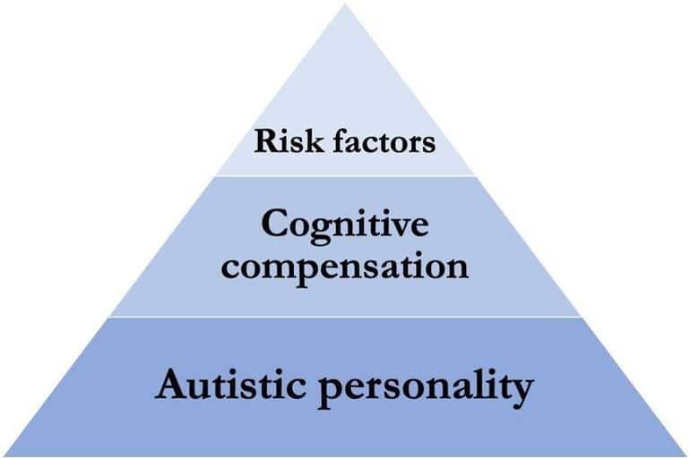
Three contributing factors

The model links three contributing factors. Together, these result in a pattern of behavior that meets the criteria for an autism diagnosis:

  1. Autistic personality—hereditary common genetic variants that give rise to an autistic personality.
  2. Cognitive compensation—intelligence and executive functions, such as the capacity to learn, understand others, and adapt to social interactions.
  3. Exposure to risk factors—for example, harmful genetic variants, infections, and other random events during gestation and early childhood that adversely affect cognitive ability.

“The autistic personality is associated with both strengths and difficulties in cognition but does not, as such, mean that diagnostic criteria are fulfilled. Still, exposure to risk factors that inhibit people’s cognitive ability may affect their capacity to tackle difficulties, which contributes to individuals being diagnosed with autism,” says Darko Sarovic, physician and postdoctoral researcher at Sahlgrenska Academy, University of Gothenburg, who wrote the thesis.

The model makes it clear that it is the many different risk factors combined that bring about the major differences among individuals on the spectrum. The various components of the model are supported by results from previous research.

Adaptive ability

High executive functioning skills may enable people to compensate for their impairment in such a way as to mitigate the symptoms, which reduces their risk of meeting the diagnostic criteria for autism. This may explain why, at group level, researchers observe a lower degree of intelligence among people diagnosed with autism, as well as other neurodevelopmental conditions.

This shows a pyramid diagram
The three factors have different weights, and jointly build up to the diagnosis of autism. Credit: University of Gothenburg

It also affords an understanding of why intellectual disability is more common among these groups. Thus, the model indicates that low cognitive ability is not part of the autistic personality but, rather, a risk factor that leads to diagnostic criteria being met.

“The autistic personality is associated with various strengths. For example, parents of children with autism are overrepresented among engineers and mathematicians. The parents themselves have probably been able to compensate for their own autistic personality traits and thus not met the criteria for an autism diagnosis.

“The impact of the disorder has then become more noticeable in their children owing, for instance, to an exposure to risk factors and relatively low cognitive ability,” Sarovic says.

Difference between girls and boys

The diagnosis of autism is more common among boys than girls, and girls often get their diagnosis later in life. Some girls reach adulthood before being diagnosed, after many years of diffuse personal difficulties.

“Girls’ symptoms are often less evident to other people. It’s well known that girls generally have more advanced social skills, which probably means that they’re better at compensating for their own difficulties. Girls also tend to have fewer autistic traits and be less susceptible to the effects of risk factors. Accordingly, the model can help to answer questions about the gender gap,” Sarovic says.

Research and diagnostics

The model also proposes ways of estimating and measuring the three factors (autistic personality, cognitive compensation and exposure to risk factors). This makes it possible to use the model in the planning of research studies and interpretation of their results.

Diagnostics is another conceivable area of use. In a pilot study in which 24 participants had been diagnosed with autism and 22 controls had not, measuring the three factors of the model enabled more than 93% to be correctly assigned to the right category. The model can also be used to explain the inception of other neurodevelopmental disorders, such as schizophrenia. Development of Theoretical Models, Classification Methods, and Biomarkers

Autism Spectrum Disorder (ASD) is an umbrella term for a group of neurodevelopmental disorders (NDD) which are behaviorally defined by the presence of difficulties with social communication, and behavioral rigidity and repetitiveness, including sensory disturbances.

The overarching aim of this thesis was to improve the categorization of autism through the development of a theoretical framework and a multivariable classification method, and identify biomarkers which together would aid in the understanding of autism and be used in ASD classification.

Paper I presents a theoretical framework for the pathogenesis of ASD and other NDDs.

The framework conceptualizes and operationalizes a three-factor model: (1) a disorder personality type that is specific for each NDD diagnostic category, but extends across the threshold for diagnosis and is not maladaptive in and of itself; (2) cognitive capacity as the ability of the individual to compensate for issues that may arise from of a “pronounced” personality type; (3) neuropathological burden which is conceptualized as the inhibition of neural and cognitive development resulting from the presence of neurodevelopmental risk factors.

It is concluded that such a framework may contribute to an improved understanding of pathogenetic mechanisms underlying NDDs, including ASD.

Papers II-IV are based on a structural and functional brain imaging study of a group of adult males with ASD, and an age- and IQ-matched group of neurotypical controls.

Paper II is a morphometric study that presents a multivariable classification method which showed up to 79% accuracy for diagnostic status, and which outperformed machine learning algorithms on the same dataset.

Paper III investigated the source space magnetoencephalographic activation in the right fusiform gyrus in response to faces and face-like objects and found only late post-stimulus group differences, potentially relating to differences in top-down cognitive mechanisms.

Paper IV compared the change in occipital magnetoencephalographic power in the gamma range in response to moving stimuli and showed a relationship with self-reported sensory sensitivity across both the ASD and control groups.

In summary, the thesis presents a theoretical framework that proposes pathogenetic mechanisms for ASD and other NDDs, a simple classification method for multivariable categorization using quantitative data, and biomarkers for face processing and sensory sensitivity.

Brain Area Necessary for Fluid Intelligence Identified


Summary: Researchers have identified and mapped regions of the brain associated with fluid memory, or the human ability to solve problems without prior experience.

Source: UCL

A team led by UCL and UCLH researchers have mapped the parts of the brain that support our ability to solve problems without prior experience – otherwise known as fluid intelligence.

Fluid intelligence is arguably the defining feature of human cognition. It predicts educational and professional success, social mobility, health, and longevity. It also correlates with many cognitive abilities such as memory.

Fluid intelligence is thought to be a key feature involved in “active thinking” – a set of complex mental processes such as those involved in abstraction, judgment, attention, strategy generation and inhibition. These skills can all be used in everyday activities – from organising a dinner party to filling out a tax return.

Despite its central role in human behaviour, fluid intelligence remains contentious, with regards to whether it is a single or a cluster of cognitive abilities, and the nature of its relationship with the brain.

To establish which parts of the brain are necessary for a certain ability, researchers must study patients in whom that part is either missing or damaged. Such “lesion-deficit mapping” studies are difficult to conduct owing to the challenge of identifying and testing patients with focal brain injury.

Consequently, previous studies have mainly used functional imaging (fMRI) techniques – which can be misleading.

The new study, led by UCL Queen Square Institute of Neurology and National Hospital for Neurology and Neurosurgery at UCLH researchers and published in Brain, investigated 227 patients who had suffered either a brain tumour or stroke to specific parts of the brain, using the Raven Advanced Progressive Matrices (APM): the best-established test of fluid intelligence.

The test contains multiple choice visual pattern problems of increasing difficulty. Each problem presents an incomplete pattern of geometric figures and requires selection of the missing piece from a set of multiple possible choices.

The researchers then introduced a novel “lesion-deficit mapping” approach to disentangle the intricate anatomical patterns of common forms of brain injury, such as stroke.

This shows a brain
Despite its central role in human behaviour, fluid intelligence remains contentious, with regards to whether it is a single or a cluster of cognitive abilities, and the nature of its relationship with the brain.

Their approach treated the relations between brain regions as a mathematical network whose connections describe the tendency of regions to be affected together, either because of the disease process or in reflection of common cognitive ability.

This enabled researchers to disentangle the brain map of cognitive abilities from the patterns of damage – allowing them to map the different parts of the brain and determine which patients did worse in the fluid intelligence task according to their injuries.

The researchers found that fluid intelligence impaired performance was largely confined to patients with right frontal lesions – rather than a wide set of regions distributed across the brain. Alongside brain tumours and stroke, such damage is often found in patients with a range of other neurological conditions, including traumatic brain injury and dementia.

Lead author, Professor Lisa Cipolotti (UCL Queen Square Institute of Neurology), said: “Our findings indicate for the first time that the right frontal regions of the brain are critical to the high-level functions involved in fluid intelligence, such as problem solving and reasoning.

“This supports the use of APM in a clinical setting, as a way of assessing fluid intelligence and identifying right frontal lobe dysfunction.

“Our approach of combining novel lesion-deficit mapping with detailed investigation of APM performance in a large sample of patients provides crucial information about the neural basis of fluid intelligence. More attention to lesion studies is essential to uncover the relationship between the brain and cognition, which often determines how neurological disorders are treated.”

Fluid intelligence is arguably the defining feature of human cognition. Yet the nature of its relationship with the brain remains a contentious topic.

Influential proposals drawing primarily on functional imaging data have implicated ‘multiple demand’ frontoparietal and more widely distributed cortical networks, but extant lesion-deficit studies with greater causal power are almost all small, methodologically constrained, and inconclusive.

The task demands large samples of patients, comprehensive investigation of performance, fine-grained anatomical mapping, and robust lesion-deficit inference, yet to be brought to bear on it.

We assessed 165 healthy controls and 227 frontal or non-frontal patients with unilateral brain lesions on the best-established test of fluid intelligence, Raven’s Advanced Progressive Matrices, employing an array of lesion-deficit inferential models responsive to the potentially distributed nature of fluid intelligence.

Non-parametric Bayesian stochastic block models were used to reveal the community structure of lesion deficit networks, disentangling functional from confounding pathological distributed effects.

Impaired performance was confined to patients with frontal lesions [F(2,387) = 18.491; P < 0.001; frontal worse than non-frontal and healthy participants P < 0.01, P <0.001], more marked on the right than left [F(4,385) = 12.237; P < 0.001; right worse than left and healthy participants P < 0.01, P < 0.001].

Patients with non-frontal lesions were indistinguishable from controls and showed no modulation by laterality. Neither the presence nor the extent of multiple demand network involvement affected performance. Both conventional network-based statistics and non-parametric Bayesian stochastic block modelling heavily implicated the right frontal lobe.

Crucially, this localization was confirmed on explicitly disentangling functional from pathology-driven effects within a layered stochastic block model, prominently highlighting a right frontal network involving middle and inferior frontal gyrus, pre- and post-central gyri, with a weak contribution from right superior parietal lobule. Similar results were obtained with standard lesion-deficit analyses.

Our study represents the first large-scale investigation of the distributed neural substrates of fluid intelligence in the focally injured brain. Combining novel graph-based lesion-deficit mapping with detailed investigation of cognitive performance in a large sample of patients provides crucial information about the neural basis of intelligence.

Our findings indicate that a set of predominantly right frontal regions, rather than a more widely distributed network, is critical to the high-level functions involved in fluid intelligence. Further they suggest that Raven’s Advanced Progressive Matrices is a useful clinical index of fluid intelligence and a sensitive marker of right frontal lobe dysfunction.

Comparative efficacy between Fexofenadine and Chlorpheniramine maleate in the treatment of allergic rhinitis.


Allergic rhinitis is a hypersensitivity disease of the nasal mucous membrane mediated by immunoglobulin E (IgE), which is characterised by itching, sneezing, watery nasal discharge, and nasal obstruction. Antihistamines have been proven to be highly effective in providing symptomatic relief of allergic diseases. They are competitive blockers of histamine at the H1 receptor.

A study was conducted to compare the efficacy of Chlorpheniramine maleate (first-generation antihistamine) and Fexofenadine (second-generation antihistamine) in patients with allergic rhinitis.

  • The mean total symptom score was reduced with both treatments. Patients receiving  Fexofenadine experienced a complete change in median symptoms score, whereas those receiving Chlorpheniramine reported a significant reduction.
  • In comparison to Chlorpheniramine, Fexofenadine was found to be relatively more effective in relieving sneezing, nasal obstruction, and nasal discharge- the three main symptoms of allergic rhinitis. Any sedative activity was not documented with the use of Fexofenadine.
  • As far as adverse effects are concerned, Fexofenadine is thought to be better tolerated than Chlorpheniramine maleate. 
  • Fexofenadine and Chlorpheniramine did not produce any significant impact on absolute eosinophil count (AEC).


Therefore, Fexofenadine is more effective in alleviating the symptoms of allergic rhinitis as compared to Chlorpheniramine. Both the antihistamines had no significant effect on the AEC

Recent updates on intranasal vaccine for COVID-19 and its benefits.


Coronavirus disease 2019 (COVID-19) is a highly contagious viral disease caused by severe acute respiratory syndrome coronavirus 2 (SARS-CoV-2) that has emerged as the most consequential global health emergency.[1] The World Health Organisation (WHO) is committed to maintaining the momentum for expanding access to COVID-19 vaccinations and supporting countries in accelerating vaccine distribution to save lives and prevent individuals from becoming critically ill. The Indian government has announced strict precautionary measures in response to the virus’s widespread distribution worldwide and has encouraged individuals to receive the full course of vaccinations, including the booster dose.

Recently, the Central Drugs Standard Control Organisation (CDSCO) has approved world’s first intranasal vaccine (BBV154) for COVID-19 developed by India for restricted use in emergency situations for patients aged ≥18 years, for heterologous booster doses.

BBV154 is a recombinant replication-deficient adenovirus vectored vaccine with a pre-fusion stabilised spike protein. It has been designed specifically to facilitate intranasal delivery via nasal drops. The nasal delivery system has been formulated and developed to be cost-effective in low- and middle-income countries.[2]

Benefits of intranasal vaccine:

  • An intranasal vaccine induces a broad immune response by neutralising immunoglobulin G (IgG), mucosal IgA, and T cell responses.
  • The prevention of COVID-19 infection and transmission depends on immune responses at the infection site (in the nasal mucosa).
  • Due to the well-organised immune systems of the nasal mucosa, the nasal route provides excellent potential for vaccination.
  • It is non-invasive and needle-free. There are no risks of needle-associated injuries and infections.
  • It is easy to administer and does not require trained healthcare workers.
  • It has high compliance.
  • It is able to meet global demand.
  • This vaccine is stable at 2-8°C for easy storage and distribution.
  • This vaccine facilitates the rapid development of variant-specific vaccines and simple nasal delivery that allows mass immunisation to protect against emerging variants of concern.

This vaccine has produced positive results in clinical trials. Therefore, it has the potential to be a crucial tool in mass vaccinations during pandemics and endemics.

Efficacy and safety of Azithromycin and Amoxicillin/Clavulanate for otitis media in children


Otitis media is a middle ear infection which can exist in an acute or chronic state. The condition may or may not manifest any symptoms. Bacteria or viruses are the aetiological agents. The majority of children suffer from acute otitis media (AOM) once by the age of 3 years, but some of them may even experience six or more recurrences before the age of 3 years.

Amoxicillin and other beta-lactam antibiotics that have been considered standard drugs for the treatment of otitis media are becoming less effective due to the emergence of resistance. Bacteria and viruses are the aetiological microorganisms causing otitis media, and beta-lactams are effective against bacteria only. However, Azithromycin possesses antiviral, antibacterial, as well as anti-inflammatory activity. 

A clinical study was conducted in order to evaluate the efficacy and safety of Azithromycin and Amoxicillin/Clavulanate for treating otitis media in children.

  • The efficacy evaluation of both these drugs was based on clinical or bacteriological efficacy. The efficacy of Azithromycin was comparable to that of Amoxicillin/Clavulanate. 
  • Azithromycin was found to be safer and more tolerable by children compared to Amoxicillin/Clavulanate. 
  • The clinical adverse events found in both Azithromycin and Amoxicillin/Clavulanate-treated groups were similar. The most common clinical adverse events observed in both treatment groups were gastrointestinal problems such as diarrhoea, vomiting, nausea, and abdominal cramp.

Therefore, Azithromycin can be considered a drug of choice for treating otitis media in children.

World’s first vaccine for honeybees approved for use by United States.


https://www.wionews.com/world/worlds-first-vaccine-for-honeybees-approved-for-use-by-united-states-report-549625

Is chashew nuts good in Diabetes?


https://www.healthifyme.com/blog/cashews-for-diabetes/