COVID Causes Personality Changes


Long-COVID can lead to a variety of brain and personality-altering symptoms. (Shutterstock)

Long-COVID can lead to a variety of brain and personality-altering symptoms.

Published in 2022, a PLOS ONE research study performed longitudinal assessments of personality changes in 7,109 Americans, aged 18 to 109, during the COVID-19 pandemic. 

It is reported that, compared to the pre-pandemic period, people became less extroverted, open, agreeable, or conscientious. What is more astonishing is that these changes were equivalent to about one decade of normative personality change if occurring during a non-COVID period. 

Epoch Times Photo

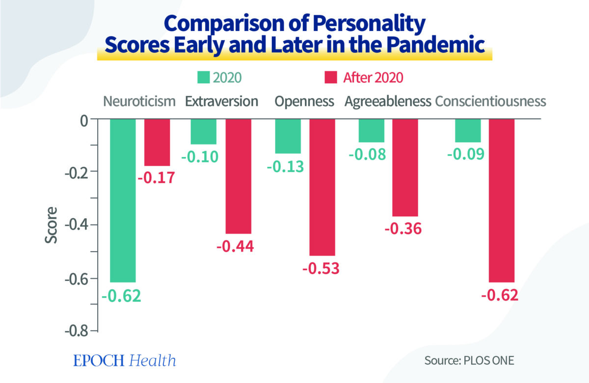
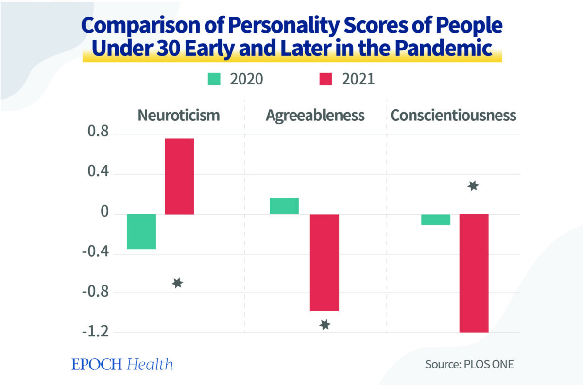
Strikingly, younger adults showed disrupted maturity by a significant increase in neuroticism and a significant decline in agreeableness and conscientiousness. 

Epoch Times Photo

The 5 Factors of Personality Traits

Personality is the solidified reaction mode of one’s thoughts, emotions, and actions. As people mature, their personality is relatively stable regardless of the environmental simulations.

There are many classification systems for personalities. The five-trait personality system was defined by five factors: namely, Openness, Conscientiousness, Extraversion, Agreeableness, and Neuroticism (OCEAN). 

Openness is the likelihood of being creative and unconventional. 

Conscientiousness describes a person’s qualities of organization, discipline, and responsibility.

Extraversion is not only the level of a person’s likelihood to interact with others but also indicates the main source of one’s energy. 

Agreeableness refers to whether a person is empathetic, helpful, and friendly.

Neuroticism measures if someone is inclined to experience negative emotions and is vulnerable to stress. 

In summary, except for neuroticism, the higher scores indicate positivity and maturity. As a person ages, the scores should be higher. For neuroticism, a low score is more desirable. 

Reasons for the Decreased Personality Scores During the Pandemic

The research did not provide data for the COVID infection and vaccination. We would like to discuss potential contributory factors causing these personality changes.

First, if people were infected by COVID-19, the SARS-CoV-2 virus itself has been proven to change one’s brain structure. This research found that those infected with SARS-CoV-2 experienced cognitive deficits equivalent to about 20 years of aging.

A study found that COVID-19 can cause neurological and psychiatric issues, such as brain fog, anxiety, and depression. People experienced higher risks of developing memory loss or inability to think coherently for as long as two years. 

Long-COVID can lead to a variety of brain and personality-altering symptoms. These include traumatic brain injury and neurodegenerative conditions such as Parkinson’s and Alzheimer’s. It can change the way that people experience, interpret, and understand the world; it can make emotions unstable. 

COVID-19 inflammatory cytokine storms trigger an uncontrolled immune response that may permanently destroy brain cells.

The National Geographic magazine reported a case in early 2020. Lorna Breen, a respected 49-year-old New York emergency room physician and former medical director of New York-Presbyterian Allen Hospital in Manhattan, was considered brilliant and energetic with no history of mental illness. After she was infected with the virus, Breen became confused and hesitant. She had to be hospitalized in a psychiatric ward. Shortly after discharge, Breen committed suicide.

Secondly, long-lasting emotional stress can alter one’s immune system. Even without the virus, persistent stress can affect physical health. 

Similar to viruses inducing changes in our immune system, the body reacts to emotions as strongly as immune cells react to antigens. Invisible, persistent emotions can cause persistent, chronic inflammation, so as to affect people’s personalities.

Robert Ader, professor of psychiatry at the University of Rochester Medical Center and founder of psychoneuroimmunology, proposed a theory that the human mind can influence the immune system’s ability to fight disease.

Our immune system is very responsive to our emotions, thoughts, and beliefs. These factors dictate whether or not we will get sick. This is the direct manifestation of the mind-body connection. 

The connection is mediated via multiple “currencies,” such as neurotransmitters, hormones, and cytokines produced by immune cells (e.g., interleukins, Tumor Necrosis Factor).

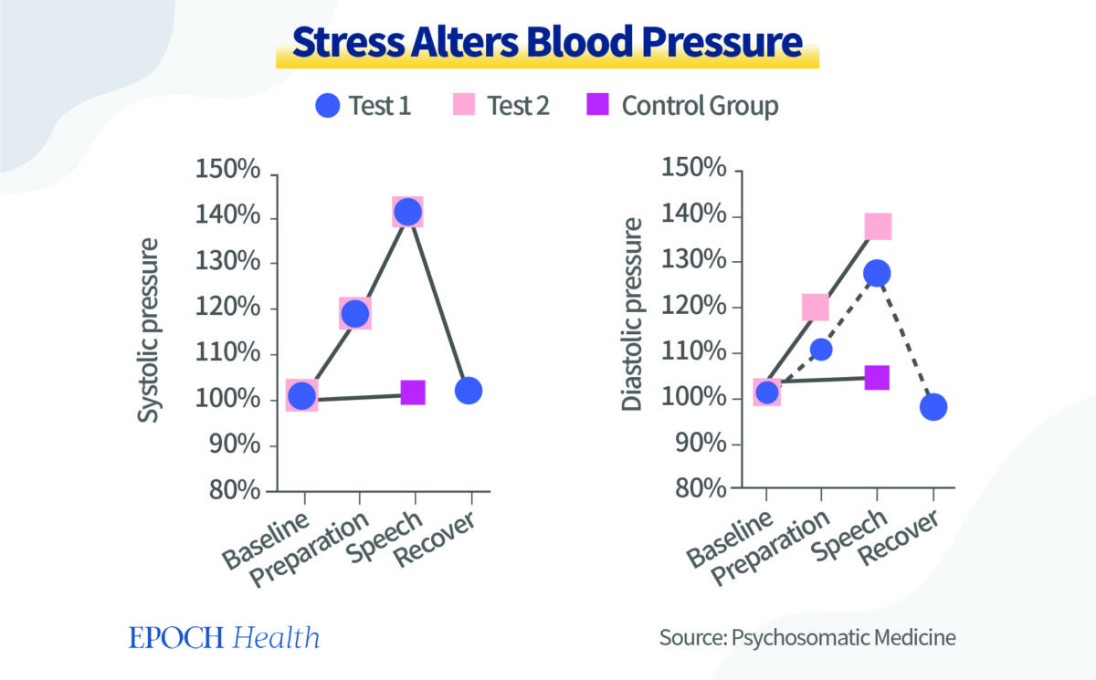
A study published in 1996 in Psychosomatic Medicine conducted a classic psychological test. One group of 79 healthy people was asked to speak publicly in front of an audience to produce a kind of social fear. Another group of 30 healthy people was asked to speak privately.

Half of the people being tested were male and half were female. The average age was 20 to 23. They were college freshmen and majored in psychology. Two tests were done after the public speech. One test had a recovery stage and the other one had no recovery stage.

The result showed that the social fear induced by public speaking caused hypertension and increased heartbeat. 

Epoch Times Photo

Levels of cortisol, the stress hormone, are also elevated during stress. When cortisol is elevated, immunity is affected accordingly. 

During the time from 9 a.m. to noon, when the experiment was done, normally there should be a steady decline in the stress hormone level. And indeed, for the control group, it decreased by 25 percent. But in the experimental group, it did not decline but increased by five percent. 

The tension brought on by short-term stress affects blood pressure and heart rate through the sympathetic-adrenal glands and affects the immune system through stress hormones. It can also affect or even change the functional state of the brain’s immune cells, the way they respond to external stimuli, and thus influence personality changes. 

This study found that repeated social defeat stress caused the increase of pro-inflammatory cytokines in adult male rats.  

Even though the cytokines themselves are too big to enter the brain, the signal from the cytokines can reach the brain and affect the function of immune cells in the nervous system, thus worsening the chronic inflammation of the nervous system, and resulting in a vicious cycle. 

When the brain is in a persistent inflammation state, it can change a person’s mental state and eventually lead to personality changes.

When Under Stress, Rationality Is Better Than Venting

Many people are used to letting out their negative emotions when under stress. 

Because stress and depression are also substances, if they accumulate inside our body too much, these negative substances make us feel uncomfortable and our body naturally wants to get rid of them. 

Releasing emotions is our natural reaction, but it is not necessarily rational and does not necessarily solve the issue, either.

The difference between human beings and other forms of life is that humans have rationality. There are rational ways to remove negative emotions. When you feel angry or frustrated, try the following tips first:

  1. Take a break: Stop talking, and walk away from the situation that makes us angry.
  2. Try a few simple movements, take a deep breath, or hold a fist.
  3. Find a quiet place and talk to yourself by heart: “What happened? Are my negative feelings helpful to the situation? Can it really help solve the problem? What’s the source of my negative emotion? What should I do to fundamentally solve the problem?”

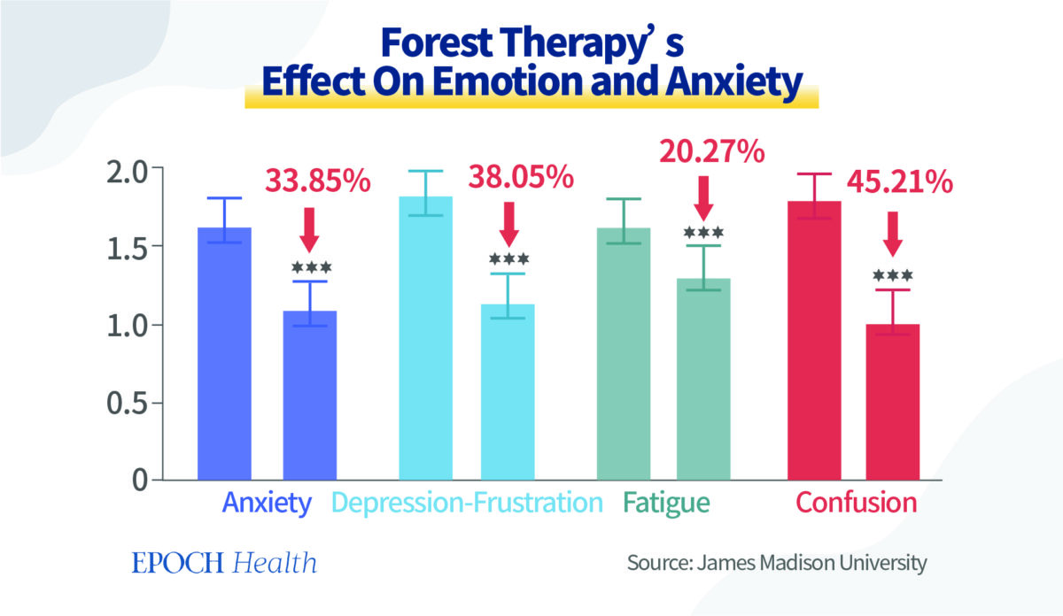
Forest Therapy Has Positive Effect on All Scores

Our minds and spirit are made of microscopic substances. If we want to improve the level of our mental health, we should nourish our minds or spirit with an equivalent level of substances. 

In the summer and fall of 2018, in a psychiatric hospital in northern Poland, forest therapy was practiced by walking in the forest for one hour and 45 minutes, accompanied by a therapist.

Forest therapy had a positive effect on almost all scores, with the greatest improvement in “Confusion” and “Depression-Frustration.” Anxiety or tension also decreased significantly. Vitality improved by 40 percent. No significant change was seen in the “Anger – Hostility” score.

Epoch Times Photo

We’ve mentioned before that horticultural therapy lowers cortisol levels, and reduces stress and negative emotions.

One of the theories to explain the effects of forest therapy and horticultural therapy is that the energy field of plants could nourish the positive energy level of our human mind and spirit. 

Light Therapy and Photobiomodulation Improves SAD 

For some people, mood changes come with the change of seasons, called seasonal affective disorder (SAD). 

When the days become shorter in the fall and winter (called “winter blues”), one may begin to feel “down” and start to feel better in the spring. The symptoms include sleep disorders or oversleeping, overeating, weight gain, social withdrawal, anxiety, and violence.

Bright light therapy (BLT) is recognized as a first-line therapy for SAD. For this treatment, patients sit in front of a very bright light box for 30 minutes to two hours a day, usually first thing in the morning, from fall to spring. 

Two meta-analyses of eight randomized blind and controlled studies (n=703 total) were consistent in demonstrating that BLT showed efficacy in the treatment of SAD when compared to a control condition.

In a placebo-controlled study, the majority of patients with SAD (61 percent of 33 patients) who received bright light therapy during the four-week study period reached symptom remission versus only 32 percent of the 31 patients receiving the placebo who achieved remission during the study, and the difference was statistically significant (p<0.05). 

Thus, BLT is proven not only to be an effective treatment for SAD but also powerful enough to bring patients with depression into remission.

Since the destructive UV rays are filtered out, it is relatively safe for most people. However, people with certain eye conditions or those taking certain medications that increase sensitivity to sunlight may need to be careful.

Psychological therapy, antidepressant therapy, and vitamin D can also be used in a combined way.

Ordinarily, people can simply rely on sunlight. Sunny weather makes one feel better. Sunlight enters the hypothalamus, where our pineal gland secretes melatonin, serotonin, and dopamine, improving day and night rhythms, boosting energy, elevating mood, and improving sleep.

There is always more sunlight outdoors than indoors, even if it’s a cloudy day. 

Scientific reasons for beneficial health effects associated with sunlight could perhaps be explained by photobiomodulation (PBM), by infrared and near-infrared light from sunlight. 

Infrared and near-infrared (NIR) radiation has profound effects on human physiology, especially as a mitochondrial stimulant, which increases ATP—cellular energy production; enhances the activity of cytochrome c oxidase as part of the mitochondrial respiratory chain; reduces lung inflammation; improves cell survival; causing reduction of TLR-4 dependent inflammatory response pathway

As most of these pathological patterns have been manifested in SARS-CoV-2 infection, accordingly, PBM has been suggested as a potential method for the treatment and prevention of COVID-19

Furthermore, transcranial PBM has beneficial effects on a range of neuropsychiatric diseases, including depression and anxiety, Alzheimer’s disease, Parkinson’s disease, stroke, and traumatic brain injury

Of all the waves in sunlight, near-infrared radiation (NIR-A), in the power range of 10 to 15 W at 810 and 980 nm, can be biologically beneficial at 3 cm depth.

Since 2020, COVID-19 has caused far-reaching negative effects on humans at physical, mental, and spiritual levels. It could even worsen our personality regardless of whether we are infected by the virus directly or not. 

The shocking impact of personality decline by more than 10 years during the COVID pandemic is enough for us to reflect on the current measures that we have taken to prevent or treat COVID-19. 

Humans are holistic beings of the physical body, mind, and spirit. To heal the trauma in our personality level, we must turn to the natural ways again—embrace the trees and enjoy the sunlight. Nature is a great source of enormous healing power. 

Autopsies Show COVID-19 Vaccination Likely Caused Fatal Heart Inflammation: Study


Syringes with COVID-19 vaccines in Berlin, Germany, on Feb. 28, 2022. (Carsten Koall/Getty Images)

Syringes with COVID-19 vaccines in Berlin, Germany, on Feb. 28, 2022.

A serious side effect linked to COVID-19 vaccines can lead to death, according to a new study.

Post-vaccination myocarditis, a form of heart inflammation, was identified in a subset of people who died “unexpectedly” at home within 20 days of receiving a COVID-19 vaccine. Researchers analyzed autopsies that had been performed on the people and conducted additional research, including studying tissue samples.

Researchers started with a group of 35, but excluded 10 from further analysis because other causes of death were identified. Of the remaining 25, researchers identified evidence of myocarditis in five.

All of the five people received a Moderna or Pfizer vaccine within seven days of their death, with a mean of 2.5 days. The median age was 58 years. None of the people had COVID-19 infection prior to being vaccinated and nasal swabs returned negative.

Autopsy findings combined with the lack of evidence of other causes of death and how the vaccination happened shortly before the deaths enabled researchers to say that for three of the cases, vaccination was the “likely cause” of the myocarditis and that the cardiac condition “was the cause of sudden death.”

In one of the other cases, myocarditis was believed to be the cause of death but researchers detected a herpes virus, an alternative explanation for the incidence of heart inflammation. The remaining case did not include an alternative explanation for the myocarditis but the researchers said the impact of the inflammation was “discrete and mainly observed in the pericardial fat.” They classified the two cases as possibly caused by vaccination.

“In general, a causal link between myocarditis and anti-SARS-CoV-2 vaccination is supported by several considerations,” the researchers said, including the “close temporal relation to vaccination”; the “absence of any other significant pre-existing heart disease”; and the negative testing for any “myocarditis-causing infectious agents.”

Limitations included the small cohort size.

The study (pdf) was published by Clinical Research in Cardiology on Nov. 27. The researchers all work for Heidelberg University Hospital. They were funded by German authorities.

Moderna and Pfizer did not respond to requests for comment.

The meticulous ruling out of possible causes apart from vaccination signals that the cases are “the tip of the iceberg,” Dr. Andrew Bostom, a heart expert based in Rhode Island, told The Epoch Times.

“If there’s a seemingly healthy person that dies suddenly in their sleep, essentially, these are typically the cases that are autopsied, and clearly the most common finding is some form of atherosclerotic coronary heart disease. But they basically ruled that out in these cases. And then they came up with the most plausible proximate cause being vaccination,” he said. “And so it suggests that the phenomenon could actually be broader than it’s been suspected to be.”

Myocarditis

Myocarditis is a serious heart condition that can manifest as chest pain and typically leads the sufferer to seek hospital care.

Doctors usually advise against all or most physical activity for a period of time.

Causes include bacteria, viruses, and fever.

Acute myocarditis resolves in about half of cases in the first two to four weeks, researchers have found, but another quarter feature longer-term problems and many of the rest lead to death or heart transplantation.

The incidence of myocarditis among COVID-19 vaccine recipients was higher than expected, researchers in the United States, Israel, and other countries have found. The highest rates have been detected in young people, particularly young males.

Estimates of the typical myocarditis incidence rates are 0.2 to 2.2 per million persons within seven days. Reports to the Vaccine Adverse Event Reporting System show higher rates for males aged 5 to 49 and females aged 12 to 29. The highest rate was 75.9 per million second doses administered. Reports to the system don’t prove causality but the system suffers from severe underreporting, according to studies, indicating the rates are even higher.

The U.S. Centers for Disease Control and Prevention (CDC) continues to recommend vaccination for virtually all people aged 6 months and older, asserting that the benefits of the vaccines outweigh the risks. Some experts disagree, saying side effects like myocarditis tilt the calculus to the risks being higher in some age groups.

Government officials have repeatedly said that most of the myocarditis cases resolve within weeks, but CDC researchers found in September that many youths who experienced post-vaccination myocarditis still had abnormal MRI results months later.

The incidence has been much lower among older people, according to U.S. authorities, which have refused to make public the autopsy results of people who die after vaccination, and various studies.

The new study “suggests we’ve been missing some severe myo[carditis] cases in our studies,” Dr. Tracy Høeg, an epidemiologist who advises the Florida Department of Health, said on Twitter.

Causality

Several vaccines have been linked to myocarditis and a related condition, pericarditis. They are made by Moderna and Pfizer and are the two most widely administered in the United States and Germany.

Both vaccines utilize messenger RNA (mRNA) technology.

Causality means that a vaccine causes a condition.

Top CDC researchers have said (pdf) the current evidence shows a causal link between the mRNA shots and heart inflammation. Other researchers have also reached that conclusion.

The U.S. Food and Drug Administration warns potential vaccine recipients that “postmarketing data demonstrate increased risks of myocarditis and pericarditis, particularly within 7 days following the second dose.”

Bostom said the evidence he’s reviewed shows a causal link.

“It’s as certain as most associations that we say are confirmed in medicine,” he said.

Some studies have identified COVID-19  as another cause of myocarditis and pericarditis, but others have indicated it might not be associated.

Other Autopsy Findings

Before the German study, other researchers around the world had reported findings from autopsies of people who died suddenly after vaccination.

In 2021, U.S. researchers reported two adults developed myocarditis within two weeks of COVID-19 vaccination, and they were unable to find causes other than vaccination.

In 2021, South Korea researchers reported that after examining the death of a 22-year-old man who died five days after receiving the Pfizer vaccine, they determined the primary cause was “myocarditis, causally-associated” with the vaccine.

In January, New Zealand researchers reported that the Pfizer vaccine was probably responsible for sudden myocarditis that led to the death of a 57-year-old woman, writing that “other causes have been discounted with reasonable certainty.”

In February, researchers in several U.S. states reported that two teenage boys who died shortly after receiving Pfizer’s vaccine experienced heart inflammation and that the inflammation was the primary cause of death.

In May, CDC researchers reported that a young boy died after experiencing post-vaccination heart inflammation, with myocarditis being pegged as the cause of death.

In September, a German researcher reported that a 55-year-old who died four months after receiving the Pfizer vaccine died of myocarditis and said “these findings indicate that myocarditis, as well as thrombo-embolic events following injection of spike-inducing gene-based vaccines, are causally associated with a[n] injurious immunological response to the encoded agent.”

And just recently, Japanese researchers reported on results from a 27-year-old man who died 28 days after admission following vaccination.

Fatty liver disease conditions ‘change the healthy heart to a failing heart’


CVD risk increases with the severity of nonalcoholic fatty liver disease, and both CV and liver events are “highly related” to the degree of hepatic fibrosis present, according to a speaker.

CVD remains the leading cause of death in adults with nonalcoholic steatohepatitis (NASH) and liver fibrosis, Bart Staels, PhD, professor at the Faculty of Pharmacy, University of Lille, France, said during a presentation at the World Congress on Insulin Resistance, Diabetes & Cardiovascular Disease. Data also show adults with biopsy-proven NAFLD have increased incidence of major adverse cardiac events, caused by atherosclerosis as well as valvular calcification and myocardial remodeling.

liver
CVD risk climbs as the severity of nonalcoholic fatty liver disease increases, and CVD remains a leading cause of death for people with nonalcoholic steatohepatitis and liver fibrosis. Source: Adobe Stock

“Patients with NASH are at elevated risk to develop CVD, but when the disease progresses — and especially when fibrosis kicks in — the risk goes much higher,” Staels said. “It is important when we discuss a link between NAFLD and the heart that we split NAFLD/NASH as one group, and [people with] fibrosis as another group.”

Connections between liver, heart

Many mechanisms could explain how the pathology of fatty liver disease has such an impact on CVD, Staels said during the presentation. People with NAFLD are often obese and have type 2 diabetes, comorbidities that further increase CV risk.

“It is no surprise that in the presence of insulin resistance and type 2 diabetes, these patients are at higher CV risk,” Staels said. “The liver plays an important role in this part of the mechanism, because hepatic insulin resistance is important with respect to the development of fatty liver, steatosis and type 2 diabetes.”

The liver also plays a role in progression of atherogenic dyslipidemia, Staels said. In the settings of insulin resistance, metabolic syndrome and type 2 diabetes, dyslipidemia is characterized by high production of VLDL particles and low HDL, further contributing to increased CV risk, Staels said.

Role of genetics

Several genes are believed to play a role in NASH and NAFLD; however, all genes linked with development of fatty liver also play a role in lipid metabolism, Staels said.

“Depending on your genetic constellation … you can have less VLDL production, meaning the lipids stay in the liver, but when you have a different [genetic] isoform, it enhances hepatic VLDL production, promoting atherosclerosis,” Staels said. “The link between the liver, lipid production and the risk it confers to the heart can be quite complicated if one tries to interpret it by identifying how genetic variants contribute.”

Emerging research also suggests apolipoprotein F (ApoF), a liver-secreted protein present on HDL and LDL particles, reduces plasma triglycerides through enhanced remnant clearance by the liver.

“We think the downregulation of ApoF in NAFLD is somehow a protection mechanism, to protect the liver from cholesterol overload, but that this happens at the expense of atherogenic dyslipidemia and possibly CVD risk,” Staels said. “We are working on this connection. This link between lipid metabolism in the liver and CV risk may also be quite complicated.”

Targeting inflammation

As in other metabolic conditions, chronic inflammation is also present in NAFLD and NASH, Staels said. Immune phenotyping in people with diagnosed NASH shows lower levels of regulatory anti-inflammatory T cells; new research aims to target immune cells as a potential NASH treatment, Staels said.

“What becomes quite clear is the metabolic components of these diseases also touch the immune cells,” Staels said. “This is due to the fact that they are also overloaded with fatty acid, inducing an inflammatory response.”

The liver-induced immune response also affects cardiac remodeling, Staels said.

In a cohort of patients who underwent catheter ablation for atrial fibrillation, Staels and colleagues observed patients presenting with metabolic associated fatty liver disease at risk for liver fibrosis exhibited increased left atrial remodeling with impaired hemodynamic, electrophysiological and histopathological properties, including altered myeloid cells and an altered immune profile.

“There really is a connection, tied to alterations in circulating immune cells,” Staels said. “We think conditions of NAFLD, NASH pathogenesis can change the healthy heart into a potentially failing heart and that immune cells and interaction with cardiac myocytes and fibroblasts play an important role.”

Liver fibrosis may drive AF

CV and liver events are highly related to the degree of liver fibrosis and liver fibrosis is likely associated with occurrence of AF, Staels said. Data also show higher liver fibrosis scores are associated with adverse atrial remodeling and AF recurrence after catheter ablation.

“This is quite an interesting observation that links NAFLD and fibrosis not only to the classical atherosclerotic model, but directly to cardiac muscle,” Staels said. “This may spawn more research to go further in that direction,” Staels said.

As Healio previously reported, eight professional societies issued a joint report on the dangers associated with NAFLD and NASH in 2021, calling on clinicians to work together across specialties and align treatment strategies.

Perspective

Scott Isaacs, MD, FACE, FACP)

Scott D. Isaacs, MD, FACE, FACP

The global prevalence of NAFLD has risen substantially, together with the obesity pandemic affecting one in three middle-aged adults in the U.S. NAFLD is considered the hepatic manifestation of the metabolic syndrome and is closely linked to visceral adiposity, insulin resistance, chronic inflammation and lipotoxicity.

Type 2 diabetes and NAFLD have a reciprocally injurious relationship mediated through common pathophysiology magnifying the risk for CV events. NAFLD is associated with hypertension and an especially atherogenic dyslipidemia with high VLDL, hypertriglyceridemia, small dense LDL particles and low HDL. As such, the leading cause of death in patients with NAFLD is CVD, not liver disease. The increased risk of both fatal and nonfatal CV events has been correlated with the severity of hepatic steatosis, inflammation, or fibrosis.

The FIB-4 score predicts CV risk, where those with FIB-4 score greater than 2.67 have a 4-fold increased risk of having a CV event compared with those with a FIB-4 score < 2.67. There is a correlation with NAFLD and atrial fibrillation and ventricular arrhythmias as well as other cardiac complications, such as heart failure with preserved ejection fraction, cardiomyopathy, and cardiac valvular calcification. However, it is difficult to establish a causal relationship between NAFLD and CVD, given the entanglement of overlapping metabolic disturbances in these individuals.

Scott D. Isaacs, MD, FACE, FACP

Adjunct Associate Professor of Medicine

Emory University School of Medicine

Source: http://www.healio.com

Critical shortcoming of NordICC trial is in interpretation of ‘screening’


“Should you still get a colonoscopy?” “A landmark study…found only meager benefits for the group of people invited to get the procedure.” “Doctors push back against European study that casts doubt.”

These are just some of the media reports surrounding the Nordic-European Initiative on Colorectal Cancer (NordICC) Study Group paper, “Effect of colonoscopy screening on risks of colorectal cancer and related death,” published in The New England Journal of Medicine in October.

HOT_WiO1222Bhatt_Blog_Graphic_01

Having witnessed the recent explosion of medical misinformation, especially during the COVID-19 pandemic, I initially wondered why this study would be circulated by one of the most respected medical journals in the world. After reading the study design in more detail, I realized that the critical shortcoming of the study is the terminology employed by the authors; specifically, the interpretation of the word “screening.”

Screening saves lives

As a gastroenterologist with a primary focus in colorectal cancer screening and prevention, I recognize that colorectal cancer is the second leading cause of cancer death among American men and women combined. Colorectal cancer affects more than 150,000 Americans every year, with 52,580 expected deaths in 2022 alone. It is estimated that 60% of these deaths could be prevented with screening.

The good news is that colorectal cancer screening rates in the U.S. have steadily increased since the 1980s, with a subsequent decrease in colorectal cancer incidence. As of 2020, 71.6% of adults aged 50 to 75 years reported being up to date with colorectal cancer screening using various modalities. However, the lifetime risk for developing colorectal cancer for an average-risk American is still one in 24. In comparison, the lifetime risk of dying in a car crash is estimated to be one in 107.

Invitation does not equal screening

The NordICC trial is a randomized controlled trial involving healthy men and women aged 55 to 64 years from population registries in Poland, Norway, Sweden and the Netherlands between 2009 and 2014. Researchers randomly assigned individuals in a 1:2 ratio to receive an invitation to undergo a colonoscopy or to receive no invitation. The invitation consisted of a letter with the appointment date and time for a prescheduled colonoscopy, educational pamphlets, and contact information. This letter was mailed about 6 weeks before the scheduled date. No other colorectal cancer screening tests were offered.

Colonoscopy was performed in 42% of the invited group, with a 50% decrease in colorectal cancer-associated death. The study estimated a 31% decrease in colorectal cancer incidence if all study participants had undergone colonoscopy. However, the risk for death from any cause in the invited group (11.03%) and the usual care group (11.04%) showed no significant difference.

The results of this study clearly show that screening colonoscopy reduces colorectal cancer — when it is performed. This translates to decreased cancer treatment and surveillance costs, as well as a diminished emotional burden. In the U.S., the conversation about completing a colorectal cancer screening test may occur over the course of several clinic visits, as patients are often hesitant to discuss “a scope going up there.” If a patient refuses screening, this decision is documented in their medical record and can be reassessed at a future appointment. Mailing a letter with an assigned procedure date and time overlooks an opportunity for patients to participate in the decision-making process. As evidenced by the study’s colonoscopy completion rate, most invitation letters likely ended up in the trash; 58% of the invited study group did not undergo screening.

In actuality, the NordICC trial assessed the response to mailed invitation, rather than the effectiveness of screening colonoscopy. Regrettably, the title wording invites misinterpretation because “screening” is used to indicate the invited study group as a whole, rather than the individuals who actually completed colonoscopy. In addition, 29% of endoscopists had an adenoma detection rate — a surrogate marker for colonoscopy quality — less than the minimum recommended threshold of 25%; as such, the decrease in colorectal cancer-associated death may have been even more pronounced with high-quality colonoscopies. Of note, the Netherlands, one of the countries involved in the NordICC trial, implemented a national screening program in 2014 with clear reduction in colorectal cancer.

Read more than the headline

Unfortunately, we live in a time when many people read headlines rather than the details. Frequently, news media is sensationalized before the facts are fully understood. Having seen the physical and emotional damage that colorectal cancer can cause, I strongly believe in colorectal cancer screening — using both invasive and noninvasive screening tests — as a preventive measure. The controversy surrounding the findings of the NordICC trial highlights the need for careful evaluation before making judgements. In an age where medical misinformation is widespread, this caveat is important for all of us to remember.

The Strong Need to Be Seen and Why It Matters


The-Need-to-Be-Seen-and-Why-It-Matters

“One of the greatest human needs is the need to be SEEN, not for who we pretend to be, but for who we truly are underneath it all.”  ~ Luminita D. Saviuc

Today I’m thinking about how most of the time we pass by one another without looking into each other’s eyes and recognizing who we truly are.

The Need to Be Seen

We walk around pretending the people around us are not people. And we pass by one another as if none of us actually exists. 

What has happened to us? 

The Need to Be Seen and Why It Matters.

We need to be seen!

Believe it or nor, we all want to be SEEN. I guess that’s one of the reasons why social media is so popular. But the kind of seeing we want to experience has nothing to do with this whole social media charade where life is presented as if it were a reality TV show and all of us are superstars. 

No. That isn’t it. 

We want to be SEEN. But SEEN for who we truly are, not for who we pretend to be.

We want people to look deep into our Core and recognize the part of us that is Sacred and Eternal. Because let’s be honest, deep down inside, you and I know that most of us are tired of pretending to be something we are not.

We want the whole world to recognize in us the part of us that is Unlimited, Eternal, and Divine. And treat us as if that’s all there is to us – Beauty, Innocence, Kindness, Purity, Light, Love, and Truth.

People need to be SEEN…

They want the whole world to look deep within their Being and discover the Greatness of who they truly are.

But who’s going to give that to them?

People on social media? Their friends and family? The strangers they can’t even look them in the eye while walking on the streets? Their bosses and co-workers?

Gabor Mate: Protecting Children from Social Media and Other Addictive Devices The Need to Be Seen and Why It Matters.

Who’s going to quench this thirst and need to be seen?

Patience. Love. And Courage.

It takes time, patience, and a great deal of love and compassion to know what human beings are truly all about; It takes courage and it takes love. And I guess that’s one of the reasons why people feel more alone and disconnected than ever. 

Cuz none of us ‘have the time’ to KNOW one another.

We’re all ‘so busy’ with being busy and becoming ‘the best of the best’ that we have no time left to open our hearts and feel the invisible love and connection that links us all together.

“On the surface, we might all look very different from one another, but at the core level, we are all the same. At the core level, we are all ONE, connected with one another in a very deep and powerful way. At the core level, we are essentially the same, all members of one human race. There is no separation except the separation we create in our minds because of our attachment to fear.” ~ Luminita D. Saviuc, 15 Things You Should Give Up to Be Happy: An Inspiring Guide to Discovering Effortless Joy

We are not machines., we need to be seen.

Yes. We have tons of virtual friends. And we exchange likes, shares, and so many great comments with all of them. But what does that have to do with anything?

We are not machines. 

We are human beings who long for deep, meaningful, and loving connections; Human Beings who long for the world to stop for a moment and realize that we exist…

Can you just breathe and look around you?

I know you are busy. And I know you have a trillion things to do. But can you stop for a moment? 

Can you just breathe and look around you? Can you notice where you are going and how fast life is passing you by?

The Need to Be Seen and Why It Matters.

All that you deem important in life always seems to be out there, never right here – never where you are, never who you are, and never with who you are.

Have you ever wondered why? Or are you too busy for that as well?

Yes, the world is in a hurry. And there are a trillion things you need to do. But none of them are more important than getting to know the Real You.  None of them are more important than learning to SEE yourself as you would want the world to SEE you.

You are your most valuable possession. The most precious gift you have is YOU. 

Do not let one day pass you by without paying attention to your Self and moving closer to your Truth. 

Let the world do what the world wants to do. And you do You. Because in the end, that’s all that truly matters.

Be true to yourself. Walk your path with integrity. And trust that in doing so, life will bless you with people and experiences that will be true to you as well.

Exposure to solid fuel for cooking linked to lung cancer mortality risk


Exposure to household air pollutants, especially solid fuel, appeared linked to lung cancer death among individuals who have never smoked, according to a study published in American Journal of Respiratory and Critical Care Medicine.

Elvin S. Cheng

“Since lung cancer patients can present with a wide range of clinical symptoms before a proper diagnosis is made, ranging from asymptomatic, some common chest symptoms (eg, cough or hemoptysis) or systemic symptoms (eg, lethargy or weight loss), to atypical presentations often due to metastasis, it remains a challenge for clinicians to make a timelier diagnosis,” Elvin S. Cheng, PhD, MPH, BScMed, MBBS, research scientist from the Daffodil Centre at the University of Sydney, told Healio. “As early detection would allow better prospect of curative treatments, a clinical history of strong exposure to solid fuels could be considered as one of the criteria for early diagnostic or screening intervention (using low-dose CT scan) for lung cancer in the never-smoking populations.”

Cooking fuel
Researchers observed that the most frequently reported household air pollutant was solid fuel use for cooking at 67%. 

In a prospective cohort study, Cheng and colleagues analyzed 323,794 individuals (mean age, 51.5 years; 89% women) who never smoked from the China Kadoorie Biobank between 2004 and 2008, to determine if household air pollution and secondhand tobacco smoking were related to death due to lung cancer.

Researchers used participants’ reports of domestic fuel use — including whether they used gas, coal, wood, electricity or other fuel and how often — to calculate the total time of household air pollution exposure. They assessed secondhand smoke exposure according to the participants’ exposure at home, the workplace or in public.

In order to find associations, researchers used Cox regression and adjusted for confounders.

Of the total cohort, 84.8% reported ever exposure to household air pollutants and 91% reported ever exposure to secondhand smoke, with 78.2% exposed to both and 2.4% to neither.

Researchers additionally observed that the most frequently reported household air pollutant was solid fuel use for cooking at 67%, followed by a “coal-smoky home” at 27%, which researchers defined as visible or smellable air pollution from use of coal burning for heating.

Individuals were studied until Dec. 31, 2016, with a median follow-up of 10.2 years. In that time, 979 individuals from the cohort died of lung cancer.

Although researchers did not find a significant association between household air pollution ever exposure and lung cancer mortality, they did find a significant log-linear positive relationship between cumulative duration of exposure and lung cancer death. Researchers observed that per every 5-year increment of exposure duration, risk for death due to lung cancer increased by 4% (HR = 1.04; 95% CI, 1.01-1.06).

Researchers also found that the highest risk for lung cancer mortality occurred among individuals who experienced 40.1 years to 50 years of exposure to household air pollution (HR = 1.53; 95% CI, 1.13-2.07). The hazard ratio for individuals who experienced more than 50 years of exposure was 1.27 (95% CI, 0.93-1.73).

Peter Ka Hung Chan

The latter hazard ratio was likely lower due to survivor bias, as people who used solid fuels for a long time tended to be elderly but healthy enough to survive without major disease, according to study researcherPeter Ka Hung Chan, DPhil, MSc, BSc, research fellow from the Oxford British Heart Foundation Centre of Research Excellence at the University of Oxford.

“Elderly who were already affected by the exposure would have a lower chance of being included in the study,” Chan told Healio.

When looking at individual factors such as sex, age, area (urban vs. rural), past history of respiratory disease and self-rated poor health in subgroup analysis of household air pollution exposure and lung cancer death, researchers observed no significant differences between the groups, except for a borderline significant difference between “poor” health (HR = 0.97; 95% CI, 0.91-1.04) and “not poor” health (HR = 1.05; 95% CI, 1.02-1.08; P for heterogeneity = .047).

In terms of secondhand tobacco smoking and lung cancer mortality, researchers found no significant association between the two factors, which Chan said is “consistent with the more recent prospective studies (as opposed to earlier retrospective studies).”

“We need more large prospective studies with more accurate assessment of solid fuel use and secondhand smoke exposure, examining not only risk of lung cancer death but also risk of developing newly diagnosed lung cancer,” Chan told Healio. “For example, we could combine questionnaire data with direct measurement of harmful chemicals in solid fuel and secondhand smoke within a prospective study, so we can potentially tease out the specific pollutants that are most relevant to lung cancer risk.”

The populations in most developed countries have seen an increase in the proportion of never-smokers due to stringent tobacco control, according to Cheng.

“However, the etiology of lung cancer among never-smokers is still largely unknown,” Cheng told Healio. “Research to study the major risk factors for lung cancer unrelated to tobacco smoke is greatly warranted to combat the impact of lung cancer as the leading cause of cancer mortality globally.”

Chan also told Healio about how these findings impact clinicians.

“Generally, I’d hope clinicians advise their patients to avoid solid fuel or secondhand smoke — eg, don’t stay too close to a barbecue for too long,” Chan said. “Often patients would get respiratory symptoms well before any other respiratory diseases like lung cancer, and those are warning signals about the potential harm on their lungs.”

This study by Cheng and colleagues adds to the literature indicating that exposure to household air pollutants needs to be studied further and global organizations need to take action on this issue to improve public health, according to an accompanying editorial byOm P. Kurmi, PhD, associate professor in epidemiology and health care research at Coventry University, U.K.

“It is too early to understand the full spectrum of diseases associated with exposure to HAP,” Kurmi wrote. “Some of these large, prospective cohorts have tried to provide better estimates; however, they often have raised some serious public health concerns, which suggests that we need to develop policies for prevention rather than waiting to understand the full spectrum of diseases and mechanisms of how some of the HAP and SHS act. Cardiorespiratory diseases and lung cancer control should be targeted to improve global health. This is important if we are serious about reducing one-third of premature mortality from noncommunicable diseases by 2030, one of the key goals of the United Nations Sustainable Development Goals.”

Source: http://www.healio.com

New guidelines on opioids for pain relief: What you need to know


Recommendations from the CDC emphasize safe, effective, and individualized options for pain relief.

A white pill in foreground and two white tablets against a dark background

Six years ago, the Centers for Disease Control and Prevention (CDC) created guidelines for prescribing opioids to help reduce the staggering number of lives lost from overdoses — a goal that unfortunately remains out of reach. As an unintended consequence, some people who were taking these medicines had trouble getting them prescribed, or getting a dosage sufficient to reduce their level of pain or avoid uncomfortable withdrawal symptoms.

Now, newly revised opioid guidelines from the CDC aim to reduce unnecessary barriers and build on best practices for prescribing and using opioids for pain. If you need relief for a chronic condition that causes you significant pain (such as disabling back problems, neuropathy pain, fibromyalgia, or osteoarthritis), here are several important takeaways from the guidelines.

Are best practices for opioid use in the new guidelines?

Yes. Many of these practices were carried forward from the 2016 guidelines. A few key recommendations are:

  • Other strategies for pain relief should always be tried first before opioids are prescribed. Opioids should not be first-line pain medicines.
  • Anyone prescribing opioids (such as oxycodone, hydrocodone, and hydromorphone) has a duty to carefully explain the possible benefits and risks, including the risk for addiction and overdose. Your doctor or medical team should help you consider whether benefits outweigh risks in your situation and continue to monitor this regularly over time. You should also discuss how to discontinue opioids if risks begin to outweigh benefits, or if these medicines don’t improve your ability to carry out your daily activities.
  • Though useful for some people, opioids are highly addictive. So are medicines known as benzodiazepines (such as lorazepam, diazepam, and alprazolam), which are used for anxiety. If combined with opioids, benzodiazepines make the risk of overdose even higher. Whenever possible, opioids and benzodiazepines should not be prescribed together.

Are opioids the best solution for many types of pain?

Frequently, the answer is no. Nonopioid pain medicines (such as ibuprofen, acetaminophen, naproxen, or topical pain relievers applied to skin) and nondrug therapies are preferred for pain that lasts up to one month (acute pain). They’re also preferred for pain lasting one to three months (subacute pain) or longer than three months (chronic pain).

Research shows these medicines are at least as effective as opioids for many painful conditions. Opioids may be prescribed to help relieve severe acute pain, like after surgery or dental procedures. However, it’s safest to take them for the shortest possible time needed to get through the worst pain — typically just a few days — and switch over to nonopioid medicines as soon as possible.

Nondrug therapies (such as physical therapy, cognitive behavioral therapy, mindfulness techniques, massage, acupuncture, and chiropractic adjustments) also may effectively relieve pain when tailored for specific conditions and situations.

Often, when a person is dealing with chronic pain, combining these strategies can help them tackle essential tasks and improve their comfort and quality of life. Talk to your medical team about the best solutions for you. This interactive tool describing options and resources for people living with chronic pain may be helpful, too.

What are some changes in the new guidelines?

Laws passed by many states in the wake of the original guidelines and the snowballing opioid crisis further restricted the ability of prescribing clinicians to treat individual patients with opioid medicines. For example, helping people taper from a higher dose of opioids to a lower one is the right choice from a health perspective for many, but not for everyone. And tapering will take some people longer than others to manage safely. Removing flexibility in how prescribing clinicians could work with their patients may have been harmful to some people.

The new guidelines

  • explain the complex nature of pain.
  • emphasize the importance of flexibility and nuance in treating individuals suffering from chronic pain.
  • recommend starting with the lowest effective dose of opioids for the shortest possible time. Risk for addiction and other side effects rises as dosage becomes higher and with the length of time opioid medicine is taken. It’s important to avoid diminishing returns in the balance of benefit and risk.
  • allow clinicians and patients to judge what treatment is best, rather than setting strict limits on dosage.
  • encourage clinicians to offer or arrange effective treatment for people with opioid use disorder, to minimize risks for withdrawal symptoms, relapse to drug use, and overdose, which is sometimes fatal.

It’s important to note that the new guidelines for opioids are not intended for pain related to cancer, pain crises in sickle cell disease, palliative care, or end-of-life care, because less restrictive use of opioids may be appropriate in such cases.

The bottom line

If you have problems with pain, talk to your doctor about the most effective combination of pain relief strategies for your situation. For many people opioids are not necessary or helpful, though some people do benefit from these medicines despite their risks. The new CDC guidelines can help patients and prescribers find this delicate balance.

Trying to lower stubbornly high LDL cholesterol?


Expert consensus focuses on individual planning to bring down elevated LDL.

An orange plastic rack holding blood test tubes with different color tops; yellow top on tube in foreground labeled "LDL Test"

Recently I met with Nancy, a 72-year-old woman with coronary artery disease, to review her latest cholesterol results. Despite taking a statin, following a healthy diet, and exercising regularly, her low-density lipoprotein (LDL) cholesterol remained above our target. “What else can I do?” she asked. “When I increase my statin dose I get terrible leg pains. But I don’t want to have another heart attack!”

When elevated, LDL contributes to cardiovascular disease, which can cause a heart attack or stroke. Taking statin drugs can drop LDL levels in most people by about 30%, substantially lowering this risk. Usually, these commonly prescribed drugs work effectively with tolerable side effects. But what if a person’s LDL level remains too high on their maximally tolerated dose? An expert consensus report issued by the American College of Cardiology lays out a clear path for next steps.

What is a healthy target for LDL cholesterol?

Target LDL depends on many factors, including your age, family history, and personal history of cardiovascular disease. For people at intermediate risk, LDL should be lowered by 30% to 50%. For those who have already had a heart attack, target LDL is no more than 70 mg/dl (note: automatic download).

Which non-statin therapies are recommended first?

Five non-statin therapies described in this post aim to help people achieve target LDL goals while minimizing side effects. They may be combined with a statin or given instead of statins.

Each helps lower LDL cholesterol when diet and statins are not sufficient, such as when there is a family history of high cholesterol (familial hypercholesterolemia). But so far, only two options are proven to reduce cardiovascular risk — the risk for heart attack, stroke, heart failure, and other issues affecting the heart and blood vessels.

Ezetimibe (Zetia)

What it does: Lowers LDL and cardiovascular risk by reducing cholesterol absorption.

How it’s given: A daily pill

Relatively inexpensive and often given with statins.

PCSK9 inhibitors, alirocumab (Praluent) and evolocumab (Repatha)

What it does: A protein called PCSK9 controls the number of LDL receptors on cells. These medicines are monoclonal antibodies against PCSK9 that increase LDL receptors on the liver, helping to clear circulating LDL from the bloodstream.

How it’s given: A shot every two to four weeks

Highly effective for lowering LDL, but expensive and may not be covered by insurance.

Three newer non-statin therapies

Three newer, FDA-approved non-statin therapies are highly effective for lowering LDL cholesterol. Whether these lessen cardiovascular risk is not yet known.

Bempedoic acid (Nexletol)

What it does: Like statins, bempedoic acid tells the liver to make less cholesterol.

How it’s given: A daily pill

Bempedoic acid is activated only in the liver, whereas statins are activated in liver and muscle tissue. Experts hope that this difference will translate to a similar LDL lowering effect, but without the muscle aches that some people who take statins report. Indeed, early trials show this medication lowers LDL cholesterol by about 20% to 25% compared to placebo.

Potential downsides include high cost and a possible increase in the risk of tendon rupture, gout, and a heart arrythmia called atrial fibrillation. Results of larger trials are expected in late 2022.

Evinacumab (Evkeeza)

What it does: Rare individuals born without a cholesterol-processing protein called ANGPTL3 have extremely low LDL and triglyceride levels, which lowers their risk for coronary heart disease by about 40%. Taking a cue from nature, scientists developed evinacumab, a monoclonal antibody that turns off ANGPTL3, mimicking this rare condition and resulting in dramatic LDL lowering of almost 50% in one trial.

How it’s given: Monthly intravenous infusion

Currently, the FDA has only approved evinacumab for people with familial hypercholesterolemia. Evinacumab appears safe in early trials, but is very expensive and can only be given in a doctor’s office.

Inclirisan (Leqvio)

What it does: Inclirisan blocks PCSK9. However, unlike alirocumab and evolocumab, which inactivate PCSK9 after it is produced, inclirisan inhibits production of PCSK9 in the liver. Inhibition of PCSK9 leads to an increase in the number of LDL receptors on the surface of the liver, speeding clearance of LDL from the bloodstream and dropping LDL by about 50% (see here and here).

How it’s given: A shot every six months

Potential downsides include increased rate of urinary tract infection, joint and muscle pain, diarrhea, and shortness of breath. This medicine is expensive and insurance may not cover it.

What does the report recommend?

It reinforces the importance of personalizing a plan to lower LDL by accounting for individual risk, cost of medication, and genetic factors. A combination of lifestyle changes and medicine can help people achieve better control of LDL. So, if you have elevated LDL cholesterol, try to follow healthy eating patterns, exercise regularly, avoid smoking and vaping, and maintain a healthy weight.

  • Statin drugs are the first choice to treat anyone who has elevated cholesterol and cardiovascular risk factors, such as diabetes and high blood pressure.
  • If statins aren’t sufficient to help you reach your LDL target, or if side effects aren’t tolerable, ezetimibe should be added next. PSCK9 inhibitors are then considered for those who remain at increased risk after adding ezetimibe.
  • If LDL targets still cannot be achieved in people with cardiovascular disease, bempedoic acid and inclirisan are considered.
  • For those with familial hypercholesterolemia, evinacumab may be appropriate.

Cardiologists eagerly await the results of studies looking at whether the three new LDL-lowering medications also lower risk for heart attack, stroke, and other poor cardiovascular outcomes. Until then, their use is likely to be limited to people at high risk for whom proven, less costly drugs cannot achieve LDL goals.

I’m too young to have Alzheimer’s disease or dementia, right?


photo of an MRI scan of a person's brain with a hand holding a magnifying glass over a portion of it

If you’re in your 80s or 70s and you’ve noticed that you’re having some memory loss, it might be reasonable to be concerned that you could be developing Alzheimer’s disease or another form of dementia. But what if you’re in your 60s, 50s, or 40s… surely those ages would be too young for Alzheimer’s disease or dementia, right?

About 10% of Alzheimer’s disease is young onset, starting before age 65

Not necessarily. Of the more that 55 million people living with dementia worldwide, approximately 60% to 70% of them have Alzheimer’s disease. And of those 33 to 38.5 million people with Alzheimer’s disease, memory loss or other symptoms began before age 65 in 10% of them. Alzheimer’s is, in fact, the most common cause of young onset dementia. A recent study from the Netherlands found that of those with a known classification of their young onset dementia, 55% had Alzheimer’s disease, 11% vascular dementia, 3% frontotemporal dementia, 3% Parkinson’s disease dementia, 2% dementia with Lewy bodies, and 2% primary progressive aphasia.

Young onset dementia is uncommon

To be clear, young onset dementia (by definition starting prior to age 65, and sometimes called early onset dementia) is uncommon. One study in Norway found that young onset dementia occurred in 163 out of every 100,000 individuals; that’s in less than 0.5% of the population. So, if you’re younger than 65 and you’ve noticed some trouble with your memory, you have a 99.5% chance of there being a cause other than dementia. (Whew!)

There are a few exceptions to this statement. Because they have an extra copy of the chromosome that carries the gene for the amyloid found in Alzheimer’s plaques, more than half of people with Down syndrome develop Alzheimer’s disease, typically in their 40s and 50s. Other genetic abnormalities that run in families can also cause Alzheimer’s disease to start in people’s 50s, 40s, or even 30s — but you would know if you are at risk because one of your parents would have had young onset Alzheimer’s disease.

How does young onset Alzheimer’s disease differ from late onset disease?

The first thing that should be clearly stated is that, just as no two people are the same, no two individuals with Alzheimer’s disease show the same symptoms, even if the disease started at the same age. Nevertheless, there are some differences between young onset and late onset Alzheimer’s disease.

People with typical, late onset Alzheimer’s disease starting at age 65 or older show the combination of changes in thinking and memory due to Alzheimer’s disease plus those changes that are part of normal aging. The parts of the brain that change the most in normal aging are the frontal lobes. The frontal lobes are responsible for many different cognitive functions, including working memory — the ability to keep information in one’s head and manipulate it — and insight into the problems that one is having.

This means that, in relation to cognitive function, people with young onset Alzheimer’s disease may show relatively isolated problems with their episodic memory — the ability to form new memories to remember the recent episodes of their lives. People with late onset Alzheimer’s disease show problems with episodic memory, working memory, and insight. So, you would imagine that life is tougher for those with late onset Alzheimer’s disease, right?

Depression and anxiety are more common in young onset Alzheimer’s disease

People with late onset Alzheimer’s disease do show more impairment, on average, in their cognition and daily function than those with young onset Alzheimer’s disease, at least when the disease starts. However, because their insight is also impaired, those with late onset disease don’t notice these difficulties that much. Most of my patients with late onset Alzheimer’s disease will tell me either that their memory problems are quite mild, or that they don’t have any memory problems at all!

By contrast, because they have more insight, patients with young onset Alzheimer’s disease are often depressed about their situation and anxious about the future, a finding that was recently confirmed by a group of researchers in Canada. And as if having Alzheimer’s disease at a young age wasn’t enough to cause depression and anxiety, recent evidence suggests that in those with young onset Alzheimer’s disease, the pathology progresses more quickly.

Another tragic aspect of young onset Alzheimer’s disease is that, by affecting individuals in the prime of life, it tends to disrupt families more than late onset disease. Teenage and young adult children are no longer able to look to their parent for guidance. Individuals who may be caring for children in the home now need to care for their spouse as well — perhaps in addition to caring for an aging parent and working a full-time job.

What should you do if you’re younger than 65 and having memory problems?

As I’ve discussed, if you’re younger than 65 and you’re having memory problems, it’s very unlikely to be Alzheimer’s disease. But if it is, there are resources available from the National Institute on Aging that can help.

What else could be causing memory problems at a young age? The most common cause of memory problems below age 65 is poor sleep. Other causes of young onset memory problems include perimenopause, medication side effects, depression, anxiety, illegal drugs, alcohol, cannabis, head injuries, vitamin deficiencies, thyroid disorders, chemotherapy, strokes, and other neurological disorders.

Here are some things that everyone at any age can do to improve their memory and reduce their risk of dementia:

Source: health.harvard.edu

Tumor-Infiltrating Lymphocyte Therapy or Ipilimumab in Advanced Melanoma


Abstract

Background

Immune checkpoint inhibitors and targeted therapies have dramatically improved outcomes in patients with advanced melanoma, but approximately half these patients will not have a durable benefit. Phase 1–2 trials of adoptive cell therapy with tumor-infiltrating lymphocytes (TILs) have shown promising responses, but data from phase 3 trials are lacking to determine the role of TILs in treating advanced melanoma.

Methods

In this phase 3, multicenter, open-label trial, we randomly assigned patients with unresectable stage IIIC or IV melanoma in a 1:1 ratio to receive TIL or anti–cytotoxic T-lymphocyte antigen 4 therapy (ipilimumab at 3 mg per kilogram of body weight). Infusion of at least 5×10⁹ TILs was preceded by nonmyeloablative, lymphodepleting chemotherapy (cyclophosphamide plus fludarabine) and followed by high-dose interleukin-2. The primary end point was progression-free survival.

Results

A total of 168 patients (86% with disease refractory to anti–programmed death 1 treatment) were assigned to receive TILs (84 patients) or ipilimumab (84 patients). In the intention-to-treat population, median progression-free survival was 7.2 months (95% confidence interval [CI], 4.2 to 13.1) in the TIL group and 3.1 months (95% CI, 3.0 to 4.3) in the ipilimumab group (hazard ratio for progression or death, 0.50; 95% CI, 0.35 to 0.72; P<0.001); 49% (95% CI, 38 to 60) and 21% (95% CI, 13 to 32) of the patients, respectively, had an objective response. Median overall survival was 25.8 months (95% CI, 18.2 to not reached) in the TIL group and 18.9 months (95% CI, 13.8 to 32.6) in the ipilimumab group. Treatment-related adverse events of grade 3 or higher occurred in all patients who received TILs and in 57% of those who received ipilimumab; in the TIL group, these events were mainly chemotherapy-related myelosuppression.

Conclusions

In patients with advanced melanoma, progression-free survival was significantly longer among those who received TIL therapy than among those who received ipilimumab.

source: NEJM