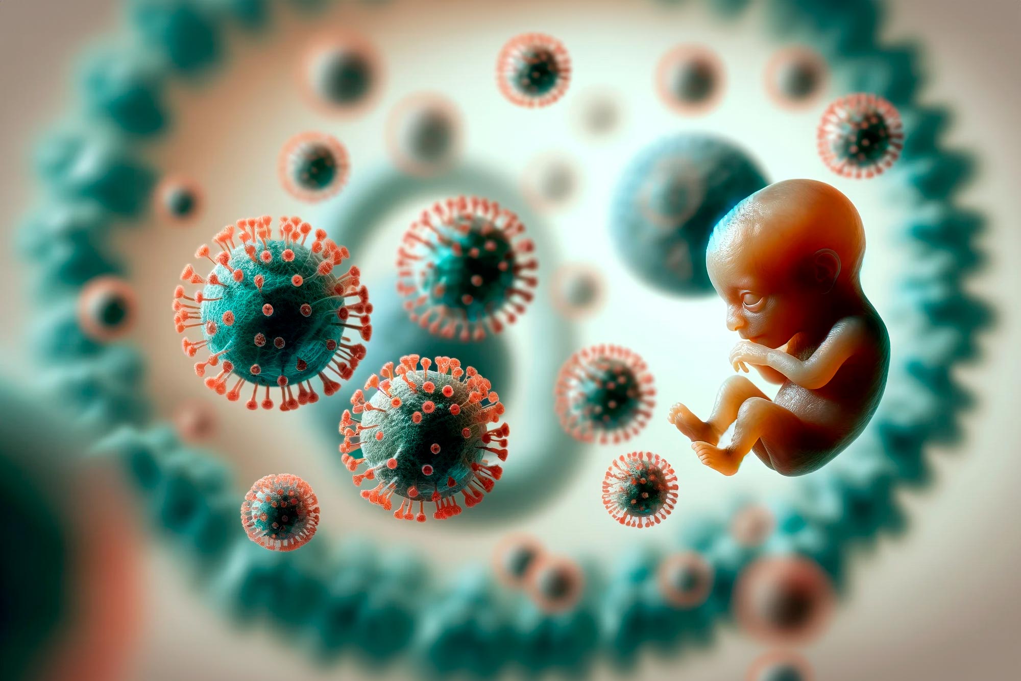
Neurologic damage following acute viral infections may be attributed to an excessive immune response to the infection, according to a new study.
Many viral infections that don’t directly infect the central nervous system (CNS) have been associated with severe neurologic disease. For these viruses, including newer viruses such as SARS-CoV-2 and Zika, the mechanism behind the association is poorly understood. To gain a better picture of how viruses may cause neurologic disease, researchers used a mouse model of Zika virus infection and identified a population of T cells that may be responsible for the damage.
“Our study ultimately finds that it isn’t the virus itself alone that causes the damage. Instead, we find that it’s a very excessive immune response to the virus,” study author Elizabeth Balint, a PhD student at McMaster University in Hamilton, Ontario, Canada, told Medscape Medical News. Balint’s research could serve as the first step toward eventually developing targeted therapies to prevent neurologic damage.
Cytotoxic T Cells
Traditionally, experts have thought that a high viral load results in more severe damage to the CNS. But surprisingly, the investigators found that the viral load did not correspond with the severity of neurologic damage. This finding suggests that a dysregulated immune response is likely at fault.
The researchers then identified a specific population of cytotoxic T cells called CD8+ T cells that became overactivated during the infection and likely caused the damage. T cells are generally thought to be antigen-specific immune cells that only attack certain pathogens. “What we found, which was quite surprising, was that there were a number of T cells that seem to be nonspecifically activated,” said Balint. The T cells appeared to be activated by a nonspecific receptor during a cytokine storm. “Lots of inflammation leads to excessive T cell activation, and then those T cells are what we’re calling ‘bystander activated’ and able to kill nonspecifically,” explained Balint.
The investigators chose to model Zika because of its status as an emerging virus that was linked to severe neurologic outcomes during the 2015-2016 epidemic. The disease is still circulating, and the mechanism for this association is poorly understood, said Balint. While the findings have yet to be tested in other models, Balint believes they may apply to viruses beyond Zika. She hopes to test other models in the future.
Specific Treatments
With a better understanding of the basic science behind how viral infections cause neurologic damage, researchers may be able to begin developing therapies to prevent and treat disease. “We’re really hoping that the more that we know about these T cells, the more specific treatments we can develop,” said Balint.
In the study, for example, the investigators experimented with an antibody that blocked a particular receptor called NKG2D to avoid activating the cytotoxic T cells. The antibody reduced cell death and prevented Zika-associated paralysis in the mice that received treatment. This antibody has been tested in clinical trials for other uses, such as for the treatment of Crohn’s disease, according to Balint.
She acknowledged that the research is in the early stages for such clinical implications but added that “it does show some promise for developing more targeted therapies.”
Difficult to Isolate
Commenting on the study for Medscape Medical News, Karl Weiss, MD, a professor of medicine at McGill University and chief of infectious diseases at Jewish General Hospital, Montreal, Quebec, Canada, said that the study advances basic science but may or may not produce applications for treatment in future research.
“In the past, whenever we tried to isolate one single element of a very complex immune system,” said Weiss, “we never were able to isolate the culprits very effectively.” That’s because the immune system includes many connected pathways and redundancies in case of failure. “If we find a way of blocking this pathway, we will prevent neurologic problems. The problem with this is that you might block this pathway, but there is probably an alternative.”
Preventing neurologic damage is an important goal, Weiss said. But because of these complexities, the research is still far from the development of any drug. Applying the science in clinical trials is a crucial next step and often the sticking point where potential treatments fail. Weiss also noted that further research using other viruses and models is needed before the findings can be generalized beyond Zika.
When considering clinical implications, Weiss said, “I think we have to be very prudent.”
A study has identified 70 virus lineages with the highest potential for causing a global pandemic, emphasizing the importance of monitoring viruses related to known human pathogens. This research supports the preparation for future pandemics by informing vaccine and diagnostic development and refining surveillance efforts to focus on the most threatening RNA viruses.
Understanding the ancestry of virus families may assist researchers in pinpointing which variants possess the potential to become Disease X, the elusive pathogen responsible for the next worldwide pandemic.
A study has identified 70 virus lineages – groups of related viruses – that pose the biggest risk. Viruses from other genetic backgrounds are unlikely to cause a high number of infections in humans, the research shows.
The findings will support ongoing efforts to monitor and prepare for future pandemics, including guiding vaccine and diagnostic development, experts say.
Understanding Disease X and RNA Viruses
Disease X is the generic term used by the World Health Organization to represent a hypothetical, unidentified pathogen that could pose a significant threat to people.
RNA viruses carry their genetic information as RNA, a structure similar to DNA. They cause many diseases, including the common cold, Covid-19 and measles, and have been responsible for most epidemics, or global pandemics, in recent history.
Monitoring RNA viruses in animal populations could help to identify those that are most likely to emerge and spread rapidly in humans. However, the huge number in circulation makes this extremely challenging and expensive.
Research Findings and Epidemic Potential
The University of Edinburgh-led research team traced the lineage, or family tree, of 743 distinct RNA virus species to track how they evolved, including all species currently known to infect humans.
Researchers compared the development of strictly zoonotic viruses – those that spread from animals to humans, but not between people – with human-transmissible viruses, which can spread within human populations.
The findings showed that viruses that can spread within human populations typically evolve separately from strictly zoonotic viruses.
Human-transmissible viruses often emerge when related viruses from the same lineage can already spread between humans.
Strictly zoonotic viruses have historically not led to epidemics in human populations. Having a close relative that can infect humans, but not spread between them, does not appear to increase the risk of epidemic potential.
Implications for Pandemic Preparedness
The research team cautioned that there is still a chance the next pandemic could come as the result of a strictly zoonotic virus – such as bird flu – or an entirely new virus. However, the findings offer a route to help streamline surveillance for Disease X among the vast number of RNA viruses in existence.
Professor Mark Woolhouse, Professor of Infectious Disease Epidemiology at the University of Edinburgh, said: “Viruses without the right ancestry don’t seem to cause epidemics. Out of potentially huge numbers of mammal and bird viruses in circulation, we should concentrate on the ones that are related to existing human viruses with epidemic potential. This research narrows the search for the next Disease X enormously.”
CNIO researchers have discovered the essential role of endogenous retroviruses in embryonic development, specifically in the transition from totipotency to pluripotency. This study, which challenges the previous notion of ‘junk DNA’, sheds light on the symbiotic relationship between viral genes and early embryonic development, with implications for regenerative medicine. Credit: SciTechDaily.com
At least 8% of the human genome is genetic material from viruses. It was considered ‘junk DNA’ until recently, but its role in human development is now known to be essential
Researchers at the Spanish National Cancer Research Centre (CNIO) describe for the first time the role of these viruses in a key process in development, when cells become pluripotent few hours after fertilization
The finding, published in Science Advances, is relevant for regenerative medicine and for the creation of artificial embryos
All animals have evolved thanks to the fact that certain viruses infected primitive organisms hundreds of millions of years ago. Viral genetic material was integrated into the genome of the first multi-cellular beings and is still in our DNA today. Researchers from the CNIO (Spanish National Cancer Research Centre) describe now in the journal Science Advances for the first time the role played by these viruses in a process that is absolutely vital for our development, and which occurs a few hours after fertilization: the transition to pluripotency, when the oocyte goes from having two to four cells.
Before this step, each of the two cells of the embryo is totipotent, i.e. it may develop inside an independent organism; the four cells of the next stage are not totipotent but are pluripotent, because they can differentiate into cells of any specialized tissue of the body.
For Sergio de la Rosa and Nabil Djouder, first author and senior author respectively, the finding is relevant for the field of regenerative medicine and the creation of artificial embryos, as it opens up a new way to generate stable cell lines in the totipotency phases. Djouder leads the Growth Factors, Nutrients, and Cancer Group at the CNIO.
We are 8% retrovirus
Genetic material from the now so-called ‘endogenous retroviruses’ was integrated into the genomes of organisms that may have been drivers of the Cambrian explosion, a period more than 500 million years ago when the world’s seas underwent a biodiversity boom. Over the past decade, genetic sequences from these viruses have been found to make up at least 8-10% of the human genome.
“Until recently, these viral remnants were considered to be ‘junk DNA’, genetic material that was unusable or even harmful,” explains De la Rosa. “Intuitively, it was thought that having viruses in the genome could not be good. However, in recent years we are starting to realize that these retroviruses, which have co-evolved with us over millions of years, have important functions, such as regulating other genes. It’s an extremely active field of research
The transition from totipotency to pluripotency, a question of pace
The research published in Science Advances shows that the MERVL endogenous retrovirus sets the pace in embryo development, especially during the specific step of the transition from totipotency to pluripotency, and explains the mechanism that makes this happen.
“It is a totally new role for endogenous retroviruses,” says Djouder. “We discovered a new mechanism that explains how an endogenous retrovirus directly controls pluripotency factors.”
This new action mechanism involves URI, a gene that Djouder’s group is researching in depth. Years ago, it was discovered that if URI is deleted in laboratory animals, embryos do not even get to develop. De la Rosa wanted to find out why, and which is how its link to the MERVL retrovirus was discovered.
A smooth transition
The findings show that one of the functions of URI is to enable the action of molecules essential for acquiring pluripotency; if URI does not act, neither do the pluripotency factors, and the cell remains in a state of totipotency. It turns out to be an endogenous retrovirus protein, MERVL-gag, which modulates the action of URI.
The researchers found that during the totipotency phase, when there are only two cells in the oocyte, expression of the MERVL-gag viral protein is high; this protein binds to URI and prevents it from acting. However, the levels gradually change, so that the levels of MERVL-gag viral protein go down and URI can enter into action: pluripotency appears.
As De la Rosa explains, “It’s a smooth transition. When there is a high expression of viral protein, there are fewer pluripotency factors; as ERV expression decreases, URI stabilizes such factors.”
Symbiotic co-evolution
“Our findings reveal symbiotic co-evolution of endogenous retroviruses with their host cells in order to guarantee the smooth and timely progression of early embryonic development,” explain the authors in Science Advances.
In other words, the three-way relationship between the viral protein, URI, and pluripotency factors is finely modulated, “to allow sufficient time for the embryo to adjust and coordinate the smooth transition from totipotency to pluripotency and cell lineage specification during embryonic development,” concludes Djouder.
This winter’s “tripledemic” is one of several recent odd trends among respiratory virus infections. Viruses, it turns out, can block one another and take turns to dominate
Three years into the pandemic, Covid-19 is still going strong, causing wave after wave as case numbers soar, subside, then ascend again. But this past autumn saw something new – or rather, something old: the return of the flu. Plus, respiratory syncytial virus (RSV) – a virus that makes few headlines in normal years – ignited in its own surge, creating a “tripledemic”.
The surges in these old foes were particularly striking because flu and RSV all but disappeared during the first two winters of the pandemic. Even more surprising, one particular version of the flu may have gone extinct during the early Covid-19 pandemic. The World Health Organization’s surveillance programme has not definitively detected the B/Yamagata flu strain since March 2020. (Read more from BBC Future about how viruses go extinct.)
“I don’t think anyone is going to stick their neck out and say it’s gone just yet,” says Richard Webby, a virologist at St Jude Children’s Research Hospital in Memphis. But, he adds, “we hope it got squeezed out.” Such an extinction would be a super rare event, Webby says.
But then, the past few years have been highly unusual times for human-virus relations, and lockdowns and masks went a long way toward preventing flu and RSV from infiltrating human nostrils. Still, Webby thinks another factor may have kept them at bay while Covid-19 raged. It’s called viral interference, and it simply means that the presence of one virus can block another.
Looking back over the past couple of years, I’m pretty confident in saying that Covid can certainly block flu and RSV – Richard Webby
Viral interference can happen in individual cells in the laboratory, and in individual animals and people exposed to multiple viruses – but it can also play out across entire populations, if enough people get one virus for it to hinder the flourishing of others at scale. This results in waves of infections by individual viruses that take turns to dominate. “Looking back over the past couple of years, I’m pretty confident in saying that Covid can certainly block flu and RSV,” Webby says.
It may be a while before we can be sure, but it looks likely that Covid-19 could block flu and RSV
It wouldn’t be the first time that scientists have observed such patterns. Back in 2009, for example, the virus to fear was swine flu, which had jumped from pigs to people in spring of that year. It looked poised to ramp up as autumn arrived – but suddenly, in some parts of Europe, it stagnated. The rhinovirus, responsible for the common cold and likely spread by children returning to school, took centre stage for a series of weeks before swine flu recaptured dominance. That flu strain then delayed the typical autumn rise of RSV by as much as two and a half months.
Running interference
There are a number of ways that interference can happen in the body. One occurs when two viruses use the same molecule to gain entry into host cells. If virus A gets there first, and grabs on to all those molecular doorknobs, then virus B will have no opportunity to enter.
Another kind of interference might happen if two viruses compete for the same resources inside the cell, such as the machinery to make new viral proteins or the means to escape that cell to infect others. “Think of it as a race between two viruses,” Webby says.
Don’t go counting on a cold granting you temporary immunity from other viruses. Interference isn’t guaranteed
But the best-understood method of interference concerns a defensive molecule called interferon that’s made by cells of all animals with backbones (and possibly some invertebrates too). Indeed, viral interference is the reason interferon got its name to begin with. When a cell senses a virus, any virus, it starts making interferon. And that, in turn, activates a slew of defensive genes. Some of the products of those genes work inside the cell or at its boundaries, where they prevent additional viruses from entering and block viruses already present from replicating or exiting the cell.
Cells secrete interferon into their surroundings, warning other cells to put up their guard. The result of all this: If a second virus then comes along, cells have their defences already activated, and they may be able to shut it out.
This “beware” message can spread throughout the body. So, in theory, getting a respiratory virus such as the rhinovirus could activate defences in, say, the gut, protecting the same person from an entirely different virus, such as norovirus. But the situation will vary depending on the viruses involved, the amount of interferon produced, and other factors. “Most of the viruses themselves have ways to neutralise the interferon system,” says Ganes Sen, a virologist at the Cleveland Clinic in Ohio, who wrote about the interactions between interferon and viruses for the Annual Review of Virology in 2015. “It’s a tug of war.”
Scientists study that back-and-forth in animals and other systems in the laboratory. For example, Ellen Foxman, an immunologist at Yale School of Medicine, investigates viral interactions in lab-grown tissues made from real human airway cells. In one experiment, she studied swine flu and a typical representative of the rhinovirus family. When Foxman and her co-authors Anchi Wu, Valia Mihaylova and Marie Landry infected the human tissue first with the rhinovirus, and then with swine flu, interferon prevented the flu from getting a foothold. In similar studies, Foxman found that rhinovirus infection also interfered with subsequent Sars-CoV-2 infection (the virus that causes Covid-19).
While some viruses can block others, it is still possible to get two viruses at once
It’s not always reliable to extrapolate from tissues in the laboratory to people or populations, but Foxman thinks the studies reflect biological truth. “It’s probable that if you get a rhinovirus infection, that’s going to make you relatively resistant to another virus for some period of time,” she says. Foxman speculates that the protective effect probably lasts days or weeks.
But don’t go counting on a cold granting you temporary immunity from other viruses. Interference isn’t guaranteed. It’s certainly possible to catch more than one virus at the same time. And interferon isn’t always beneficial, either – sometimes, it can make people more susceptible to infection, not less. A well-known example is that the flu makes people more susceptible to a secondary bacterial infection.
In the ongoing pandemic, it’s still hard to say how much of a role, if any, interference played in shutting down RSV and flu in populations around the globe. During the first Covid wave in 2020, Foxman thinks that not enough people had Covid for it to be interfering with other viruses on a grand scale. (RSV underwent an unusual summer peak in 2021 as people eased up on masking and other precautions.)
But by the second Covid winter, in 2021-22, Webby thinks he sees population-level evidence for interference. Influenza was starting to pick up in the fall, he says, but then the omicron variant of Covid burst onto the scene. Flu rates fell – even though people were back at work and school and traveling for the holidays. The coronavirus had a big advantage that season, he says, because many people still lacked immunity to it. It doesn’t mean Covid will always edge out influenza in the future.
In the third Covid winter now underway in the Northern Hemisphere, conditions are different yet again. Many people now have immunity to Covid, from a recent bout or from vaccination, but fewer have experienced RSV or flu in recent memory. That set the scene for flu and RSV to stage a massive dual comeback, hitting early and hard.
Any potential interference during the present tripledemic winter will become more obvious once epidemiologists can look back on the season and see if each virus took its turn. Already, there are indicators that the fall surges of RSV and flu might have peaked, while Covid is on the upswing after the winter holidays. But there are still several cold months to come, providing ample opportunity for any of the trio to rise again.
Question Is use of tecovirimat within 7 days of mpox symptom onset associated with lower rates of mpox disease progression among people with HIV (PWH)?
Findings In this cohort study including 112 PWH after propensity matching, those treated with tecovirimat within 7 days of mpox symptom onset compared with those who were treated after 7 days or who did not receive tecovirimat were 13 times less likely to progress to severe mpox disease.
Meaning The findings of this study support the use of tecovirimat in all PWH as soon as mpox is suspected.
Abstract
Importance Despite a lack of effectiveness data in humans, tecovirimat was widely prescribed to people with HIV (PWH) with mpox during the 2022 mpox epidemic, particularly PWH with low CD4+ T-cell counts or severe mpox clinical manifestations.
Objective To evaluate if PWH with mpox who were treated with tecovirimat within 7 days of symptom onset were less likely to have mpox disease progression.
Design, Setting, and Participants This cohort study included PWH diagnosed with mpox at 4 hospitals in Atlanta, Georgia, between June 1 and October 7, 2022. Patients were grouped according to whether they were treated with tecovirimat within 7 days of mpox symptom onset (early tecovirimat cohort) or they did not receive tecovirimat or received the drug 7 or more days after symptom onset (late or no tecovirimat cohort). Multivariable logistic regression models were used to identify factors associated with progression of mpox disease. The 2 cohorts were then matched 1:1 using propensity scores based on the identified factors, and mpox disease progression was compared.
Exposures Treatment with tecovirimat within 7 days of mpox symptom onset.
Main Outcome and Measures Progression of mpox disease, defined as the development of at least 1 severe mpox criterion established by the US Centers for Disease Control and Prevention, after symptom day 7.
Results After propensity score matching, a total of 112 PWH were included in the analysis; 56 received tecovirimat within 7 days of mpox symptom onset (early tecovirimat group) and 56 were either treated later or did not receive tecovirimat (late or no tecovirimat group). In the early tecovirimat group, the median (IQR) age was 35 (30-42) years; 54 individuals (96.4%) were cisgender men, 46 (82.1%) were Black individuals, and 10 (17.9%) were individuals of other races (American Indian or Alaska Native, Asian, Native Hawaiian or Other Pacific Islander, or White) or unknown race. In the late or no tecovirimat group, the median (IQR) age was 36 (32-43) years; 54 (96.4%) were cisgender men, 49 (87.5%) were Black individuals, and 7 (12.5%) were individuals of other races or unknown race. Mpox disease progression occurred in 3 PWH (5.4%) in the early tecovirimat group and in 15 PWH (26.8%) in the late or no tecovirimat group (paired odds ratio, 13.00 [95% CI, 1.71-99.40]; P = .002).
Conclusion and Relevance Results of this cohort study support starting tecovirimat in all PWH as soon as an mpox diagnosis is suspected. Additional research is warranted to confirm these findings.
Introduction
The 2022 global mpox outbreak disproportionately affected people with HIV (PWH).1,2 This population may develop more severe mpox disease manifestations and worse clinical outcomes,3,4 especially individuals with lower CD4+ T-cell counts5 and nonsuppressed HIV viremia,6 highlighting the urgent need for effective therapeutic agents for this population.
Tecovirimat (ST-246), an antiviral agent developed to treat smallpox, has antiviral activity against other orthopoxviruses, including mpox virus. In animal models, tecovirimat was shown to prevent morbidity and mortality associated with mpox, especially when started within 5 days of mpox inoculation.7–9
Based on these data, the US Food and Drug Administration approved tecovirimat for mpox treatment using expanded access for an investigational new drug. Data showing the effectiveness of tecovirimat for treating mpox disease in humans are lacking.10,11 Meeting enrollment goals for a randomized controlled trial assessing the efficacy of tecovirimat for mpox infection (the STOMP [Study of Tecovirimat for Human Monkeypox Virus] trial12) has been challenging due to declining number of mpox cases. We aimed to perform a matched cohort analysis to examine the association between early tecovirimat treatment (started within 7 days of mpox symptom onset) and progression of mpox disease among PWH.
Discussion
In this prospective matched cohort analysis, PWH with mpox disease who were prescribed tecovirimat within 7 days of symptom onset were significantly less likely to have progression of mpox disease compared with PWH who were not prescribed tecovirimat within 7 days of symptom onset. Results of the present study suggest that tecovirimat treatment should be started early at the time of suspected mpox diagnosis in all PWH, especially in those with nonsuppressed HIV viremia or mucosal site involvement.
Limitations
This study has limitations. First, it is possible that unmatched confounding variables could have contributed to fewer cases of severe mpox disease being observed in the early tecovirimat cohort. Second, this study has a small sample size and is inadequately powered to examine specific mpox complications or mortality. Third, most of our population (84.8%) were Black individuals, which reflected the population affected by the 2022 mpox outbreak in Atlanta but may limit generalizability to populations with different races and ethnicities. Fourth, the timing of tecovirimat initiation was determined based only on the time that the tecovirimat prescription was written, and we could not confirm if individuals took the medication or for how long. Fifth, we did not evaluate adverse events associated with tecovirimat. A large cohort study previously reported that tecovirimat is well tolerated.11
Conclusions
To our knowledge, this cohort study represents the first controlled analysis of tecovirimat for the treatment of mpox disease in PWH. The findings support starting tecovirimat in all PWH as soon as an mpox diagnosis is suspected. Additional research is needed to confirm these findings.
The year 2023 marked the 25th anniversary of the first detected outbreak of Nipah virus disease. Despite Nipah virus being a priority pathogen in the WHO Research and Development blueprint, the disease it causes still carries high mortality, unchanged since the first reported outbreaks. Although candidate vaccines for Nipah virus disease exist, developing new therapeutics has been underinvested. Nipah virus disease illustrates the typical market failure of medicine development for a high-consequence pathogen. The unpredictability of outbreaks and low number of infections affecting populations in low-income countries does not make an attractive business case for developing treatments for Nipah virus disease—a situation compounded by methodological challenges in clinical trial design. Nipah virus therapeutics development is not motivated by commercial interest. Therefore, we propose a regionally led, patient-centred, and public health-centred, end-to-end framework that articulates a public health vision and a roadmap for research, development, manufacturing, and access towards the goal of improving patient outcomes. This framework includes co-creating a regulatory-compliant, clinically meaningful, and context-specific clinical development plan and establishing quality standards in clinical care and research capabilities at sites where the disease occurs. The success of this approach will be measured by the availability and accessibility of improved Nipah virus treatments in affected communities and reduced mortality.
The increase in mpox (formerly known as monkeypox) cases in west Africa in 2017 should have served as a warning sign for potential outbreaks elsewhere. However, it was not until the multicountry outbreak in 2022, when WHO declared a public health emergency of international concern, that the disease received substantial global attention.
This mpox outbreak highlighted two issues in global health that must be addressed: high-consequence infectious diseases receive attention only when they affect high-income countries; and even when interventions are available, they might not reach those who need them most,
We must avoid repeating the same pattern with Nipah virus disease. Recurrent outbreaks in economically disadvantaged rural communities in south and southeast Asia have resulted in few attempts to understand and mitigate the disease. We should not wait until an extensive outbreak affects high-income countries before acting towards developing better treatment options and deploying them when and where they are needed.
Nipah virus is a highly lethal zoonotic paramyxovirus, which can infect humans through contact with infected bats, pigs, or humans or by consuming contaminated raw date palm sap.
The virus causes a rapidly progressive illness that affects the respiratory system and CNS, causing respiratory distress and encephalitis, with a very high case-fatality ratio. Since its discovery in 1998 and 1999 in Malaysia and Singapore, respectively, Nipah virus has caused sporadic outbreaks in Bangladesh, India, and the Philippines. Concerns exist about future outbreaks in other regions where Pteropus fruit bats, the zoonotic reservoir of Nipah virus, are found (table 1). The wide geographical distribution of these bats across Africa, south Asia, and Oceania, combined with the virus’ capacity for person-to-person transmission and evidence of ongoing viral evolution, underscores the potential for larger epidemics.
In 2018, WHO listed Nipah virus as a priority pathogen for urgent research and development and produced a roadmap to address the research needs for Nipah virus, including developing diagnostics, therapeutics, and vaccines.
Encouragingly, the Coalition for Epidemic Preparedness Innovations, primarily funded by public and philanthropic organisations, with some contributions from the private sector, has invested in the development of multiple vaccine candidates for Nipah virus. Notable progress has been made, with at least two candidates having reached in-human (phase 1) clinical trials.
In contrast, investment and progress in therapeutics development is less advanced; there are no approved therapies available for Nipah virus disease.
Clinical outcomes in patients with Nipah virus disease
Nipah virus mortality varies widely, from 9% in Singapore (the only high-income country reporting Nipah virus cases), to 100% in some transmission clusters,
The reasons for the differences in mortality rates between countries, which require further investigation, are presumably multifaceted and might include variations in the infecting Nipah virus strains (Nipah virus Malaysia vs Nipah virus Bangladesh),
patient characteristics, and access to adequate supportive care. In 2023, Bangladesh had the largest Nipah virus outbreak since 2015, with a case-fatality ratio of 71% with ten (71%) deaths out of 14 reported cases.
The continued high mortality is possibly a result of several factors, including delays in diagnosis, absence of specific therapies, and poorly effective supportive care.
The absence of laboratory infrastructure and diagnostic capabilities in rural areas where Nipah virus outbreaks occur creates challenges in early diagnosis, thereby impeding timely treatment.
Additionally, the non-specific initial signs and symptoms of Nipah virus infection, combined with the absence of point-of-care rapid diagnostic tests, often lead to delays in diagnosis in endemic areas, where many patients present with encephalitis of various causes. Distinguishing Nipah virus infection, representing only about 3% of all encephalitis cases in Bangladesh, from other causes of encephalitis is, therefore, challenging.
Treatment involves administration of oxygen, fluids, and nutritional support, and resuscitation as needed, alongside symptomatic treatment, including the use of antipyretics, anticonvulsants, medication for raised intracranial pressure, hypoglycaemia, and shock. Empirical treatment is typically initiated with antibiotics (intravenous ceftriaxone), antivirals (intravenous aciclovir), and steroids.
Patients with worsening conditions, such as deteriorating levels of consciousness, uncontrolled seizures, haemodynamic instability, and multiorgan failure are assessed for transfer to an intensive care unit or tertiary centre.
Improving patient outcomes in ongoing Nipah virus outbreaks remains a major challenge because there is no standardised supportive care available to patients in endemic areas, including timely access to intensive care units. Advancing knowledge to guide clinical care and improve outcomes for isolated Nipah virus infections and small clusters will be good preparation for a potential larger epidemic in the future.
Why is there so little progress in improving patient outcomes?
There are several key barriers to identifying and evaluating therapeutic interventions for Nipah virus disease.
Epidemiological challenges
Nipah virus disease is characterised by low prevalence even in endemic areas, with sporadic cases and unpredictable outbreaks in terms of location, size, and timing.
These characteristics present enormous methodological and operational challenges for clinical trials. Bangladesh, where the epidemiology of Nipah virus is well understood, and which has had the highest number of reported outbreaks, is a suitable site for potential Nipah virus therapeutic trials.
However, even in Bangladesh, Nipah virus infections remain infrequent and geographically dispersed, with an average of 14 confirmed cases reported each year.
The current epidemiological conditions make doing traditional phase 3 efficacy trials impossible.
Operational challenges
Nipah virus disease outbreaks occur in areas with little research infrastructure and clinical research capacity, leading to poor understanding of the clinical characteristics and outcomes of the disease and hindering the implementation of effective clinical trials. In addition, the shortage of laboratory and clinical infrastructure in rural communities in Bangladesh and India creates challenges for timely diagnosis and enrolment into research studies.
Moreover, the absence of biosafety level 4 facilities in Bangladesh to manage the high-level biocontainment requirements for handling Nipah virus, impedes locally led research on disease pathogenesis and the in-vitro and in-vivo evaluation of therapeutics.
In Bangladesh, where Nipah virus is endemic, surveillance of Nipah virus disease relies on a central laboratory located in the capital city, Dhaka. As a result, confirming a diagnosis can take several days or weeks.
Therefore, the development of rapid, ideally point-of-care, diagnostic tests and improved laboratory infrastructure and diagnostic capabilities is key for the accurate diagnosis and timely treatment of patients and prevention of disease spread.
Market and policy challenges
Apart from one inconclusive open-label trial with ribavirin during the Malaysian outbreak and a phase 1 study with the monoclonal antibody m102.4 done in healthy volunteers in Australia, no other human clinical trials have been done for any potential Nipah virus therapeutic candidates.
Little progress in therapeutic development reflects both a market failure and a global health policy failure. The small number of Nipah virus infections in countries unable to provide the substantial financial resources required to invest in the research and development process or to pay for stockpiling therapeutics, means that there is no financial incentive for profit-driven private sector investments. Equally, there have been few public investments made both at the local level and globally to support the discovery and development of new therapeutics and attract private sector actors into the types of public–private partnerships that have pushed drugs through the research and development pipeline in other disease areas, ideally creating affordable products and a sustainable local market.
There are potential therapeutic candidates for henipaviruses, including Nipah virus, currently progressing through the research and development pipeline, including at least eight small molecules and four monoclonal antibodies, which are mainly in the preclinical stage (table 2).
Only the monoclonal antibody m102.4 has phase 1 data available, and both m102.4 and ribavirin have been used on a compassionate basis during outbreaks.
To effectively evaluate potential interventions for improving patient outcomes in light of the aforementioned challenges, a coordinated, public health-focused approach must be adopted that maximises the chance of identifying and progressing the best therapeutic options in a timely way, that emphasises fairness, transparency, and equitable access to interventions for Nipah virus-affected communities.
We propose a model, similar to the West Africa Lassa fever Consortium, to generate essential elements of a pathway to develop and make available new treatments to people in need.
This model includes: laying out a clinical development plan that is regulatory-compliant, clinically meaningful, and context-specific; strengthening research sites’ capacity to meet regulatory quality standards (good clinical practices and good clinical laboratory practices); and ensuring good participatory practice for trials.
Moreover, the model recognises the need for involving key stakeholders both in Nipah virus-endemic countries and internationally, and establishing an end-to-end partnership and financing structure, in which all actors commit to the collective end goal of equitable access to effective treatments. The key aspects of this plan are summarised in the following sections.
Enhanced clinical epidemiology
A key clinical difference between Nipah virus outbreaks is that despite a consistently high proportion of patients presenting with encephalitis (about 97%), in Malaysia, fewer people had concomitant respiratory symptoms (14–29%),
The variation in pulmonary involvement is possibly caused by differences in Nipah virus strains and the route of infection. Experimental evidence indicates that the Nipah virus-B strain replicates more efficiently in human tracheal and bronchial epithelium than the Nipah virus-M stain.
Whether early ribavirin treatment during the Malaysian outbreak might have had a role in controlling virus replication in the lung, thus accounting for lower mortality, is unclear.
To gain a comprehensive understanding of the clinical characteristics and natural history of Nipah virus disease, including both encephalitis and pulmonary presentations, prospective observational clinical research is essential. This research can form the foundation for developing standardised clinical trial methodologies.
Such enhanced clinical epidemiology studies should also document current clinical care practices to identify gaps and inform the development of context-specific standard-of-care guidelines. During the Malaysian outbreak in 1998–99, ten (91%) of 11 patients cared for in a Singapore hospital survived,
suggesting that improving elements of supportive care can greatly improve patient outcomes.
Defining use cases and target product profile (TPP) for Nipah virus therapeutics
Developing safe and effective therapeutic agents to treat acute Nipah virus disease and acceptance of these agents by clinicians and patients is crucial for improving survival rates and reducing Nipah virus-associated morbidity and long-term disability. However, to design and select therapeutics that meet these objectives, we need to define use cases and a clear criterion for down-selection and prioritisation of candidate products for clinical trials.
Defining use cases of potential therapeutic options (figure) is essential to support the development of the TPP that serves as a guide for drug developers and other actors in the clinical development pathway, indicating the necessary and suitable characteristics for future therapeutic candidates after consultation with key stakeholders and end users.
The methodology for developing a TPP for Nipah virus disease should adhere to WHO principles, encompassing a major public-health need, considering end users’ perspectives, promoting access and equity, and fostering consensus among stakeholders for ethical research and drug development.
The TPP development process should also incorporate an end-to-end perspective connecting product development and regulatory, policy, and financing considerations.
This process should involve relevant stakeholders from national and international regulatory agencies, ethics boards, ministries of health, and clinicians experienced in treating Nipah virus disease. The needs and preferences of end users, clinicians, and survivors should also be incorporated into the TPP development process to define ideal treatment characteristics.
Rapid diagnostic capability, including point-of-care testing, is needed for early case detection and optimal deployment strategies for diagnostics in different geographical areas. Local laboratories must be able to do proficiency testing to monitor the reproducibility and performance of diagnostic field assays. National laboratory strategies for Nipah virus diagnosis and detection in the countries most affected by the virus should also be established.
Clinical trial design
To progress effectively, we need therapeutic candidates to advance through preclinical research, early human safety testing, and manufacturing, so that they can be tested clinically. Clinical trial protocols are also essential to ensure a coordinated and standardised assessment of the candidate drugs’ clinical safety and efficacy.
Pharmacometric trial
Repurposing existing marketed compounds for treating Nipah virus disease is a potential alternative or complement to developing new drugs, as this strategy capitalises on the existing compounds’ established safety profiles and reduces costs and time. Similar approaches were successfully used for COVID-19.
Pharmacometric modelling and simulation can be used to model in-vitro activity of potential repurposed therapeutic candidates targeting Nipah virus disease to evaluate the likelihood of clinically significant antiviral activity in vivo.
With potential preclinical data in animal models for Nipah virus therapeutic candidates (eg, remdesivir and favipiravir), pharmacometric evaluation of Nipah virus clearance rate from serial oral swabs quantitative PCR viral density estimates might offer a cost-effective and time-efficient method to prioritise potential candidates for further evaluation in clinical trials.
Core outcome sets, data variables, and phase 2/3 trial protocol
To establish a basis for the development of clinical trial methods, a core set of data variables and outcome measures should be developed and incorporated into trial protocols.
Consultation and consensus with stakeholders, such as clinicians, statisticians, clinical researchers, patients and public health representatives, regulators, and ethics boards are crucial to obtain their views on the trial design. The goal should be to use tools that are freely and publicly available, and ready ahead of time to be implemented in phase 2/3 therapeutic trials for Nipah virus disease. Although containing a recommendation for a core method, the tools should allow for context-specific adaptation and use by any member of the Nipah virus research community.
Given the challenges of phase 3 Nipah virus-specific trials in the current epidemiological conditions, a patient-centred syndromic approach appears to be pragmatic. By concentrating on encephalitis, the primary and predominant clinical presentation of Nipah virus disease patients (97% in India, 90% in Bangladesh, 88% in Singapore, 64% in the Philippines, and 55% in Malaysia), and doing trials to assess therapeutic approaches for all-cause encephalitis in endemic regions, we could substantially improve the management and clinical outcomes of encephalitis at large (figure). Previous encephalitis treatment trials, although low in number, have not succeeded in improving clinical outcomes. This two-pronged approach integrating Nipah virus-specific interventions into a broader syndromic strategy does not only address encephalitis in a patient-centred manner, but also provides a practical solution for dealing with low Nipah virus caseloads and builds clinical research capabilities that could respond to a change in epidemiology.
Creating a clinical trial platform that evaluates therapeutics to treat all-cause encephalitis, complete with a network of strategically positioned sites that have enhanced capacity, capability, and essential infrastructure required for trials, would be essential to empower local actors to assess clinically the candidate treatments selected. Given the low number of patients and available sites where trials can be done, the trials should adopt a portfolio approach, in which multiple drug candidates can be prioritised and evaluated under a single protocol.
An adaptive design should be used to allow inclusion of new drugs as they become available, either sequentially or simultaneously, and to drop that fail to meet efficacy and safety criteria and are not accepted by the patient community.
Capacity strengthening plan for clinical trials in endemic countries
To ensure effective clinical trials for Nipah virus disease, adequate research, clinical care, and diagnostic capabilities at participating sites are essential. The trial capacity assesment would involve mapping out potential study sites and assessing their capacity, including facilities, equipment, human resources, research infrastructure, and background information on Nipah virus disease case management. Capacity gaps should be identified, and capacity strengthening requirements should be defined to assess individual, organisational, and environmental capacity needs, along with the required investments to upgrade. These assessments should be done using standardised methods.
Investments should be made to boost sustainable research capacity across the Nipah virus research landscape, including training of clinicians and researchers, development of data platforms for observational and multicounty research, outreach, education, and enhancing capacity for data sharing and analysis. Meeting good clinical practice, good laboratory practice, and good participatory practice for trials will be a prerequisite for research centres to participate in these trials.
Community outreach and engagement
Understanding the expectations of the Nipah virus-affected communities and involving their members in the research is essential for successful and impactful research. Furthermore, as research interventions aim to improve the outcomes of people affected by Nipah virus, patients and Nipah virus survivors should be involved in the decision-making processes, the results of which will ultimately affect them and future patients. Examples of this engagement could include community involvement in the TPP development process of new therapeutics, where factors such as acceptable drug administration and side-effects can be advised from a community standpoint.
These engagement initiatives will also lead to a greater chance of acceptance and implementation of any potential therapeutic interventions. However, social and behavioural barriers might also affect the implementation of interventions through preventing patients from accessing health care. Nipah virus-related stigma is one such barrier. Tools are being developed in collaboration with the International Centre for Diarrhoeal Disease Research, Bangladesh to measure the types and extent of stigmatisation associated with Nipah virus disease to reduce this issue.
The International Centre for Diarrhoeal Disease Research in Bangladesh in collaboration with the Institute of Epidemiology Disease Control and Research, the Ministry of Health and Family Welfare, and Government of the People’s Republic of Bangladesh, has developed a Nipah virus survivor support programme. This programme involves following up a cohort of Nipah virus survivors to monitor the long-term consequences of Nipah virus disease. By involving these survivors in research design and gaining insights into their lived experiences, valuable information can be gathered to inform strategies aimed at helping communities access the health care they need while promoting the uptake of potential treatments.
Developing a viable value proposition and ensuring access to potential therapeutics
To effectively respond to the urgent public health needs for Nipah virus therapeutics, a comprehensive approach is required that includes securing funding from multiple sources, advocacy to policy makers and global stakeholders, and the development of a clinical trial platform embedded in the local health system that serves as the central asset around which value is created.
The value proposition should be informed by a robust assessment of the risk of future Nipah virus outbreaks and the economic, societal, and health effects that such outbreaks could generate. A partnership between drug developers and the trial sites platform should be defined upfront to serve public health goals, leveraging different capabilities and financing opportunities towards ultimate availability and access to potential Nipah virus treatment. Having pre-established agreements in place for drug availability and pricing will avoid repeating the inequalities seen in cases, such as Ebola virus disease and COVID-19.
Ebola virus disease, COVID-19, and mpox epidemics have underscored the importance of a coordinated, public health-centred, and end-to-end research and development ecosystem that can deliver appropriate countermeasures to address outbreaks where and when they occur.
This approach will help to ensure equitable access to adequate health care for neglected populations, and to prevent onward wider transmission and spillover.
In addressing the challenges posed by Nipah virus, a coordinated public health approach is essential to prioritise fairness, transparency, and equitable access to interventions for Nipah virus-affected communities. The clinical development plan for Nipah virus therapeutics should be co-developed with the leadership and ownership of the Nipah virus-endemic country. Collaboration among researchers and institutions within affected nations should empower local communities and ensure they are the primary beneficiaries of research outcomes when needed. To address concerns about equitable access to potential interventions in Nipah virus-endemic countries, especially where trials are done, post-trial access and benefit-sharing commitments must be integrated into trial protocols, financing contracts, and collaboration agreements upheld by local authorities and ethics committees. International research institutions and funding bodies must design funding mechanisms with clear conditionalities that prioritise the development of suitable treatment and enable their availability and equitable access.
Fecal matter transplants may transfer nonpathogenic viruses along with beneficial bacteria, scientists show.
Fecal matter transplantation can help to boost populations of “good” gut bacteria in patients undergoing treatment for persistent infections by pathogenic bacteria such as Clostridium difficile, and is being trialed as a therapy for other gastrointestinal problems like irritable bowel syndrome and ulcerative colitis. But bacteria are not the only microbes being transplanted, according to a study led by researchers at the University of Pennsylvania’s Perelman School of Medicine. The findings, published today (March 29) in mBio, show that nonpathogenic viruses also make the trip.
“Donors are screened very extensively for gastrointestinal diseases and other infectious diseases,” study coauthor Frederic Bushman of Penn said in a statement. “However you worry about the unknown unknowns, infectious agents that might be bad, but [are] not screened for.”
To investigate what else might be transplanted by the technique, the researchers analyzed fecal matter administered to three children with chronic ulcerative colitis from a single healthy donor. Each child received up to 30 of these transplants over two to four months.
“We could see bacterial viruses moving between humans and we were able to learn some things about transmission,” said Bushman in the statement. However, he added, “we did not see any viruses that grow on animal cells that may be of concern for infecting and harming patients. We saw mostly temperate bacteriophages.”
These phages are of relatively little concern as a health risk, although they could conceivably carry toxins or contribute to antibiotic resistance in a recipient. In their paper, the authors concluded that further characterization of the microbes being transferred during fecal transplantation could help to “guide development of safest practices.”
Earlier this week, the FDA approved the first-ever virus as a medical treatment for melanoma. The strain has the mouthful of a name — talimogene laherparepvec (T-VEC) — but the virus’s debut into the realm of medical science has been long awaited. According to Nature, as far back as the 19th century, scientists noticed that viruses mysteriously shrunk certain tumors and currently in China there is a booming medical tourism industry for the treatment of head and neck cancer with oncolytic adenovirus H101.
So how exactly does virus therapy work? When we get sick from a virus, it invades our bodies and changes the DNA of certain cells, essentially making them zombie cells that do the virus’ bidding. With virus therapy, the same mechanism is at work, except the virus switches off the DNA that is expressing cancer.
While using viruses to treat cancer may sound like science fiction, virology is definitely pushing the boundaries of pathogen study into the realm of fascinating paradox.
What’s fascinating about using virus therapy to cure cancer is the propensity for viruses to cause cancer. In fact, 15 percent of all cancer deaths are caused by a virus, with the most common being human papillomavirus (HPV). What’s even more fascinating is that most of cancer research is done with HeLa cells, or immortal cancer cells that were taken from a woman in the 1950s who contracted the HPV virus.
While using viruses to treat cancer may sound like science fiction, virology is definitely pushing the boundaries of pathogen study into the realm of fascinating paradox.
Could deadly viruses’ rapid evolution be turned against them? And could we ever control the pace of our own evolution?
It is possible the virus is spread in droplets when an infected person coughs or sneezes.
Experts believe the virus is not very contagious – if it were, we would have seen more cases.
Globally, since September 2012, there have been 153 laboratory-confirmed cases of infection with Mers coronavirus.
The Saudi government statement said “preliminary” laboratory checks had proved positive.
The health ministry said it was working with the ministry of agriculture and laboratories to “isolate the virus and compare its genetic structure with that of the patient’s”.
If the virus carried by the camel and that of the patient “prove to be identical, this would be a first scientific discovery worldwide, and a door to identify the source of the virus”, it added.
The World Health Organization, which has been monitoring the global situation, says there is currently no reason to impose any travel restrictions because of the virus.

")


00707-7&id=gr1.jpg)