Could viral infections over a lifetime influence Alzheimer’s risk?


Epigenetic changes in blood immune cells suggest that viral infections and other external factors may play a role in Alzheimer’s risk.

  • Scientists are continuing to look for new ways to treat Alzheimer’s disease as rates are expected to increase.
  • Researchers from Northwestern University Feinberg School of Medicine found that immune cells in the blood of people with Alzheimer’s disease are epigenetically altered.
  • The study also discovered several genes that may be therapeutic targets for manipulating the body’s peripheral immune system.

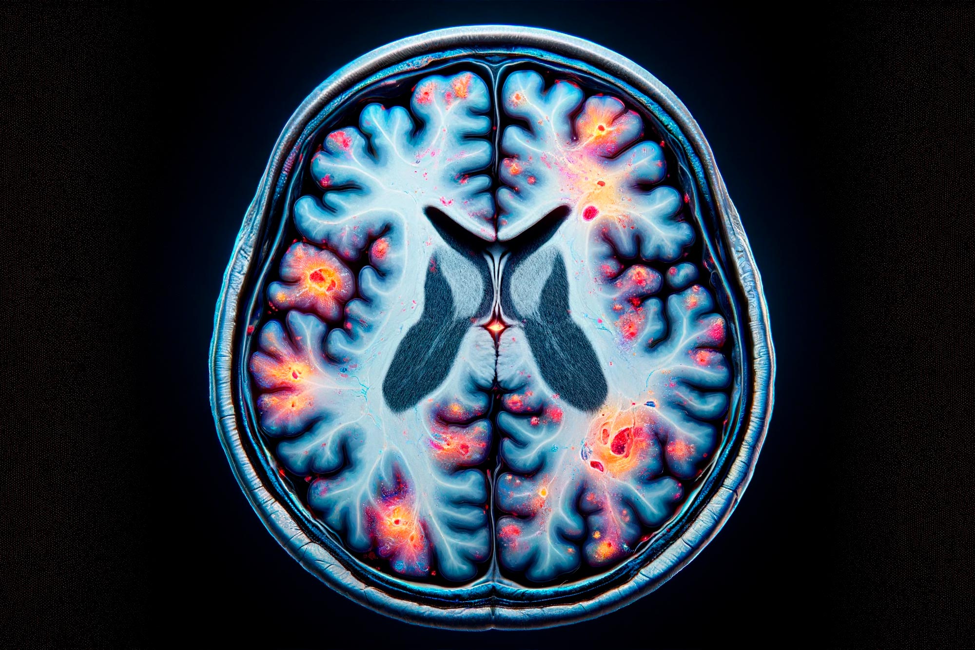
With rates of Alzheimer’s disease expected to riseTrusted Source and still no cure for this type of dementia, finding new ways to treat this disease has been at the forefront of research over the past few years.

Adding to this research is a new study from Northwestern University Feinberg School of Medicine. The findings recently published in the journal NeuronTrusted Source found that immune cells in the blood of people with Alzheimer’s disease are epigenetically alteredTrusted Source.

This alteration, the researchers say, is potentially caused by a previous viral infection, environmental pollutants, or other lifestyle factors.

The research revealed several genes that may be therapeutic targets for manipulating the body’s peripheral immune systemTrusted Source.

What is the peripheral immune system?

The body’s immune system can be considered as comprising two parts — the central immune system and the peripheral immune system, a term used to describe immune responses that happen outside the brain.

The peripheral immune system includes circulating white blood cells that detect antigens, such as bacteria or viruses, when they enter the body. This part of the immune system acts as the first wave of attack against any foreign substance.

According to Dr. David Gate, assistant professor of neurology at Northwestern University Feinberg School of Medicine and senior and corresponding author on this study, there is mounting evidence that the peripheral immune system plays a role in Alzheimer’s disease.

“In recent years, we have shown that immune cells of the cerebrospinal fluid — a fluid that flows in and around the brain — are clonally expanded and activated,” Dr. Gate told Medical News Today. “This means that they have previously responded to some type of immune stimulus.”

Previous researchTrusted Source has connected the peripheral immune system to neurodegenerative diseases, and studiesTrusted Source have shown an association between types of peripheral immune cells and cognition, brain structure, and Alzheimer’s disease pathology.

How epigenetics may play a role in Alzheimer’s disease

For this study, Dr. Gate said the team wanted to find out whether there might be epigenetic changes within the immune system of Alzheimer’s disease patients that might promote the trafficking of these changes to the cerebrospinal fluid and the brain.

“Epigenetics essentially reflects changes to our DNA that have occurred in the past,” he explained. “There are many influences on epigenetics, such as the environment, pollutants, viral infections, lifestyle factors, and behaviors. It is possible that these influences work in concert, or in isolation, to promote inflammation that puts one at risk of developing Alzheimer’s disease.”

Dr. Gate and his team examined immune cells from peripheral blood samples taken from people with Alzheimer’s disease. When compared to healthy controls, researchers found that every immune cell type in the participants with Alzheimer’s disease had epigenetic changes indicated by open chromatinTrusted Source.

Additionally, scientists looked for which genes were more open in the immune cells and found more exposure in the protein CXCR3Trusted Source on T cellsTrusted Source.

“Epigenetic changes alter the way our genes are translated into proteins,” Dr. Gate explained. “In this study, we observed an epigenetic change in a gene that encodes the protein CXCR3. CXCR3 is a signal receptor on the surface of immune cells called T cells. This receptor essentially serves as an antenna that we believe allows them to traffic signals put out by the Alzheimer’s brain.”

Researchers also found epigenetic changes in a type of white blood cell called monocytesTrusted Source.

“Monocytes are very important to immune defense. They secrete inflammatory proteins that protect your body in the case of infection. In this study, we found that there are epigenetic changes to genes that encode these inflammatory proteins. This is significant because it could signal that Alzheimer’s disease patients have a more pronounced pro-inflammatory immune system.”

– Dr. David Gate

Viral infections may increase Alzheimer’s risk

As epigenetics only provides a snapshot into the past, Dr. Gate said we can only speculate on what might have caused these discovered epigenetic changes.

“However, in the past decade, we have grown to appreciate the fact that viral infections are a risk factorTrusted Source for the development of dementias like Alzheimer’s disease,” he continued.

“While our data do not provide evidence that epigenetic changes in Alzheimer’s disease patients’ immune systems were caused by viral infections, this is certainly a tantalizing possibility. In this scenario, viral infections promote inflammatory responses over the course of one’s life that promote Alzheimer’s disease risk via mechanisms that we do not yet understand,” suggested Dr. Gate.

“Our ultimate goal is to design immune cell therapies for Alzheimer’s disease,” Dr. Gate added. “Using information from this study, we can potentially target the genes that harbor epigenetic changes.”

‘Implications in prevention and treatment of Alzheimer’s’

After reviewing this study, Dr. Manisha Parulekar, chief of the Division of Geriatrics at Hackensack University Medical Center in New Jersey, not involved in the research, told MNT she was not surprised by its results.

“Basically, it reinforces the idea that the patient’s behavior or environment has caused changes that affect the way their genes work,” Dr. Parulekar explained. “Many of these altered immune genes are the very same ones that increase an individual’s risk for Alzheimer’s.”

“Alzheimer’s disease is known to have complex pathophysiology, possibly involving multiple genes in combination [with] lifestyle, environmental, and other risk factors. Reading this new article, I am excited to see more evidence of changes in immune cells with changes in genes that were attributed to Alzheimer’s disease. The advancement in this science will likely have implications in [the] prevention and treatment of Alzheimer’s disease.”

– Dr. Manisha Parulekar

MNT also spoke with Dr. Karen D. Sullivan, a board-certified neuropsychologist, owner of the I CARE FOR YOUR BRAIN program, and Reid Healthcare Transformation Fellow at FirstHealth in Pinehurst, NC, about this study.

Dr. Sullivan said that as we do not know much about the peripheral immune system in Alzheimer’s disease, this study is a novel contribution to our understanding of a very complex, multi-faceted neurodegenerative disease.

“The main question I have is the directionality here,” she continued. “Is the peripheral inflammatory response in Alzheimer’s disease a cause or effect? The best thing I take away is that we may have a new therapeutic target in Alzheimer’s disease: restricting peripheral inflammation.”

“We would need to see these results replicated in larger sample sizes and to more deeply understand their relationships to the functional decline of Alzheimer’s disease,” Dr. Sullivan added.

Unmasking the Immune System’s Secret Role in Brain Damage From Viral Infections.


Recent research has revealed that neurological damage from acute viral infections, like Zika and COVID-19, is caused by the immune system’s response, particularly by a unique population of T cells. This discovery shifts the focus from the viruses to the immune system, offering new avenues for treatment.

A new study discovered that T cells, not viruses, are responsible for neurological damage in diseases like Zika and COVID-19, suggesting new treatment strategies.

For years, there has been a long-held belief that acute viral infections like Zika or COVID-19 are directly responsible for neurological damage, but researchers from McMaster University have now discovered that it’s the immune system’s response that is behind it.

The research, published today (February 5, 2024) in Nature Communications, was led by Elizabeth Balint, a PhD student at McMaster, and Ali Ashkar, a professor with the Department of Medicine and the Canada Research Chair in Natural Immunity and NK Cell Function.

The Role of T Cells in Neurological Diseases

“We were interested in trying to understand why so many viral infections are associated with neurological diseases,” says Balint. “Our evidence suggests that it’s not the virus itself that causes the damage, but a unique population of T cells, which are part of the immune system, that are actually responsible for the damage.”

To come to this conclusion, the McMaster team focused on Zika virus. During laboratory testing, researchers, as expected, found T cells that were specific for Zika and designed to eliminate infected cells. They found something else, too.

“What was interesting in our study is that although we did find some T cells specific for Zika, we identified cells that weren’t functioning like a normal T cell and were killing lots of cells that weren’t infected with Zika.”

These cells are called NKG2D+CD8+ T cells and researchers say their aggressive response is responsible for neurological damage suffered from infections beyond just Zika, like COVID-19 and even septic shock.

Immune Response and Potential Treatments

The aggressive response is the result of the body producing large amounts of inflammatory proteins called cytokines, which in moderation help to coordinate the body’s response in battling an infection or injury by telling immune cells where to go and what to do when they arrive.

“If our body’s immune cells overreact and overproduce inflammatory cytokines, this condition will lead to non-specific activation of our immune cells which in turn leads to collateral damage. This can have severe consequences if it happens in the brain,” Ashkar says.

The discovery offers researchers and scientists a new target for treatments of neurological diseases sparked by acute viral infections. In fact, Balint has already found a treatment that holds promise.

“Elizabeth has experimented with an antibody that can completely block and treat devastating neurotoxicity in the animal model, which is already in clinical trials for different uses in humans,” says Ashkar.

Balint hopes to continue her work towards finding a treatment that would be effective in humans.

“There are a few different other viruses we’re interested in studying, which will aid us in creating the best treatment options,” Balint says.

Map of thrombogenesis in viral infections and viral-driven tumours


Abstract

Viruses are pathogenic agents responsible for approximately 10% of all human cancers and significantly contribute to the global cancer burden. Until now, eight viruses have been associated with the development of a broad range of malignancies, including solid and haematological tumours. Besides triggering and promoting oncogenesis, viral infections often go hand-in-hand with haemostatic changes, representing a potential risk factor for venous thromboembolism (VTE). Conversely, VTE is a cardiovascular condition that is particularly common among oncological patients, with a detrimental impact on patient prognosis. Despite an association between viral infections and coagulopathies, it is unclear whether viral-driven tumours have a different incidence and prognosis pattern of thromboembolism compared to non-viral-induced tumours. Thus, this review aims to analyse the existing evidence concerning the association of viruses and viral tumours with the occurrence of VTE. Except for hepatitis C virus (HCV) and human immunodeficiency virus (HIV) infection, which are associated with a high risk of VTE, little evidence exists concerning the thrombogenic potential associated with oncoviruses. As for tumours that can be induced by oncoviruses, four levels of VTE risk are observed, with hepatocellular carcinoma (HCC) and gastric carcinoma (GC) associated with the highest risk and nasopharyngeal carcinoma (NPC) associated with the lowest risk. Unfortunately, the incidence of cancer-related VTE according to tumour aetiology is unknown. Given the negative impact of VTE in oncological patients, research is required to better understand the mechanisms underlying blood hypercoagulability in viral-driven tumours to improve VTE management and prognosis assessment in patients diagnosed with these tumours.

Conclusion

Viruses are well-known pathogenic agents responsible for the direct and indirect development of approximately 10% of human cancers. Several viruses linked to oncogenesis in humans have been identified, including HBV, HCV, EBV, HPV, KSHV, HTLV-1, HIV and MCPyV, which are associated with a vast diversity of malignant diseases. Beyond tumorigenesis, viruses are major inducers of blood coagulability. Indeed, haemostasis is frequently disturbed by the pro-inflammatory environment imposed by viral infections, which supports the idea that a viral infection can trigger thromboembolic events.

Conversely, over the years, the association between cancer and thrombosis has been a matter of discussion among researchers. This cardiovascular disease is commonly diagnosed in patients with malignancy and constitutes their second cause of death after cancer. Indeed, there is a bidirectional and complex interplay between both conditions, with tumour cells interacting with haemostatic components and, in parallel, promoting cancer progression and aggressiveness, which worsens the patients’ clinical outcomes.

Although seemingly evident, the literature has not discussed venous thrombosis in viral-induced tumours. Since cancer patients may harbour the oncovirus for their whole life (e.g., chronic infections), and given the remarkably complex interplay of viral infections with both cancer development and haemostasis, it would be of interest to identify a potential pattern of VTE incidence across viral-driven tumours, as well as the putative underlying mechanisms. Except for HCV and HIV, which are associated with an increased VTE risk with several underlying mechanisms proposed, most viruses (HBV, EBV, HPV, HTLV-1 and KSHV) have only a putative associated VTE risk, and few to no mechanisms currently described. On the other hand, for MCPyV, no association was found. Conversely, among the various tumours that can be induced by oncoviruses, GC and HCC were associated with the highest VTE risk, HL, NHL and CC were included in the intermediate VTE risk, while NPC was included in the lowest VTE risk category. Furthermore, to the best of our knowledge, data regarding KS, ATL and MCC association with venous thrombosis was not available in the literature, therefore, considered unknown. Unfortunately, whether the incidence of thrombosis in cancer patients is influenced by tumour aetiology is unexplored, at least to the best of our knowledge, and it is unclear to what extent the presence of viral infections in oncological patients can induce thrombogenesis, being further studies required to fulfil the current gaps in our knowledge.

Overall, the present review summarized the data concerning VTE risk and the associated mechanisms across several viral-induced tumours. This is relevant given the need to better predict VTE development, allowing for better thromboprophylaxis and ultimately improving the prognosis of patients diagnosed with these tumours. Furthermore, the data exposed can be useful to identify potential therapeutic targets that could improve VTE treatment efficacy and decrease the associated side effects, the major one being haemorrhage, which is more common among oncological patients. Beyond the influence of tumour aetiology on thrombosis development, another point to be explored is whether thromboembolic events may facilitate viral infection, reinfection, and viral carcinogenesis.

Latent HIV Reservoir May Be Larger Than Previous Estimates


The sleeping giant of HIV infection—a reservoir of viral DNA that lies dormant in human immune cells—could be far larger than previously believed.

A study published today in the journal Cell shows that the reservoir, consisting of proviruses integrated into resting CD4 immune cells, may be 60 times larger than scientists had estimated. Antiretroviral therapy (ART) kills replicating HIV but not the latent proviruses, which pose a major barrier to eradicating the virus from the body and curing infection.

The latent HIV reservoir in people who are infected could be 60 times larger than previously estimated, according to a new study. Image:  JAMA, ©AMA

Recent research had indicated that fewer than 1% of proviruses become infectious when resting CD4 cells are activated in a test tube. Without activation, proviruses can’t replicate. The proviruses that don’t cause infection have been considered defective, but investigators hadn’t described specifics about the deficiencies.

In the new study, a team of investigators used a more detailed method to study proviruses that didn’t switch on and become infectious when the resting CD4 cells they inhabited were activated in the laboratory. They cloned the genomes of 213 inactive proviruses from 8 HIV-infected patients treated with ART for more than 6 months. Their genetic analyses showed that about 88% of proviruses that didn’t turn on had obvious defects that prevented them from replicating. But nearly 12%—a far greater percentage than previously estimated—were capable of replicating and causing infection.

The investigators said their study suggests that there are enough proviruses that don’t turn on but are capable of replicating to boost the size of the latent reservoir by 60-fold. “These results indicate an increased barrier to cure, as all intact noninduced proviruses need to be eradicated,” senior author Robert Siliciano, MD, PhD, of Johns Hopkins University School of Medicine in Baltimore, said in a statement.

“We would like to use these findings by developing better ways to measure the size of the latent reservoir in patients who are participating in future trials of potentially curative strategies,” Siliciano added. “In this way, we think our analysis will contribute to HIV eradication efforts.”

New test uses a single drop of blood to reveal entire history of viral infections .


Cheap and rapid test allows doctors to access list of every virus that has infected or continues to infect a patient, and could transform disease detection
The new test uses a single drop of blood, and draws on advances in synthetic biology and rapid gene sequencing to analyse more than 1000 strains of virus.


Researchers have developed a cheap and rapid test that reveals a person’s full history of viral infections from a single drop of blood.

The test allows doctors to read out a list of the viruses that have infected, or continue to infect, patients even when they have not caused any obvious symptoms.

The technology means that GPs could screen patients for all of the viruses capable of infecting people. It could transform the detection of serious infections such as hepatitis C and HIV, which people can carry for years without knowing.

“Normally, when a doctor wants to know if someone’s been infected with a virus, they have to guess what the virus is, and then look specifically for that virus,” said Stephen Elledge, who led the project at Brigham and Women’s Hospital in Boston.

“This could lead to a diagnostic where people go annually to their doctor and get their viral history recorded. It could certainly discover viral infections that are serious and that a patient didn’t know they had,” he said.

The $25 (£16) test draws on advances in synthetic biology and rapid gene sequencing to analyse more than 1000 strains of human viruses in one pass. Until now, most tests have looked for only a single virus at a time. Elledge estimates that the latest test, called VirScan, can process 100 samples in two to three days.

The test exploits the fact that the immune system makes antibodies to fight viruses whenever the body becomes infected. These antibodies can live on in the bloodstream for years and even decades.

To develop the test, Elledge engineered batches of harmless viruses to carry bits of proteins from human viruses on their surfaces. In total, they carried proteins from more than 1000 strains of the 206 kinds of viruses known to infect people. Antibodies use these protein fragments to recognise invading viruses and launch their attacks.


When a droplet of blood from a patient is mixed with the modified viruses, any antibodies they have latch on to human virus proteins they recognise as invaders. The scientists then pull out the antibodies and identify the human viruses from the protein fragments they have stuck to.

“It’s the first time we’ve been able to look in a completely unbiased manner at what viruses are infecting people, and we can do it for all known viruses,” said Elledge. The test picks up the antibodies a person produces from vaccinations, but these can be discarded from the test results. Details are reported in the journal Science.

In a demonstration of the technology, the team analysed blood from 569 people in the US, South Africa, Thailand and Peru. The test found that, on average, people had been infected with 10 species of viruses, though at least two people in the trial had histories of 84 infections from different kinds of viruses.

The most common infections were herpes viruses, which cause cold sores, enteroviruses that upset stomaches, influenza, and rhinoviruses that trigger common colds. Those in the US had experienced fewer infections than those in the other countries, and as expected, older people had richer viral histories than youngsters.

The test could bring about major benefits for organ transplant patients. One problem that can follow transplant surgery is the unexpected reawakening of viruses that have lurked inactive in the patient or donor for years. These viruses can return in force when the patient’s immune system is suppressed with drugs to prevent them rejecting the organ. Standard tests often fail to pick up latent viruses before surgery, but the VirScan procedure could reveal their presence and alert doctors and patients to the danger.

“This could be very valuable,” said Iwijn De Vlaminck, a biomedical engineer at Cornell University in New York, who was not involved in the study. “What this allows you to do is look into the past and measure a person’s exposure to previous infections. That has important advantages, because you can detect these infections that go to latency. You could screen blood from patients and organ donors in this very broad manner and predict potential future issues with viral reactivation.”

Scientists believe the test will also cast light on how certain viral infections can predispose people to seemingly unrelated diseases later in life. Some infections can cause permanent damage to body tissues, or alter the immune system, in ways that leave people more at risk of medical problems when they are older. For example, infection with Epstein-Barr virus can raise the risk of cancer. But how other viruses affect long term health is far murkier. “That kind of analysis is something this really makes possible,” said Elledge.