Abstract
Modern cancer therapy has successfully cured many cancers and converted a terminal illness into a chronic disease. Because cancer patients often have coexisting heart diseases, expert advice from cardiologists will improve clinical outcome. In addition, cancer therapy can also cause myocardial damage, induce endothelial dysfunction, and alter cardiac conduction. Thus, it is important for practicing cardiologists to be knowledgeable about the diagnosis, prevention, and management of the cardiovascular complications of cancer therapy. In this first part of a 2-part review, we will review cancer therapy–induced cardiomyopathy and ischemia. This review is based on a MEDLINE search of published data, published clinical guidelines, and best practices in major cancer centers. With the number of cancer survivors expanding quickly, the time has come for cardiologists to work closely with cancer specialists to prevent and treat cancer therapy–induced cardiovascular complications.
Introduction
Heart diseases and cancer are the top 2 leading causes of death in the United States. Because these maladies share several common risk factors, many of our patients, especially the elderly, are afflicted by both cancer and heart diseases. Furthermore, cancer therapies, either radiation treatment or chemotherapy, can cause cardiovascular complications. Thus, it is important for practicing cardiologists to be familiar with the prevention, diagnosis, and management of cardiovascular complications of cancer therapy. This topic was reviewed in the Journal in 2009.[1] The purpose of this new State-of-the-Art Review is to provide an update in this emerging discipline that abounds with exciting new developments. Cardiovascular complications covered in this 2-part review are heart failure (HF), myocardial ischemia, myocarditis, hypertension (HTN), pulmonary HTN, pericardial diseases, thromboembolism, QT prolongation and arrhythmias, and radiation-induced cardiovascular diseases. A MEDLINE search for each of these complications was performed; clinically relevant complications were selected based on experiences at the MD Anderson Cancer Center and centers affiliated with authors of this review. Diagnostic and treatment recommendations are based on the best practices developed at MD Anderson Cancer Center and recent guidelines.[2–6]
Heart Failure
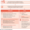
HF due to chemotherapy has been long recognized as a serious side effect of daunorubicin, the first anthracycline used clinically.[7]Although the anthracycline class of chemotherapy agents remains the major cause of chemotherapy-induced cardiomyopathy (CIMP), newer cancer therapy, such as trastuzumab or proteasome inhibitors, can also cause cardiomyopathy (Central Illustration, Table 1). It should also be recognized that patients can develop signs and symptoms of clinical heart failure during chemotherapy; however, the cause of cardiac decompensation may be due to fluid overload, stress-induced cardiomyopathy, or primary cancer, but not chemotherapy.[2]CIMP has been described in 1% to 5% of cancer survivors[8,9] and portends one of the worst survivals among cardiomyopathies.[10]Early diagnosis and timely intervention has been shown to result in a superior clinical outcome in cancer patients treated with cardiotoxic chemotherapy.[11]
Management of Cancer Therapy–Induced Cardiovascular Complications
Best practices in the management of cancer therapy–induced cardiomyopathy and ischemia. ACC = American College of Cardiology; ACEI = angiotensin-converting enzyme inhibitor; AHA = American Heart Association; CV = cardiovascular; EF = ejection fraction; VSP = vascular endothelial growth factor signaling pathway.
Definition
In the initial report by Von Hoff et al.,[12] HF was defined as the presence of tachycardia, shortness of breath, neck vein distention, gallop rhythms, ankle edema, hepatomegaly, cardiomegaly, and pleural effusion.[12] With the advance of cardiac imaging, echocardiography or multigated acquisition radionuclide ventriculography-based evaluation of left ventricular ejection fraction (EF) has recently been included in the diagnostic criteria.[4,13] In the trastuzumab clinical trials, drug-associated cardiotoxicity is defined as 1 or more of the following: 1) cardiomyopathy characterized by a decrease in EF globally or due to regional changes in interventricular septum contraction; 2) symptoms associated with HF; 3) signs associated with HF, such as S3 gallop, tachycardia, or both; and 4) decline in initial EF of at least 5% to <55% with signs and symptoms of HF or asymptomatic decrease in EF of at least 10% to <55%.[14] This definition does not include subclinical cardiovascular damage, such as diastolic dysfunction and changes in LV strain, which may occur earlier in response to some of the chemotherapeutic agents. Common Terminology Criteria for Adverse Events has also defined cardiomyopathy and/or heart failure for the purposes of uniform reporting. In Common Terminology Criteria for Adverse Events 4.03, echocardiography and biomarkers were included to provide a more precise definition of cardiotoxicity.
Incidence and Pathogenesis
Anthracyclines. In a retrospective review of 3 trials, the incidence of doxorubicin-related HF was found to be 5% at a cumulative dose of 400 mg/m2, 16% at a dose of 500 mg/m2, and 26% at a dose of 550 mg/m2.[15] However, subclinical events occurred in about 30% of the patients, even at the doses of 180 to 240 mg/m2, about 13 years after the treatments.[16] Interestingly, histopathological changes, such as myofibrillar loss and vacuolization, can be seen in endomyocardial biopsy specimens from patients who have received as little as 240 mg/m2 of doxorubicin.[17] These findings suggest that there is no safe dose of anthracycline. Even doses as low as 100 mg/m2 have been associated with reduced cardiac function.[16,18] Nonetheless, some patients had no significant cardiac complications despite receiving doses as high as 1,000 mg/m2.[19] Individual susceptibility is most likely due to genetic variants in genes that regulate anthracycline cardiotoxicity.[20] Other risk factors for anthracycline toxicity include cumulative dose, intravenous bolus administration, higher single doses, history of prior irradiation, use of concomitant agents known to have cardiotoxicity, female sex, underlying CV disease, age (young and elderly), delayed diagnosis, and increase in cardiac biomarkers such as troponins during and after administration.[9,21–23]
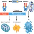
Doxorubicin poisons topoisomerase 2 to cause deoxyribonucleic acid (DNA) double-strand break and demise of cancer cells. In the cardiomyocytes, doxorubicin targets topoisomerase 2β to induce DNA double-strand breaks, and doxorubicin-bound topoisomerase 2β binds promoters of antioxidative and electron-transport genes to reduce their transcripts and protein expression[24,25] (Figure 1). Doxorubicin-treated cells have a marked increase in reactive oxygen species and are defective in mitochondria biogenesis. Thus, topoisomerase 2β accounts for the 3 hallmarks of anthracycline-induced cardiotoxicity: myocyte death, reactive oxygen species generation, and mitochondriopathy. Reduced topoisomerase 2β expression has been linked to a coding variant in the retinoic receptor γ gene, which predicts susceptibility to anthracycline-induced cardiotoxicity in childhood cancer.[26]
Figure 1.
Mechanism of Doxorubicin-Induced Cardiotoxicity
Doxorubicin inhibits topoisomerase 2β to induce DNA double-strand break, leading to p53 activation and death of cardiomyocytes. Doxorubicin-bound topoisomerase 2β binds to promoters of antioxidative genes and PGC-1 that are required for expression of antioxidative enzymes and electron transport chains. Thus, topoisomerase 2β is able to account for the 3 hallmarks of doxorubicin-induced cardiotoxicity: cardiomyocyte death, generation of reactive oxygen species (ROS), and mitochondriopathy.[24,25]
Alkylating agents. Alkylating agents add an alkyl group to the DNA of rapidly dividing cells and, in the case of bifunctional alkylating agents, cross-link the 2 DNA strands, thereby inhibiting DNA replication and cell proliferation.[27] Symptoms may include arrhythmias, conduction disorders, and fulminant HF.[28,29] Alkylating agents such as cyclophosphamide induce electrocardiogram (ECG) alterations in the form of low QRS voltage and nonspecific T-wave or ST-segment abnormalities.[28,30,31] Acute symptoms usually occur within 1 to 2 weeks, last for a few days, and in some patients, resolve without any late consequences.[28,29,31] The incidence of high-dose cyclophosphamide-induced HF has been reported to be as high as 28%.[32] Autopsies of patients with cyclophosphamide-induced cardiotoxicity show hemorrhagic myocardial necrosis with interstitial edema and fibrin deposition. The risk of complications is higher in elderly patients and in those exposed to anthracycline or mediastinal irradiation.[1,29,31]
Ifosfamide is an alkylating nitrogen mustard used for the treatment of lymphomas; sarcomas; and testicular, breast, and lung carcinoma.[33]There is a dose-dependent increase in HF associated with ifosfamide administration.[33,34] Autopsy studies demonstrated increased heart weight, small pericardial effusions, subendocardial hemorrhage, and petechial lesions in the epicardium.[29]
Targeted Therapies Against HER-2 Pathway
Trastuzumab is a monoclonal antibody against the human epidermal growth factor receptor tyrosine kinase (HER2 or ErbB2), which regulates cell growth and repair.[35] Overexpression of HER2 occurs in approximately 25% of breast cancers and confers increased proliferative and metastatic potential. HER-2 is expressed in cardiomyocytes and is required for survival of cardiomyocytes.[36]Mice deficient in HER-2 develop dilated cardiomyopathy and are more sensitive to doxorubicin.[36]
Trastuzumab administration in HER2-positive breast cancers led to significant reductions in recurrence rate and overall mortality. A pivotal study in the metastatic setting demonstrated a 33% reduction in mortality at 1 year and an increase in median survival by 5 months.[37] HF occurred in 8% of patients who received an anthracycline with cyclophosphamide; however, the incidence of HF increased to 27% with the addition of trastuzumab.[37] Subsequently, several large trials confirmed efficacy of trastuzumab in increasing disease-free survival from cancer, but also established trastuzumab’s association with HF.[38,39] In these trials, 1.7% to 4.1% of trastuzumab-treated patients developed HF when anthracycline was not part of the therapeutic regimen.[38,39] Trastuzumab-related cardiotoxicity includes various degrees of LV systolic dysfunction, occasionally leading to HF.[40]Symptoms are usually mild or moderate and improve following medical management and termination of drug administration.[41,42]Improvement is usually seen in about 4 to 6 weeks after trastuzumab withdrawal.[43] After symptomatic improvement, reinstitution of trastuzumab treatment is usually possible.[41–43]
Other targeted therapies against HER-2 are relatively less cardiotoxic.[44–46] Perez et al.[46] reviewed cardiac safety data in 44 clinical trials that used lapatinib. Of these study patients, only 1.6% experienced a cardiac event, and of those, only 0.2% was symptomatic. The rate of cardiac events in the lapatinib group was similar to those who did not receive lapatinib.[44,46] Similarly, there was no significant increase in left ventricular dysfunction with the addition of pertuzumab to trastuzumab in the NeoSphere study.[47,48]In the CLEOPATRA (Clinical Evaluation of Pertuzumab and Trastuzumab Trial in Human Epidermal Growth Factor Receptor 2-positive Metastatic Breast Cancer) trial, the incidence of cardiac adverse events was 14.5% in the pertuzumab plus trastuzumab plus docetaxel arm compared with 16.4% in the control-placebo plus trastuzumab plus docetaxel arm.[49,50] However, in a head-to-head comparison between lapatinib and trastuzumab, patients treated with lapatinib have shorter disease-free survival but more noncardiac toxicity, such as rash and diarrhea.[51] Thus, one must consider both efficacy and toxicity in choosing drugs.
Vascular endothelial growth factor signaling pathway inhibitors.Vascular endothelial growth factor signaling pathway (VSP) inhibitors include antibodies such as bevacizumab, which binds to vascular endothelial growth factor (VEGF), and small molecule tyrosine kinase inhibitors such as sunitinib and sorafenib, which inhibit the downstream kinase involved in VEGF receptor signaling.[52]Cardiovascular side effects of VSP inhibitors include HTN, cardiomyopathy, conduction abnormalities, acute coronary syndromes (ACS), and arterial thromboses. Several VSP inhibitors also block receptors that are involved in the compensatory response to stress in the cardiomyocytes. When the heart is unable to compensate for HTN induced by VSP inhibitors, it could lead to HF.[53] Therefore, maintaining good blood pressure control during VSP inhibitor therapy can prevent HF.[54]
Bevacizumab is the first- or second-line chemotherapy for advanced solid tumors.[55–61] Its use has been associated with HTN, thromboembolism, and cardiomyopathy.[62] Approximately 2% to 4% of patients treated with bevacizumab will develop HF.[29]Predisposing factors include previous therapy with cardiotoxic chemotherapy drugs, such as anthracyclines[63] and capecitabine,[64]as well as radiation to the mediastinum.[29] In a meta-analysis of VSP inhibitors and their role in cardiovascular complications, the odds ratio for cardiac dysfunction caused by VSP inhibitors was 1.35.[65]More than one-half of the clinical trials included in the analysis (40 of 77) utilized bevacizumab as the drug of treatment.[65] Another meta-analysis showed that bevacizumab resulted in a significant decrease in EF with a relative risk index of 3.4.[66]
In a large trial of sunitinib for the treatment of metastatic renal cell carcinoma, 13% of patients developed LV dysfunction and 3% had severe HF.[67] Severe HF was more common in a single-center study of 48 patients treated for metastatic renal cell carcinoma or gastrointestinal stromal tumor.[68] In a meta-analysis of 6,935 patients treated with sunitinib, the incidence of HF was 4.1% with a relative risk of 1.81 compared with placebo.[69] In a retrospective analysis of a phase 1/2 trial of sunitinib for the treatment of gastrointestinal stromal tumor, there was a 2.5-fold increase in cardiotoxicity with higher doses.[70] Sorafenib has been associated with a 4% risk of mild LV dysfunction and a 1% risk of clinical HF.[71] HF due to VSP inhibitors is reversible with cessation of therapy.[72]
Proteasome inhibitors. Proteasome is a protein complex present in all cells that degrades other proteins. Inhibition of proteasome blocks cell proliferation and induces apoptosis in tumor cells, especially in multiple myeloma. Bortezomib, a reversible proteasome inhibitor, is not known to cause HF.[73–75] However, 7% of patients treated with carfilzomib developed new-onset or worsening of pre-existing HF or myocardial ischemia.[76] In a phase 2 trial of 266 patients treated with carfilzomib monotherapy for relapsed myeloma, 10 experienced HF (3.8%), 4 (1.5%) had a cardiac arrest, and 2 (0.8%) had a myocardial infarction (MI) during the study.[77] The risk did not appear to be cumulative, at least through 12 cycles of therapy. Cardiotoxicity appears to be largely reversible with cessation of therapy and the initiation of HF treatment.[78]
Screening, Risk Stratification, and Early Detection Strategies
Baseline risk assessment. Patients undergoing chemotherapy should have careful clinical evaluation and assessment of CV risk factors, such as coronary artery disease, diabetes, and hypertension (Table 2).[2–6] These risk factors should be managed according to the American College of Cardiology (ACC)/American Heart Association (AHA) guidelines.[79] This is especially important if cancer therapy is known to cause HF. Aggressive HTN management is advised for patients treated with VSP inhibitors. Physical exercise has been shown to reduce cardiotoxicity in mouse models.[80]
EF assessment is mandatory to establish baseline cardiac function before cardiotoxic cancer treatment.[3] Echocardiography is the preferred modality for assessment of cardiac structure and function. A multigated acquisition radionuclide ventriculography scan has less interobserver variability; however, radiation exposure limits its utility as a cardiac monitor. Cardiac magnetic resonance imaging can be used to obtain a precise EF; however, the use of cardiac magnetic resonance imaging is limited by its cost. Cardiac biomarkers, such as troponins and brain natriuretic peptides (BNPs), can be used to monitor for development of cardiac dysfunction.[3,5,6] However, it is not known whether routine monitoring of biomarkers is useful in changing clinical outcomes. A number of composite scores have been designed to risk-stratify cancer patients;[81–83] however, they have not been validated in prospective studies.
Echocardiogram. The echocardiogram is the most important tool for serial evaluation of the heart during cancer therapy. EF should be determined using the biplane method of discs according to the American Society of Echocardiography guideline.[3] If the endocardial border is not distinct, ultrasonic contrast should aid in endocardial border definition and subsequent volume calculations. However, temporal EF variability may be up to 10%, with a confidence interval of 95% in 2-dimensional EF readings.[84] Three-dimensional echocardiography has a temporal variability of 6% and is considered the most accurate EF measurement by echocardiography. Because CIMP is defined as a drop of EF of ≥10% or ≥5% or more in the presence of HF symptoms, an accurate measurement of EF is paramount.
Myocardial strain. Tissue Doppler imaging (TDI) and speckle-tracking strain imaging have emerged as 2 quantitative techniques for estimating global and regional myocardial mechanical function and have the potential to detect early signs of LV dysfunction.[85]However, TDI is both user- and angle-dependent and is unable to differentiate translational motion or tethering effects from myocardial contractility. Speckle-tracking echocardiography is an angle-independent technique that uses an image-processing algorithm for analyzing motion of ”speckles” or ”fingerprints” within a 2-dimensional echocardiography image, and it has replaced TDI strain as the preferred method for quantitative assessment of cardiac deformation.[86,87]
Several studies have evaluated the utility of strain imaging for the detection of chemotherapy-associated cardiotoxicity. Fallah-Rad et al.[88] evaluated 42 patients with breast cancer overexpressing HER-2 who received trastuzumab in the adjuvant setting after anthracycline therapy. Within 3 months, peak global longitudinal and radial strain detected preclinical changes in LV systolic function before a decrease in EF was observed several months later. A recent prospective multicenter study by Sawaya et al.[89] demonstrated that global longitudinal strain (GLS) <19% was predictive of subsequent cardiotoxicity and was present in all patients who later developed symptoms of HF. Negishi et al.[90] similarly showed that a ≥11% relative reduction in GLS was predictive of subsequent trastuzumab-associated cardiotoxicity.
Abnormalities in strain parameters can also be seen several years after a cardiotoxic exposure. In 75 asymptomatic breast cancer survivors who received anthracycline with or without adjuvant trastuzumab, GLS was significantly decreased in the chemotherapy group 6 years after therapy compared with control subjects.[91] In another meta-analysis, GLS consistently detected early myocardial changes during therapy.[92] A 10% to 15% early reduction in GLS during therapy appears to be the most useful parameter for the prediction of cardiotoxicity. In late cancer survivors, global radial and circumferential strain are consistently abnormal, even with normal EF, but their clinical value in predicting subsequent ventricular dysfunction or HF has not been explored.[92]
Biomarkers. The use of EF in the diagnosis of CIMP has important limitations. First, the measurement of EF is subject to technique-related variability, which can be higher than the thresholds used to define cardiotoxicity.[14,84] Second, the reduction in EF is often a late phenomenon.[11,88,93,94] Hence, there is a growing interest in identifying markers of early myocardial damage to predict the development of HF. Biomarkers are an economic and effective way of detecting myocardial dysfunction in apparently asymptomatic patients. The assessment of troponin and BNP has shown incremental utility in identifying patients at increased risk for adverse outcomes.[95–97]
Troponins. Troponin is an important biomarker for ACS and other myocardial damage.[98–100] In an early study, cardiac troponin I (cTnI) was elevated in 32% of 204 patients receiving high-dose chemotherapy, and an increase in cTnI occurred in more than 50% of patients soon after drug administration.[101] A follow-up study showed that patients with negative cTnI (<0.08 ng/ml), immediately and 1 month after chemotherapy, showed no EF reduction and had low incidence of cardiac events.[9] In contrast, patients with positive cTnI had a higher incidence of adverse cardiac events, including HF and asymptomatic LV dysfunction.[9]
Serial troponin measurements in patients with hematologic malignancies treated with anthracycline showed troponin elevation correlated with EF reduction.[102] A persistent release of cTnI was associated with a probability of major cardiac events within the first year of follow-up.[9,22] In children treated with high-dose doxorubicin for acute lymphoblastic leukemia, cTnT increased in approximately 30% of cases, and the amount of elevation was predictive of cardiac dysfunction during follow-up.[103,104] Thus, most studies showed a good correlation of elevated enzymes with LV dysfunction, especially in patients who were treated with high-dose anthracycline.
Dodos et al.[105] performed a series of cardiac troponin T (cTnT) measurements on the 3rd to 5th day following the first course of anthracycline and after the last course. They did not observe troponin elevation followed by EF deterioration. In their study, cTnT levels did not exceed the upper limit of the normal range in all patients. Only 7% of patients had low-level elevation of cTnT, and only 1 of these patients developed a decrease in EF. McArthur et al.[106] studied a group of patients treated with bevacizumab, doxorubicin, and cyclophosphamide followed by paclitaxel in early-stage breast cancer. A total of 7 patients (9%) experienced either a symptomatic or asymptomatic EF decline. There was no association between EF change and troponin elevation.[106] These authors speculate that cTnI release could be missed because samples were drawn prior to chemotherapy.[106] Thus, the utility of using troponins in predicting EF changes depends on the timing of blood drawn relative to chemotherapeutic administration.
In patients with breast cancer who receive anthracycline or trastuzumab, an elevation of high-sensitivity cTnI with a decrease in GLS of at least 19% is highly specific in predicting CIMP.[89] Based on these data, an expert panel proposed that cTnI should be measured at baseline and every 3 weeks during trastuzumab therapy accompanied by echocardiography and GLS at baseline and every 3 months.[3] A small study concluded that an increase in high-sensitivity troponins is a good predictor of LV dysfunction.[107] A high baseline level of high-sensitivity troponins is also a predictor of adverse outcomes.[107]
Brain Natriuretic Peptide
Natriuretic peptides are produced by splitting a prohormone into the amino-terminal inactive form and the carboxy-terminal biologically active hormone. The ventricle secretes biologically active BNP and inactive amino-terminal pro-BNP in response to increased ventricular volume and pressure.[108] Clinical studies have utilized BNP and amino-terminal pro-BNP as biomarkers of CIMP, and although results are mixed, several studies have indicated that these peptides could be good early indicators of cardiac damage.[108–111]
In an early anthracycline study, an increase in BNP level correlated with E/A ratio increase, suggesting that BNP level may be predictive of diastolic dysfunction. BNP increase during anthracycline treatment is usually transient and, in most cases, is not predictive of clinical outcome. Only patients with persistent elevation of BNP developed overt HF, which also suggests a potential use of BNP in long-term follow-up.[112] Nousiainen et al.[113] found no significant correlations between echocardiography parameters and natriuretic peptides until the cumulative doxorubicin dose reached 500 mg/m2. Meinardi et al.[114] showed that during chemotherapy, concentrations of BNP in plasma increase as EF decrease. Daugaard et al.[115] also found that neither baseline levels of N-terminal pro-atrial natriuretic peptide nor BNP nor changes in these variables during therapy were predictive of a change in EF.[115] However, persistent elevation of BNP may be an indication of adverse cardiac outcomes.[114,115] C-reactive peptide,[116–118] myeloperoxidase,[116] galacetin-3,[116,119,120] and ST-2[120,121]have been investigated as potential biomarkers; however, they cannot be recommended as routine tests at present.
Preventive Strategies for Anthracycline-induced Cardiotoxicity
Selecting a nonanthracycline regimen. A randomized study of 3,222 women with HER-positive early breast cancer found that a nonanthracycline-containing regimen has equal efficacy and less cardiotoxicity.[122] With a nonanthracycline regimen containing docetaxel, carboplatin, and trastuzumab (TCH), patients had a 5-year disease-free survival rate of 81% compared with 84% in the anthracycline-containing regimen (ACT [doxorubicin, cyclophosphamide, docetaxel] or ACT-H [doxorubicin, cyclophosphamide, docetaxel, trastuzumab]). Importantly, the TCH regimen has much lower cardiotoxicity than ACT or ACT-H. Thus, TCH was proposed as an alternative nonanthracycline-containing regimen for HER-positive early breast cancers (Table 2).
Substituting doxorubicin with less-cardiotoxic anthracyclines. Over the past 5 decades, more than 2,000 modified anthracycline chemicals have been tested in an attempt to reduce cardiotoxicity while retaining tumoricidal efficacy. Although several anthracycline derivatives have been evaluated in clinical trials, only epirubicin[123]and idarubicin[124] received approval for clinical use. However, a critical analysis by the Cochrane database identified no difference in cardiotoxicity between epirubicin and doxorubicin at equipotent doses.[125]
Continuous infusion. Replacing bolus administration with slow infusions does not significantly affect anthracycline area under the curve, but it diminishes anthracycline Cmax and anthracycline accumulation in the heart.[126] A Cochrane review[127] showed a significantly lower rate of clinical HF with an infusion duration of 6 h or longer compared with a shorter infusion duration in the adults. In the pediatric populations, the results of infusion of anthracycline have been disappointing. A randomized trial in children with high-risk acute lymphocytic leukemia found that continuous infusion offered no additional cardiac protection over bolus administration in a median follow-up of 8 years post-diagnosis.[128] A follow-up at 10 years also revealed no incremental therapeutic efficacy for infusion.[129] Thus, continuous infusion cannot be recommended in the pediatric population.[130,131]
PEGylated liposomal doxorubicin. PEGylated liposomal doxorubicin comprises an aqueous core of doxorubicin hydrochloride encapsulated in liposomes with a protective hydrophilic outer coating of surface-bound methoxypolyethylene glycol.[132,133] Delivery of doxorubicin in a PEGylated liposomal form decreases the circulating concentrations of free doxorubicin and results in selective uptake of the agent in tumor cells. In randomized trials, PEGylated liposomal doxorubicin was as effective as doxorubicin or other traditional combination chemotherapies.[134–139] Thus, PEGylated liposomal doxorubicin is a useful option in the treatment of various malignancies.[140] However, the cost associated with administering this drug has prevented its widespread adoption.[141]
Dexrazoxane. Dexrazoxane was originally developed as an anticancer agent.[142] Using fibroblasts from topoisomerase 2β knockout mice, Lyu et al.[143] showed that dexrazoxane is protective against anthracycline-induced toxicity in a topoisomerase 2β–dependent manner, linking dexrazoxane to the topoisomerase 2β theory of anthracycline cardiotoxicity[25] (Figure 1).
The protective effect of dexrazoxane against anthracycline-induced cardiotoxicity has been demonstrated in numerous clinical trials in adults and children.[19,104,144–146] Dexrazoxane was approved in Europe and the United States for cardioprotection in patients treated with anthracyclines (Cardioxane, Clinigen Group, Burton-on-Trent, United Kingdom; and Zinecard, Pfizer, New York, New York) with several generic preparations available (procard and cardynax). In addition, dexrazoxane has been also approved for treatment of accidental extravasation of anthracyclines (Savene, Clinigen Group).
Unfortunately, 1 phase 3 trial suggested that dexrazoxane may lower the efficacy of anthracycline in treating breast cancer.[19] In this trial, a significant difference in objective response was reported (47% vs. 61%, respectively; p = 0.019). Although a high response in the placebo group was quite unusual, other endpoints (including survival or time to progression) were not affected by dexrazoxane in this study.[19,147]A careful meta-analyses of all available randomized clinical trials found no evidence that dexrazoxane lowers doxorubicin’s anticancer effect.[148,149] However, the U.S. Food and Drug Administration (FDA) has approved dexrazoxane only in patients who have received more than 300 mg/m2 of doxorubicin for metastatic breast cancer and who may benefit from continued doxorubicin treatment.[150]
Another controversy about dexrazoxane pertains to an increased risk of second malignancies in the pediatric cancer survivors. This was observed in survivors of Hodgkin lymphoma who had received dexrazoxane in combination with doxorubicin and etoposide. It was postulated that combining these drugs could exceed a threshold above which topoisomerase inhibitors caused genetic instability in normal tissues.[151] This report led the European Medicine Agency to disapprove the use of dexrazoxane in children. However, 2 studies of survivors of childhood acute lymphoblastic leukemia did not detect an increased risk of second malignancies from dexrazoxane.[152,153]Thus, the risk/benefit analysis supports a wider clinical usage of dexrazoxane with the possible exception of patients receiving etoposide or etoposide-anthracycline combinations.
Preventative Strategies Against Trastuzumab-induced Cardiotoxicity
The metastatic breast cancer trial showed that concurrent treatment of trastuzumab and anthracycline had detrimental cardiac outcomes.[14,37] However, as anthracycline and trastuzumab were not administered at the same time, the incidence of HF drastically reduced. In the N9831 trial, the incidence of New York Heart Association functional class III/IV HF or cardiac deaths was 0% in the control arm (anthracycline without trastuzumab) and 3.3% in the concurrent anthracycline/trastuzumab arm.[154] In the B-31 study, these toxic effects occurred in 0.8% of the control group (anthracycline without trastuzumab) and 4.1% of the anthracycline/trastuzumab arm. HF was not reported in the FinHer trial, in which trastuzumab was omitted during the 3 cycles of epirubicin-containing regimen.[155] These results were in marked distinction to the 27% incidence of HF in the original metastatic trials.
Treatment
Symptomatic and asymptomatic HF should be treated according to the ACC/AHA guidelines (Table 2).[156,157] We recommend HF treatment when subclinical cardiotoxicity was detected by strain imaging and biomarkers.[2,3] Many cancer patients with overt or subclinical HF can be treated with angiotensin-converting enzyme inhibitors or beta-blockers to allow completion of the chemotherapy. Anthracycline-induced cardiotoxicity was considered irreversible, whereas trastuzumab was reversible.[43,158] Thus, a different EF cutoff threshold for withholding therapy is recommended (Table 2).[2]It should be noted that cessation of cancer therapy should be considered only as the last resort. Every effort should be made to manage HF to allow chemotherapy to continue. Patients with symptomatic and/or overt HF should be treated according to the ACC/AHA HF guidelines.[156,157]
Pathophysiology
Persistent coronary spasm occurring at the level of a pre-existing plaque was observed during cardiac catheterization in a patient receiving continuous 5-FU infusion.[188] Experimental work in rabbit aortic rings showed that incremental doses of 5-FU–induced endothelium-independent vasoconstriction, secondary to protein kinase C–mediated vasoconstriction of vascular smooth muscle.[189]An alternative mechanism of cardiotoxicity postulates a direct toxic effect of 5-FU on the coronary endothelium,[190–192] with ensuing endothelial injury leading to microthrombotic occlusions.[190]Although undetectable by coronary angiography, endothelial injury and small vessel thrombosis could be key ultrastructural findings.[191,192] 5-FU–induced endovascular injury could be reduced by anticoagulants.[193,194] Little is known regarding the cardiotoxic effect of paclitaxel. Myocardial ischemia in association with paclitaxel is thought to be due to concurrent use of other drugs and pre-existing cardiac conditions.[195] The polyoxyethylated castor oil, known as Cremophor EL (BASF, Ludwigshafen, Germany), is used as a vehicle for paclitaxel in the injectable formulation and may contribute to the overall cardiotoxicity by inducing histamine release.[195]
VEGF stimulates endothelial cell proliferation to maintain endothelial viability and vascular integrity.[196] Consequently, bevacizumab administration may impair the regenerative potential of endothelial cells in response to stress. Exposed subendothelial collagen can trigger tissue factor activation, resulting in thromboembolism.[196,197]VEGF inhibition also impairs the production of nitric oxide and prostacyclin, as well as increases hematocrit and blood viscosity via overproduction of erythropoietin, all of which heighten the thromboembolic risk.[196] Carfilzomib was found to increase coronary perfusion pressure and resting vasoconstriction tone in isolated rabbit heart and aorta.[198] In addition, carfilzomib amplified the spasmogenic effect of noradrenaline and angiotensin II, curbed the antispasmogenic activity of nifedipine and nitroglycerin, and reduced the vasodilating effect of acetylcholine.
Screening, Diagnosis, and Treatment
Because pre-existing CAD is a known risk factor for the development of chemotherapy-induced ACS, ischemic workup should be initiated in all high-risk patients before administration of drugs known to cause cardiac ischemia (Table 3). Patients with suspected ACS should be treated according to ACC/AHA guidelines.[199,200] Besides statins and beta-blockers, the cornerstones of ACS treatment include percutaneous coronary intervention as well as antiplatelet and anticoagulant therapy, all of which pose an incremental bleeding risk in cancer patients with thrombocytopenia. Although prospective studies in this specific population are currently lacking, a retrospective analysis carried out in cancer patients with thrombocytopenia and ACS showed that aspirin improved the 7-day survival rate without increasing the bleeding risk.[201] Although case-specific considerations are warranted, life-saving interventions should not be denied to cancer patients with ongoing ACS because of thrombocytopenia.[202] The response to anticoagulants and antiplatelet agents in patients with platelet counts >50,000/μl seems to be comparable to that observed in patients with normal platelet counts.[202] However, reduced heparin doses, ranging from 30 to 50 U/kg, may be required for patients whose platelet counts are <50,000/μl.[202] Dual antiplatelet therapy with aspirin and clopidogrel can be used for patients with platelet counts >30,000/μl, whereas aspirin as a single agent should be given to those with platelet counts >10,000/μl. With a platelet count below 10,000/μl, bleeding risk should be carefully evaluated against the risk of leaving the thrombotic event untreated.[201] In cancer patients with ACS and thrombocytopenia, revascularization can still proceed with radial access, micropuncture kits, and closure devices for the artery entry site. When the femoral access is chosen, prolonged groin pressure of at least 30 min should be instituted to obtain hemostasis.[203]
Patients treated with 5-FU or capecitabine should be closely monitored for myocardial ischemia with serial ECGs. Preemptive use of coronary vasodilators, such as nitrates and calcium-channel blockers, should be considered. In cancer patients who develop acute chest pain while receiving 5-FU or capecitabine, the offending drugs should be withheld until diagnostic workup is completed and anti-anginal therapy is instituted. It is possible to re-challenge the patients with close monitoring; however, an alternative regimen that does not contain the offending drug is a better option.[159,163,165,172]
Temporary or permanent discontinuation of sorafenib is also advised in the management of patients developing cardiac ischemia during or following treatment.[183] There is a scarcity of data about re-challenge. Because patients who experienced a stroke or MI within 12 months from enrollment have been excluded from the trials evaluating bevacizumab, the safety of the drug in this high-risk population is unknown.[204] Treatment with bevacizumab should be promptly discontinued in patients who develop severe ATEs during treatment. The safety of restarting bevacizumab after resolution of an ATE has not been evaluated.[204] As cardiac complications caused by carfilzomib are serious, high-risk patients, including those age ≥75 years, should undergo an ischemic workup prior to starting carfilzomib treatment.[201] Prompt discontinuation of carfilzomib is warranted when chest pain develops during infusion.

