Efficacy and Safety of Trastuzumab Deruxtecan in Patients With HER2-Expressing Solid Tumors: Primary Results From the DESTINY-PanTumor02 Phase II Trial


ABSTRACT

Trastuzumab deruxtecan (T-DXd) is a human epidermal growth factor 2 (HER2)–directed antibody-drug conjugate approved in HER2-expressing breast and gastric cancers and HER2-mutant non–small-cell lung cancer. Treatments are limited for other HER2-expressing solid tumors.

METHODS

This open-label phase II study evaluated T-DXd (5.4 mg/kg once every 3 weeks) for HER2-expressing (immunohistochemistry [IHC] 3+/2+ by local or central testing) locally advanced or metastatic disease after ≥1 systemic treatment or without alternative treatments. The primary end point was investigator-assessed confirmed objective response rate (ORR). Secondary end points included safety, duration of response, progression-free survival (PFS), and overall survival (OS).

RESULTS

At primary analysis, 267 patients received treatment across seven tumor cohorts: endometrial, cervical, ovarian, bladder, biliary tract, pancreatic, and other. The median follow-up was 12.75 months. In all patients, the ORR was 37.1% (n = 99; [95% CI, 31.3 to 43.2]), with responses in all cohorts; the median DOR was 11.3 months (95% CI, 9.6 to 17.8); the median PFS was 6.9 months (95% CI, 5.6 to 8.0); and the median OS was 13.4 months (95% CI, 11.9 to 15.5). In patients with central HER2 IHC 3+ expression (n = 75), the ORR was 61.3% (95% CI, 49.4 to 72.4), the median DOR was 22.1 months (95% CI, 9.6 to not reached), the median PFS was 11.9 months (95% CI, 8.2 to 13.0), and the median OS was 21.1 months (95% CI, 15.3 to 29.6). Grade ≥3 drug-related adverse events were observed in 40.8% of patients; 10.5% experienced adjudicated drug-related interstitial lung disease (ILD), with three deaths.

CONCLUSION

Our study demonstrates durable clinical benefit, meaningful survival outcomes, and safety consistent with the known profile (including ILD) in pretreated patients with HER2-expressing tumors receiving T-DXd. Greatest benefit was observed for the IHC 3+ population. These data support the potential role of T-DXd as a tumor-agnostic therapy for patients with HER2-expressing solid tumors.

INTRODUCTIONChooseTop of pageAbstractINTRODUCTION <<METHODSRESULTSDISCUSSIONREFERENCES

Human epidermal growth factor receptor 2 (HER2) is a transmembrane tyrosine kinase receptor involved in the stimulation of cell proliferation, differentiation, and survival.1 HER2 overexpression can occur in a range of solid tumors, including breast, gastric, biliary tract, bladder, pancreatic, and gynecological tumors.2 HER2 overexpression is associated with a biologically aggressive tumor phenotype, poor prognosis, increased risk of disease recurrence, and limited benefit from chemotherapy.1,35 HER2-directed therapy is standard of care for HER2-expressing unresectable or metastatic breast cancer, HER2-positive locally advanced or metastatic gastric cancers, colorectal and gastroesophageal junction adenocarcinomas, and HER2-mutant non–small-cell lung cancer.69 However, many patients with other HER2-expressing solid tumors will progress on standard therapy, with poor prognosis and limited alternatives.5,1013 This represents an opportunity to improve outcomes for such patients with novel HER2-targeted therapeutics.

CONTEXT

  • Key Objective
  • What is the efficacy and safety of trastuzumab deruxtecan (T-DXd; 5.4 mg/kg once every 3 weeks) in previously treated patients with locally advanced or metastatic human epidermal growth factor 2 (HER2)–expressing (immunohistochemistry [IHC] 3+/2+) solid tumors?
  • Knowledge Generated
  • DESTINY-PanTumor02 demonstrated that treatment with T-DXd resulted in durable responses across multiple tumor types, alongside clinically meaningful rates of progression-free survival and overall survival, with the greatest benefit observed in the HER2 IHC 3+ population. The safety profile was consistent with the known profile for T-DXd, including the incidence of interstitial lung disease (ILD).
  • Relevance (G.F. Fleming)
  • T-DXd provides meaningful benefit for patients with multiple types of solid tumors that express HER2, particularly for those whose tumors express HER2 at the 3+ level on central review.**Relevance section written by JCO Associate Editor Gini F. Fleming, MD.

Trastuzumab deruxtecan (T-DXd) is a HER2-directed antibody-drug conjugate composed of a humanized immunoglobulin G1 anti-HER2 monoclonal antibody, a tetrapeptide-based cleavable linker, and a potent topoisomerase I inhibitor payload.14 T-DXd is currently approved in the United States and European Union for treatment of HER2-expressing breast cancer and HER2-positive gastric or gastroesophageal junction adenocarcinoma and in the United States and Japan for HER2-mutant non–small cell lung cancer.1517 In early-phase studies, T-DXd demonstrated antitumor activity in a range of HER2expressing malignancies, including colorectal, salivary gland, biliary tract, and endometrial cancer.18 In August 2023, T-DXd was granted breakthrough therapy designations in the United States for adult patients with unresectable or metastatic HER2-positive (immunohistochemistry [IHC] 3+) solid tumors that have progressed after prior treatment and have no satisfactory alternatives and for patients with HER2-positive (IHC 3+) metastatic colorectal cancer who have received ≥2 prior treatment regimens.19 The aim of this study (ClinicalTrials.gov identifier: NCT04482309) was to assess the efficacy and safety of T-DXd in patients with selected, locally advanced, metastatic, or unresectable HER2-expressing solid tumors.

METHODSChooseTop of pageAbstractINTRODUCTIONMETHODS <<RESULTSDISCUSSIONREFERENCES

Study Design and Participants

This open-label, multicenter, phase II study (ClinicalTrials.gov identifier: NCT04482309) evaluated the efficacy and safety of T-DXd 5.4 mg/kg once every 3 weeks in patients with previously treated HER2-expressing solid tumors in seven cohorts.

Eligible patients were age 18 years or older; had histologically confirmed locally advanced, unresectable, or metastatic biliary tract, bladder, cervical, endometrial, ovarian, pancreatic, or other solid cancers (excluding breast, colorectal, gastric, and non–small-cell lung cancers); who progressed after ≥1 systemic treatment or had no satisfactory alternative treatment options; Eastern Cooperative Oncology Group performance status of 0-120; HER2-overexpressing tumors with IHC 3+/2+ (local or central testing) scored using current ASCO/College of American Pathology guidelines for scoring HER2 in gastric cancer21; and had ≥1 investigator-assessed measurable lesion on the basis of RECIST 1.1.22 Patients with noninfectious interstitial lung disease (ILD)/pneumonitis requiring steroids, or if suspected ILD/pneumonitis could not be ruled out by imaging at screening, were excluded. HER2 expression for eligibility was based on local assessment, where available. Otherwise, eligibility was determined by central testing. HER2 IHC status was assessed centrally using HER2 HercepTest (DAKO) and scored according to gastric-specific criteria. Prior HER2-targeted therapy was permitted. Eligibility criteria are provided in Appendix 2, online only.

The study Protocol (online only) was approved by the institutional review board at each site and was conducted in accordance with the International Conference on Harmonisation Good Clinical Practice, the Declaration of Helsinki, and local regulations on the conduct of clinical research. All patients provided written informed consent before study participation.

Procedures

T-DXd was administered intravenously once every 3 weeks at 5.4 mg/kg of body weight. RECIST scans were performed at screening and every 6 weeks until documented disease progression (RECIST 1.1) or withdrawal of consent. Treatment continued until documented disease progression (RECIST 1.1), withdrawal of consent, or when discontinuation criteria were met. Dose interruptions and/or reduction and supportive therapy were permitted for clinically significant and/or unacceptable toxicity. For suspected ILD/pneumonitis, treatment was interrupted pending evaluation, and all events were followed until resolution (including after discontinuation) regardless of severity (Appendix 2).

End Points

The primary end point was investigator-assessed confirmed objective response rate (ORR), defined as the proportion of patients with a confirmed complete or partial response by RECIST 1.1 (Appendix 2). Secondary efficacy end points included duration of response (DOR; time from date of first documented response [complete or partial] until the date of documented progression or death in the absence of disease progression); disease control rate (percentage of patients with a best objective response of confirmed complete response or partial response, or with stable disease for at least 5 weeks after first dose); progression-free survival (PFS; time from first dose until date of objective disease progression or death regardless of withdrawal or receipt of another cancer therapy); and overall survival (OS; time from date of first dose until death due to any cause). An independent central review per RECIST 1.1 was performed and reported alongside the investigator-assessed results for secondary outcomes. Exploratory endpoints included subgroup analysis by HER2 status.

Secondary safety end points included the occurrence of adverse events (including drug-related adverse events, serious adverse events, and adverse events of special interest [ILD/pneumonitis and left ventricular dysfunction]) and changes in vital sign measurements and standard clinical laboratory parameters. Adverse events were coded and graded according to the Medical Dictionary for Regulatory Activities (version 26.0) and National Cancer Institute Common Terminology Criteria for Adverse Events (version 5.0). Potential cases of ILD/pneumonitis were evaluated by an independent adjudication committee.

Statistical Analysis

A sample size of 40 patients per cohort was determined to provide sufficient precision for the estimation of objective response in each cohort (eg, for ORR 35%, exact CI would be 20.6 to 51.7). Efficacy and safety results are presented by cohort and overall on the basis of the full analysis set (patients who received at least one dose of study medication). Outcomes are reported in all patients enrolled by local and central testing; subgroup analyses by HER2 status are reported as confirmed by central testing alone. Descriptive statistics were used to summarize each end point. Kaplan-Meier estimations were used to describe DOR, PFS, and OS. Exact 95% CIs for binomial proportions were calculated using the Clopper-Pearson method.

RESULTSChooseTop of pageAbstractINTRODUCTIONMETHODSRESULTS <<DISCUSSIONREFERENCES

Between October 7, 2020, and July 7, 2022, a total of 268 patients with HER2-expressing solid tumors were enrolled from >120 sites across 15 countries. Of them, 267 (99.6%) patients received at least one dose of study treatment and were included in the full analysis set; one patient withdrew before receiving treatment (Appendix Fig A1).

The median age was 62 (range, 23-85) years. Patients had received a median of two lines of prior therapy (range, 0-12; Table 1). Across all cohorts, 40.8% had received ≥three prior lines, and 14.2% had received prior HER2 therapy (trastuzumab [12.4%], pertuzumab [1.9%], zanidatamab [1.5%], trastuzumab emtansine [1.1%], trastuzumab duocarmazine [0.4%], and/or tucatinib [0.4%]). The other tumors cohort included patients with salivary gland cancer (n = 19), malignant neoplasm of unknown primary site (n = 5), extramammary Paget disease (n = 3), cutaneous melanoma (n = 2), oropharyngeal neoplasm (n = 2), adenoid cystic carcinoma, head and neck cancer, lip and/or oral cavity cancer, esophageal adenocarcinoma, intestinal adenocarcinoma, appendiceal adenocarcinoma, esophageal squamous cell carcinoma, testicular cancer, and vulvar carcinoma (all n = 1).

TABLE 1. Demographics and Baseline Clinical Characteristics

View larger version (996K)

In total, 202 patients were enrolled on the basis of local HER2 testing, and 65 patients were enrolled on the basis of central HER2 testing. According to HER2 testing for eligibility, 111 patients were enrolled with IHC 3+ expression, 151 with IHC 2+ expression, and five with IHC 1+ expression (Table 1). On the basis of central testing, there were 75 patients with IHC 3+ expression, 125 with IHC 2+ expression, 25 with IHC 1+ expression, 30 with IHC 0 expression, and 12 patients were unknown, owing to unavailable/unevaluable samples for central testing (Appendix Table A1).

At data cutoff (June 8, 2023), the median follow-up duration across all cohorts was 12.75 months (range, 0.4-31.6); 235 patients had discontinued treatment (progressive disease [n = 167, 62.5%], any adverse event [n = 32, 12.0%], death during study [n = 18, 6.7%], patient decision [n = 11, 4.1%], investigator decision [n = 4, 1.5%], unknown [n = 2, 0.7%], lost to follow-up [n = 1, 0.4%]), and 32 (12.0%) patients remained on treatment. The median number of 21-day treatment cycles for all patients was eight.

Among the 267 patients, 99 patients (37.1%; [95% CI, 31.3 to 43.2]) had a confirmed objective response by investigator assessment. Investigator-assessed ORRs in all patients by cohort (Fig 1 and Appendix Table A2) were 57.5% for endometrial (95% CI, 40.9 to 73.0), 50.0% for cervical (95% CI, 33.8 to 66.2), 45.0% for ovarian (95% CI, 29.3 to 61.5), 39.0% for bladder (95% CI, 24.2 to 55.5), 30.0% for other tumors (95% CI, 16.6 to 46.5), 22.0% for biliary tract (95% CI, 10.6 to 37.6), and 4.0% for pancreatic (95% CI, 0.1 to 20.4). In patients with centrally confirmed HER2 IHC 3+ expression (n = 75), investigator-assessed ORRs by cohort (Fig 1) were 84.6% for endometrial (n = 13 [95% CI, 54.6 to 98.1]), 75.0% for cervical (n = 8 [95% CI, 34.9 to 96.8]), 63.6% for ovarian (n = 11 [95% CI, 30.8 to 89.1]), 56.3% for bladder (n = 16 [95% CI, 29.9 to 80.2]), 44.4% for other tumors (n = 9 [95% CI, 13.7 to 78.8]), 56.3% for biliary tract (n = 16 [95% CI, 29.9 to 80.2]), and 0% for pancreatic cancer (n = 2). In the pancreatic cohort, no objective response was observed in the first 15 patients, and the cohort was closed for further recruitment according to prespecified futility criterion, by which time 25 patients had been enrolled. Investigator-assessed ORRs by central IHC 3+/2+ status are provided in Figure 1A.

FIG 1.Investigator-assessed responses as per RECIST 1.1. (A) ORR across tumor cohorts, according to HER2 status by central testing. aResponses in the other tumors cohort include responses in extramammary Paget disease, oropharyngeal neoplasm, head and neck cancer, and salivary gland cancer. (B) The maximum change in tumor size, according to tumor type. Patients with IHC 3+ status (central testing) are marked with a dot. The other tumors cohort includes responses in extramammary Paget disease, head and neck cancer, oropharyngeal neoplasm, and salivary gland cancer. (C) DOR in patients with an objective response, according to tumor type. DOR was defined as the time from the date of first documented response (complete response or partial response) until the date of documented progression, or death in the absence of disease progression. Response was determined by investigator assessment according to RECIST 1.1 and required confirmation after the first observed response at least 4 weeks later. Censored patients are marked with a rounded dot, patients who stopped responding are marked with a triangular dot, and patients with a complete response are marked with a square dot. BTC, biliary tract cancer; DOR, duration of response; HER2, human epidermal growth factor receptor 2; IHC, immunohistochemistry; ORR, objective response rate.

Responses were observed in patients who received (n = 38; 36.8% [95% CI, 21.8 to 54.0]) or did not receive (n = 227; 37.4% [95% CI, 31.1 to 44.1]) prior HER2 therapy. Across all tumor types, 100 patients (37.5% [95% CI, 31.6 to 43.6]) had a confirmed ORR by independent central review. By cohort, ORRs by independent central review in all patients were 57.5% for endometrial (95% CI, 40.9 to 73.0), 37.5% for cervical (95% CI, 22.7 to 54.2), 42.5% for ovarian (95% CI, 27.0 to 59.1), 41.5% for bladder (95% CI, 26.3 to 57.9), 35.0% for other tumors (95% CI, 20.6 to 51.7), 26.8% for biliary tract (95% CI, 14.2 to 42.9), and 12.0% for pancreatic (95% CI, 2.5 to 31.2).

The investigator-assessed median DOR (Fig 1C and Appendix Table A2) across all cohorts was 11.3 months (95% CI, 9.6 to 17.8), ranging from 5.7 months in the pancreatic cohort to 22.1 months in the other tumors cohort; median DOR was not reached in the endometrial cohort. In all HER2 subgroups, the longest median DOR was in patients with IHC 3+ (22.1 months [95% CI, 9.6 to not reached]).

The investigator-assessed median PFS (Fig 2 and Appendix Table A2) was 6.9 months (95% CI, 5.6 to 8.0), ranging from 3.2 months in the pancreatic cohort to 11.1 months in the endometrial cohort. In all HER2 subgroups, the longest median PFS was in patients with IHC 3+ (11.9 months [95% CI, 8.2 to 13.0]). PFS by tumor cohort and HER2 status is provided in Figure 2 and Appendix Table A2.

FIG 2.Kaplan-Meier estimates of PFS, according to tumor type. (A) Endometrial cancer, (B) cervical cancer, (C) ovarian cancer, (D) bladder cancer, (E) other tumors, (F) biliary tract cancer, and (G) pancreatic cancer. IHC, immunohistochemistry; NR, not reached; PFS, progression-free survival.

Across all cohorts, the median OS (Fig 3 and Appendix Table A2) was 13.4 months (95% CI, 11.9 to 15.5; 66% maturity), ranging from 5.0 months in the pancreatic cohort to 26.0 months in the endometrial cohort. In all HER2 subgroups, the longest median OS was in patients with IHC 3+ (21.1 months [95% CI, 15.3 to 29.6]). OS by tumor cohort and HER2 status is provided in Figure 3 and Appendix Table A2.

FIG 3.Kaplan-Meier estimates of OS, according to tumor type. (A) Endometrial cancer, (B) cervical cancer, (C) ovarian cancer, (D) bladder cancer, (E) other tumors, (F) biliary tract cancer, and (G) pancreatic cancer. IHC, immunohistochemistry; NR, not reached; OS, overall survival.

Percentage change of target lesion size from baseline and a full breakdown of efficacy in the other tumors cohort are shown in Appendix Fig A2 and Appendix Table A3, respectively.

Among 267 treated patients (median follow-up of 12.75 months), ≥1 investigator-assessed drug-related adverse event was experienced by 226 (84.6%) patients (Table 2), with the most common being nausea (55.1%), anemia (27.7%), diarrhea (25.8%), vomiting (24.7%), and fatigue (24.7%). Grade 3 or higher drug-related adverse events occurred in 109 (40.8%) patients, with the most common being neutropenia (10.9%) and anemia (10.9%). Serious drug-related adverse events occurred in 36 (13.5%) patients. Drug-related adverse events led to discontinuation in 23 (8.6%) patients and dose reduction in 54 (20.2%) patients. Drug-related adverse events and non–drug-related adverse events resulting in death occurred in four (1.5%) and 19 (7.1%) patients, respectively. Adjudicated drug-related events of ILD/pneumonitis occurred in 28 (10.5%) patients, with the majority as low grade (grade 1, n = 7 [2.6%]; grade 2, n = 17 [6.4%]). There was one (0.4%) grade 3 event and three (1.1%) fatal adjudicated drug-related cases of ILD/pneumonitis, one each in the biliary tract, endometrial, and other tumors cohorts. Non-drug–related adverse events are provided in Appendix Table A4.

TABLE 2. Incidence of Drug-Related Adverse Events

View larger version (587K)

DISCUSSIONChooseTop of pageAbstractINTRODUCTIONMETHODSRESULTSDISCUSSION <<REFERENCES

In this phase II study, T-DXd demonstrated durable responses across multiple tumor types, alongside clinically meaningful PFS and OS in pretreated patients. The highest response rates and longest DOR, PFS, and OS were observed in tumors with IHC 3+ expression. Responses were also observed irrespective of prior HER2 therapy.

HER2 protein expression, gene amplification, and gene mutation have been identified as therapeutic targets in multiple tumor types.23 However, HER2-targeted therapy is not currently approved beyond breast, gastric, colorectal, and lung cancer.5,15,24 The tumor types investigated here were predefined on the basis of epidemiological frequency, prevalence of HER2 expression, and unmet medical need.2,5 Investigations are supported by phase I clinical data of T-DXd and encouraging results from the HERALD phase II basket trial which assessed T-DXd in advanced solid tumors with HER2 amplification.18,25

Of note are the magnitudes of benefit observed in the endometrial, cervical, and ovarian cohorts; the highest ORRs were observed in these cohorts across all studied tumor types (57.5% for endometrial, 50.0% for cervical, 45.0% for ovarian). To the best of our knowledge, this is the first report of a HER2-directed antibody-drug conjugate in these gynecological tumors. In the endometrial cohort, 77.5% of patients had ≥two prior lines of therapy. The ORR in patients with HER2 IHC 3+ expression was 84.6%. In all patients with endometrial cancer, median PFS and OS were 11.1 months and 26.0 months, respectively. The clinically significant response and survival rates observed in this study are encouraging for HER2-expressing endometrial cancers, which are typically associated with high risk for progression and poor survival rates.10 In the cervical cohort, 85.0% of patients had ≥two prior lines of therapy, and the ORR in patients with HER2 IHC 3+ expression was 75.0%. The median OS in this cohort was 13.6 months in all patients, not reached in IHC 3+ patients, and 11.5 months in IHC 2+ patients. These data are promising in a cohort with few treatment options and a typically low response rate to treatment.11 The median number of prior treatments in the ovarian cohort was three, and 35.0% of patients had five or more prior lines of therapy; the median OS was 13.2 months in all patients and 20.0 months in patients with HER2 IHC 3+ expression. The results from this study further support use of a HER2 antibody-drug conjugate for treating ovarian cancer, and the outcomes are promising for a disease subgroup with a high mortality rate.12,26

Although there was only one investigator-assessed responder in the pancreatic cohort (4.0%; closed to recruitment with 25 patients enrolled), when assessed by independent central review, three responses were observed (12.0%). PFS and OS results showed potential in the late-line pancreatic cancer setting; however, it is challenging to draw conclusions from this cohort owing to the low patient numbers, particularly in the IHC 3+ group.

Biliary tract cancer (BTC) is uncommon12 but has a high mortality rate13 and limited clinical benefit from second-line chemotherapy.27 The phase II trial of T-DXd in patients with unresectable or recurrent HER2-expressing BTCs showed promising activity in patients with HER2-positive (IHC 3+ and IHC 2+/in-situ hybridization+) BTC.28 The data in the DESTINY-PanTumor02 trial further support HER2 as a therapeutic target in BTC where an ORR of 56.3% and OS of 12.4 months were observed in patients with IHC 3+ tumors.

Safety findings for T-DXd in this trial were consistent with the established safety profile.15 A risk of pulmonary adverse events, primarily ILD/pneumonitis, has been observed in patients receiving T-DXd and is an important consideration for these patients.29,30 Although most cases of adjudicated drug-related ILD in this trial were low-grade and manageable and overall incidence was consistent with that in previous studies,31 three adjudicated drug-related ILD/pneumonitis-related deaths occurred. Multidisciplinary guidelines for diagnosing and managing T-DXd–related ILD/pneumonitis have been published.29 T-DXd–related ILD/pneumonitis can be safely managed with a multidisciplinary team, who should manage the ILD/pneumonitis jointly with the medical oncologist and may include a primary care physician, nurse practitioner, pulmonologist, pathologist, pharmacist, infectious disease specialist, and radiologist. Patients should be proactively monitored for ILD/pneumonitis, and suspected cases should be actively managed by a multidisciplinary team; T-DXd treatment should be interrupted in the event of grade 1 ILD/pneumonitis, and the event must resolve before treatment may resume.29

This tumor-agnostic biomarker-driven approach represents an innovative application of the principles of precision medicine.5 Despite the prospects of the tumor-agnostic strategy, only six drugs have received US Food and Drug Administration approval on the following basis: pembrolizumab for microsatellite instability high, mismatch repair deficient, or tumor mutational burden high tumors; dostarlimab for mismatch repair deficient tumors; larotrectinib or entrectinib for tumors with NTRK gene fusions; dabrafenib plus trametinib for tumors with BRAF V600E mutations; and selpercatinib for tumors with RET gene fusions.32 As with those studies, this trial has a clear rationale on the basis of preclinical/clinical data and demonstrates meaningful antitumor activity across endometrial, cervical, ovarian, bladder, biliary tract, and other tumor cohorts.

A tumor-agnostic investigative approach has some limitations, most notably the single-arm nature of the studies. It was not possible to include a single comparator, given the range of tumor types that were included. Another potential limitation is the few patients included with HER2 IHC 1+ tumors. The protocol allowed for recruitment of patients with HER2 IHC 1+ tumors once 3 of 15 responders within a cohort had been observed in centrally confirmed HER2 IHC 3+ or IHC 2+ tumors. However, only the cervical cohort prospectively opened enrollment to patients with IHC 1+ tumors as recruitment in other cohorts was complete by the time response rate data were available on the first 15 patients. There is limited evidence available from this study in HER2-low patients, a population of growing clinical interest after the approval of T-DXd in HER2-low breast cancer.15 The few responses in patients who were determined to be IHC 1+/0 on retrospective central testing suggest that further exploration in patients with IHC 1+ tumors is warranted beyond breast cancer.

In this global, multicenter phase II study, treatment with T-DXd demonstrated robust clinical activity providing durable clinical benefit for pretreated patients with selected HER2-expressing solid tumors. The observed safety profile, including ILD, was consistent with that in previously reported studies of T-DXd. These data provide clinical evidence for antitumor activity of T-DXd across multiple tumor types, suggesting potential tumor-agnostic activity in patients with HER2-expressing solid tumors.
Using Machine Learning to Accelerate TCR-Based Therapeutics for Solid Tumors


Data-driven approaches could foster discovery of novel and efficacious TCRs across a variety of tumor indications

Therapeutics for solid tumors remain in a nascent corner of cancer research despite representing 90 percent of all cancers in adults. Thirty years after the first Chimeric Antigen Receptor (CAR) was reported, CAR-T cell therapies have seen significant breakthroughs managing liquid tumors that can affect the bone marrow, blood cells, and the lymphatic system. Yet, the CAR-T approach, where T cells are engineered against specific targets on the tumor surface to locate and fight cancerous cells more effectively, has had limited impact on solid tumors.

Another approach, T-cell receptor (TCR)-based therapies—where cancer cells are identified and eliminated through highly-specific and sensitive T-cell receptors (TCR)—represents a promising therapeutic alternative. T cell receptor–based therapies harness the unique features of TCRs to recognize a wide range of tumor-specific surface and intracellular targets in different types of cancer. However, TCR-based therapeutics are caught in a discovery bottleneck which limits its applicability. Despite the evolution of cancer target identification, too few TCR targets have been discovered to address the unmet need, and suitable treatment is currently restricted to a minor global patient population.

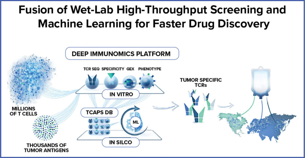
In recently published peer-reviewed research, ImmunoScape demonstrated that its cutting-edge machine learning platform can accurately predict antigen specificity of human T-cells. By probing hundreds of blood samples for T cell specificity using its unique multiplex high-throughput T cell target screening platform, ImmunoScape has built a massive database of high-quality and functionally validated TCRs against different target classes, which was leveraged for training of its machine learning models. This study shows the capabilities of machine learning and opens possibilities for accelerating the challenging discovery of novel TCRs that could be transformed into TCR-based therapeutics.

ImmunoScape Dec 2023 sponsored content illustration
Credit: ImmunoScape

Leveraging its Deep Immunomics and machine learning platforms, ImmunoScape has built a discovery engine that offers 360-degree views of lab-validated data from millions of T cells. More than 100 functionally validated TCRs have been identified to date, with many more in the discovery pipeline. With a constantly growing repertoire of high-quality and functionally validated TCR data, ImmunoScape continues to refine its machine learning augmented processes to build an emerging pipeline to enable faster discovery of tumor-specific TCRs against novel tumor targets covering a globally diverse patient population.

ImmunoScape’s diverse TCR portfolio generates opportunities beyond T cell engineering and is applicable to multiple product modalities. TCRs can be used to equip a variety of immune cells with specific tumor-targeting potential, while taking advantage of unique cell characteristics, such as distinct trafficking or enhanced cytotoxicity. For example, TCR-NK (Natural Killer) cell therapies can focus potent cytotoxic NK-cell activity toward tumors bearing targeted antigens. This approach can also be applied to multiple off-the-shelf (allogeneic) cell therapies using cost-efficient manufacturing processes.

Combination therapies, complimented with TCR-engineering, further present opportunities to overcome the known challenges associated with solid tumors, such as suppressive tumor microenvironments, or to enhance overall T-cell fitness for deep and durable anti-tumor responses.

In some cases, TCRs are best deployed as soluble proteins re-directing endogenous T cells, rather than delivered cell therapies. Currently, there is an FDA-approved TCR-based bispecific highlighting the success of this modality. ImmunoScape’s research in building novel affinity-enhanced TCR-based bispecifics for selected tumor targets from its TCR portfolio is on the horizon.

Emerging technological advances across drug development will enable and accelerate delivery of efficacious TCR-based therapies to greater numbers of patients. The prospect of multiplexing TCRs offers a powerful approach to combat HLA and tumor antigen heterogeneity. The integration of TCRs into emerging mRNA-based delivery methods pose additional intriguing possibilities for off-the-shelf manufacturing approaches to bring multi-TCR therapies to patients on a global scale.

The development of TCR-based therapeutics across modalities will not only accelerate the delivery of treatment for solid tumors, but it also holds the potential to cut off cancer before it develops.

Using Machine Learning to Accelerate TCR-Based Therapeutics for Solid Tumors


Data-driven approaches could foster discovery of novel and efficacious TCRs across a variety of tumor indications

ImmunoScape Dec 2023 sponsored content feature image
Credit: ImmunoScape

By Michael Fehlings, PhD

Sponsored content brought to you by

ImmunoScape Logo

Therapeutics for solid tumors remain in a nascent corner of cancer research despite representing 90 percent of all cancers in adults. Thirty years after the first Chimeric Antigen Receptor (CAR) was reported, CAR-T cell therapies have seen significant breakthroughs managing liquid tumors that can affect the bone marrow, blood cells, and the lymphatic system. Yet, the CAR-T approach, where T cells are engineered against specific targets on the tumor surface to locate and fight cancerous cells more effectively, has had limited impact on solid tumors.

Another approach, T-cell receptor (TCR)-based therapies—where cancer cells are identified and eliminated through highly-specific and sensitive T-cell receptors (TCR)—represents a promising therapeutic alternative. T cell receptor–based therapies harness the unique features of TCRs to recognize a wide range of tumor-specific surface and intracellular targets in different types of cancer. However, TCR-based therapeutics are caught in a discovery bottleneck which limits its applicability. Despite the evolution of cancer target identification, too few TCR targets have been discovered to address the unmet need, and suitable treatment is currently restricted to a minor global patient population.

In recently published peer-reviewed research, ImmunoScape demonstrated that its cutting-edge machine learning platform can accurately predict antigen specificity of human T-cells. By probing hundreds of blood samples for T cell specificity using its unique multiplex high-throughput T cell target screening platform, ImmunoScape has built a massive database of high-quality and functionally validated TCRs against different target classes, which was leveraged for training of its machine learning models. This study shows the capabilities of machine learning and opens possibilities for accelerating the challenging discovery of novel TCRs that could be transformed into TCR-based therapeutics.

ImmunoScape Dec 2023 sponsored content illustration
Credit: ImmunoScape

Leveraging its Deep Immunomics and machine learning platforms, ImmunoScape has built a discovery engine that offers 360-degree views of lab-validated data from millions of T cells. More than 100 functionally validated TCRs have been identified to date, with many more in the discovery pipeline. With a constantly growing repertoire of high-quality and functionally validated TCR data, ImmunoScape continues to refine its machine learning augmented processes to build an emerging pipeline to enable faster discovery of tumor-specific TCRs against novel tumor targets covering a globally diverse patient population.

ImmunoScape’s diverse TCR portfolio generates opportunities beyond T cell engineering and is applicable to multiple product modalities. TCRs can be used to equip a variety of immune cells with specific tumor-targeting potential, while taking advantage of unique cell characteristics, such as distinct trafficking or enhanced cytotoxicity. For example, TCR-NK (Natural Killer) cell therapies can focus potent cytotoxic NK-cell activity toward tumors bearing targeted antigens. This approach can also be applied to multiple off-the-shelf (allogeneic) cell therapies using cost-efficient manufacturing processes.

Combination therapies, complimented with TCR-engineering, further present opportunities to overcome the known challenges associated with solid tumors, such as suppressive tumor microenvironments, or to enhance overall T-cell fitness for deep and durable anti-tumor responses.

In some cases, TCRs are best deployed as soluble proteins re-directing endogenous T cells, rather than delivered cell therapies. Currently, there is an FDA-approved TCR-based bispecific highlighting the success of this modality. ImmunoScape’s research in building novel affinity-enhanced TCR-based bispecifics for selected tumor targets from its TCR portfolio is on the horizon.

Emerging technological advances across drug development will enable and accelerate delivery of efficacious TCR-based therapies to greater numbers of patients. The prospect of multiplexing TCRs offers a powerful approach to combat HLA and tumor antigen heterogeneity. The integration of TCRs into emerging mRNA-based delivery methods pose additional intriguing possibilities for off-the-shelf manufacturing approaches to bring multi-TCR therapies to patients on a global scale.

The development of TCR-based therapeutics across modalities will not only accelerate the delivery of treatment for solid tumors, but it also holds the potential to cut off cancer before it develops.

Immunotherapy against Solid Tumors May Be More Effective with Intact Lymph Nodes


Cancer treatment routinely involves taking out lymph nodes near the tumor in case they contain metastatic cancer cells. But new findings from a clinical trial by researchers at UC San Francisco and Gladstone Institutes have shown that immunotherapy can activate tumor-fighting T cells in nearby lymph nodes.

The team’s study of CD8+ T cell populations in human head and neck squamous cell carcinomas identified a role for lymph nodes in modulating antitumor responses in response to immune checkpoint blockade (ICB) immunotherapy. Results from the newly reported human study suggest that leaving lymph nodes intact until after immunotherapy could boost efficacy against solid tumors, only a small fraction of which currently respond to these newer types of treatments.

“This work really changes our thinking about the importance of keeping lymph nodes in the body during treatment,” said Matt Spitzer, PhD, an investigator for the Parker Institute for Cancer Immunotherapy and Gladstone-UCSF Institute of Genomic Immunology and senior author of the team’s study, which is published in Cell. In their paper, which is titled “Dynamic CD8+ T cell responses to cancer immunotherapy in human regional lymph nodes are disrupted in metastatic lymph nodes,”  the authors concluded, “These results lay a foundation for the future development of immunotherapies that optimally harness anti-tumor immunity in human LNs and inform immune-monitoring strategies for cancer patients treated with ICB.”

Immune checkpoint blockade (ICB) immunotherapy targeting the PD-1/PD-L1 axis has “revolutionized oncology,” the authors noted. “CD8+ T cells are central effector cells that mediate the efficacy of ICB and have been extensively studied in the tumor microenvironment (TME).” However, the mechanisms that underlie immunotherapy remain incompletely understood, especially in humans, they continued.

In settings such as cancer, antigen-specific CD8+ T cells can become exhausted. Studies in mice have shown that progenitor or precursor exhausted cells (Tpex) may differentiate into transitional intermediated exhausted cells (Tex-int) and subsequently into terminally exhausted cells (Tex-term), “losing proliferative capacity and effector functions as they differentiate,” the authors stated. Most immunotherapies are aimed only at reinvigorating  the potentially exhausted T cells in the tumor. But the new research shows that allowing the treatment to activate the immune response of the lymph nodes as well can play an important role in driving positive response to immunotherapy.

Lymph nodes are often removed because they are typically the first place metastatic cancer cells appear, and without surgery, it can be difficult to determine whether the nodes contain metastases. And as the team noted, “Despite the potential importance of the tdLN [tumor-draining lymph nodes] in ICB-driven CD8+ T cell responses, a lack of LN sampling in clinical datasets leaves many unanswered questions about the relationship between the immune responses in the LN and tumor in human cancer patients.”

“Immunotherapy is designed to jump start the immune response, but when we take out nearby lymph nodes before treatment, we’re essentially removing the key locations where T cells live and can be activated,” Spitzer said, noting that the evidence supporting the removal of lymph nodes is from older studies that predate the use of today’s immunotherapies.

Researchers have largely been working under the assumption that cancer immunotherapy works by stimulating the immune cells within the tumor, Spitzer continued. But in a 2017 study in mice, Spitzer showed that immunotherapy drugs are actually activating the lymph nodes. “That study changed our understanding of how these therapies might be working,” he said. Rather than the immunotherapy pumping up the T cells in the tumor, T cells in the lymph nodes are likely the source for T cells circulating in the blood. Such circulating cells can then go into the tumor and kill off the cancer cells. The authors further wrote, “… recent studies found that CD8+ T cells in the periphery, such as in secondary lymphoid organs (SLOs) including tumor-draining (td) lymph nodes (LNs), are integral for ICB response in mouse models.”

Having shown that intact lymph nodes can temper cancer’s hold in mice, Spitzer’s team wanted to know whether the same would prove true in human patients. They designed their trial to include patients with head and neck cancers because of the high number of lymph nodes in those areas. “We examined CD8+ T cells in human head and neck squamous cell carcinomas, regional LNs, and blood using mass cytometry, single-cell genomics, and multiplexed ion beam imaging.”

The study included 12 patients whose tumors hadn’t yet metastasized past the lymph nodes. Such patients would typically undergo surgery to remove the tumor, followed by other treatments if recommended. Instead, patients first received a single cycle of the anti-PD-L1 immunotherapy drug atezolizumab. A week or two later, Spitzer’s team measured how much the treatment had activated the patients’ immune systems.

As part of the study each patient’s tumor and nearby lymph nodes were surgically removed after immunotherapy and analyzed to determine how the immunotherapy had affected them. The team found that, after immunotherapy, the cancer-killing T cells in the lymph nodes began springing into action. They also found higher numbers of related immune cells in the patients’ blood. “Changes in uiLNs [uninvolved lymph nodes] correlated with an increase in proliferating Tex-int in peripheral blood.” Spitzer attributes some of the trial’s success to its design, which allowed the team to get a lot of information from a small number of patients by looking at the tissue before and after surgery, and by running detailed analyses.

“Being able to collect the tissue from surgery shortly after the patients had been given the drug was a really unique opportunity,” he said. “We were able to see, at the cellular level, what the drug was doing to the immune response.” That kind of insight would be challenging to get from a more traditional trial in patients with later-stage disease, who would not typically benefit from undergoing surgery after immunotherapy.

Another benefit of the study design was that it allowed researchers to compare how the treatment affected lymph nodes with and without metastases. “No one had looked at metastatic lymph nodes [metLNs] in this way before,” said Spitzer. “We could see that the metastases impaired the immune response relative to what we saw in the healthy lymph nodes.” The authors noted, “Patients with metLNs also experienced weaker CD8+ T cell responses in the blood following treatment, supporting an important role for LNs in generating circulating CD8+ T cell responses that associate with clinical response.” The results, they noted, “… add to a developing understanding of the impact of metastases in LN on the anti-tumor immune response.”

It could be that the T cells in these metastatic nodes were less activated by the therapy, Spitzer said. If so, that could explain, in part, the poor performance of some immunotherapy treatments. “ … regional lymph nodes with tumor metastasis (metLNs) exhibited an impairment in these responses to ICB associated with immunosuppressive cellular niches around Tpex and a reduction in the circulating CD8+ T cell response.”

Even so, the treatment prompted enough T-cell activity in the metastatic lymph nodes to consider leaving them in for a short period of time until treatment ends. “Removing lymph nodes with metastatic cancer cells is probably still important but taking them out before immunotherapy treatment may be throwing the baby out with the bathwater,” said Spitzer.

As the authors further concluded, “In summary, our data highlight the important role of CD8+ T cell responses in human LNs at steady-state and after ICB immunotherapy while also revealing the disruption of these key processes by LN metastasis … These results highlight a central role for uiLNs in mediating responses to ICB, which may create new opportunities for next-generation immunotherapies focused on optimally harnessing these responses.”

A subsequent goal of the current trial is to determine whether giving immunotherapy before surgery protects against the recurrence of tumors in the future. Researchers won’t know the answer to that until they’ve had a chance to monitor the participants for several years. “My hope is that if we can activate a good immune response before the tumor is taken out, all those T cells will stay in the body and recognize cancer cells if they come back,” Spitzer said.

Next, the team plans to study better treatments for patients with metastatic lymph nodes, using drugs that would be more effective at reactivating their immune responses.

Data Shows Promise for Interventional Oncology Treatment of Solid Tumors


Positive pre-clinical and early clinical study results using the Aliya Pulsed Electric Field (PEF) system from Galvanize Therapeutics were presented during oral sessions at the Society of Interventional Radiology (SIR) 2023 Annual Scientific Meeting.

The Aliya PEF system, which has US Food and Drug Administration clearance for soft tissue ablation, is being studied in Europe and Asia to treat solid tumors by inducing PEF-mediated cell death and neoantigen creation, designed to stimulate the patient’s own immune system to activate against the tumor.

The INCITE-ES clinical study is a treat and resect study conducted outside of the US, designed to assess safety, including impact on sensitive structures such as vessels, airways and pleura; and examine immune activation in patients with non-small cell lung cancer (NSCLC)(NCT04732520). Results demonstrated high technical success with a favorable safety profile in delivering PEF energy with a single needle with no PEF-related adverse events and no impact on planned surgical resections. PEF treatment significantly reduced malignant tissue, and 95% of the assessed sensitive structures within the PEF treatment zone remained viable and unaffected by the PEF energy. Additionally, PEF increased tertiary lymphoid structure accumulation, indicating potential immunoactivation with PEF.

A pre-clinical study, which was featured by SIR as an SIR 2023 exceptional abstract, compared tumor response with incomplete ablation using PEF vs radiofrequency ablation (RFA) in mice with breast cancer tumors. Results showed that PEF induced a greater infiltration of immune cells in the ablated zone and increased tumor recruitment of antigen cells as compared to RFA. PEF also showed greater suppression of tumor growth and had a synergistic effect in combination with immunotherapy, unlike RFA.

“Galvanize’s unique approach to improving cancer outcomes through electrosurgical immunotherapy continues to show compelling promise, and we are excited to share our newest study results with leading interventional radiologists and other medical experts around the world,” said Jonathan Waldstreicher, MD, Founder and CEO of Galvanize Therapeutics. “We are making additional investments to expand our Aliya™ clinical research program, including a US clinical study of patients with NSCLC.”

Centered on disease biology and how energy can alter cellular physiology, the Aliya PEF system delivers high-voltage, high-frequency electrical pulses through a single monopolar electrode placed in the target tissue. PEF energy destabilizes the cells, resulting in cell death. Aliya does not rely on thermal mechanisms to induce cell death.

The non-thermal modality of cell death with the Aliya PEF system preserves surrounding healthy tissue, enabling ablation near sensitive structures, such as nerves and blood vessels. The Aliya waveform and electrode are designed to maximize the potential for releasing tumor antigens and may stimulate an immune response, potentially disrupting the immunosuppressive tumor microenvironment.

In the search for therapies for solid tumors, companies are turning to a novel target: claudin-6


On target column illo cancer drug targets

On Target is a recurring feature from STAT that dives deep into the most promising drug targets in oncology. This column is adapted from the new STAT Report: Targeting cancer: the new frontier of immunotherapy and precision oncology.

Targeted immunotherapies like CAR-T have been remarkably successful in combating blood cancers like chronic lymphocytic leukemia. But malignancies that involve solid tumors have proved far more challenging for these new technologies. As yet, there has been no engineered cell therapy for solid tumors, which make up the overwhelming majority of cancers and include breast, lung, pancreatic, ovarian and prostate cancers.

One of the biggest roadblocks has been finding the right molecular targets. These therapies work by killing any cell that carries a designated marker, so scientists need to find a molecular target that exists on cancer cells but is not present in healthy tissues –  especially life-sustaining tissues like the heart or the brain.

Scientists have found few targets that fit this bill, but recently, a protein called claudin-6 or CLDN6 has been drawing the interest of researchers as one potential guide for immunotherapies. Several biotech and pharma companies have begun developing bispecific antibodies — engineered antibodies that can bind to two different antigens — or CAR-T cell therapies targeting CLDN6. BioNTech, whose founders helped discover the protein as a possible cancer antigen, is one of the most notable.

Researchers working with BioNTech presented data from a clinical trial of a CAR-T therapy targeting CLDN6 at the American Association of Cancer Research meeting in New Orleans in 2022. The trial showed that, with an immunologic boost from an mRNA vaccine, the therapy could shrink some solid tumors. BioNTech code-named the combination of the vaccine and therapy BNT211, and it remains one of the first examples of early efficacy from CAR-T cells in solid tumors, something that the cancer immunotherapy field has long struggled to achieve.

That data also helped put CLDN6 more firmly on the map as a target, said Martin Lehr, the CEO of Context Therapeutics, a biotech that is also developing drugs targeting CLDN6. “For me as an oncology drug developer, the BioNTech data was really exciting. It creates opportunities for companies like us,” he said.

The discovery

In the 1990s, Özlem Türeci and her husband, Uğur Şahin, the co-founders of BioNTech, were mulling over how to find a viable target for solid tumors. Ideally, they knew, it would be a protein that wasn’t present in healthy tissues but was highly expressed on cancer cells. That gave them an idea. Cancer cells sometimes turn on embryonic genes, or genes that are only turned on during fetal development and then silenced after birth.

“We knew cancer cells like to activate embryonic genes because they make use of proliferation,” Türeci said.

So, if Türeci and her colleagues could find activated embryonic genes in cancer cells, that might yield a useful therapeutic target. In their search, “We got this set of interesting targets,” Türeci said. “One of them was claudin-6.”

Türeci and Şahin went on to found a company called Ganymede that was focused on building antibodies to CLDN6 and other targets, including one in the same protein group, claudin 18.2. Astellas Pharma acquired that company, along with its claudin assets, but it wasn’t the end of Türeci and Şahin’s interest in CLDN6. Later, after they created BioNTech, they turned back to CLDN6 and created more therapies targeting the protein, including BNT211, the CAR-T and mRNA vaccine combination.

The biology

CLDN6 is a member of the claudin family of proteins, involved with regulating cell permeability and adhesion and helping maintain the structure and shape of cells. While scientists know that CLDN6 is present during the development of a fetus, its exact function is not entirely clear. The protein helps cancer cells spread, Türeci said,  “but we can’t explain fully how it promotes transformation of a cancer cell, for example. We do not know in-depth how CLDN6 functions. To understand this in-depth, it really is a project of a lifetime.”

Enhanced T Cell Therapy Found Effective against Multiple Solid Tumors


Results of the first-in-human trial of an enhanced T cell therapy that targets multiple solid tumors bearing the antigen MAGE-A4, showed noteworthy outcomes in several cancers, particularly synovial sarcoma. The multi-center phase I clinical trial (clinical trial ID: NCT03132922) was led by researchers at the University of Texas MD Anderson Cancer Center and sponsored by Adaptimmune, a clinical-stage biopharmaceutical company focused on developing cancer immunotherapies.

The findings were published in the journal Nature Medicine on January 9, 2023 “Autologous T cell therapy for MAGE-A4+ solid cancers in HLA-A*02+ patients: a phase 1 trial.”

The enhanced T cell therapy, afamitresgene autoleucel (afami-cel) achieved an objective response rate (ORR) of 44% in patients with synovial sarcoma while the overall response rate across all types of cancers tested in this trial was 24%. (ORR, defined as the proportion of patients with a complete or partial response to treatment is a common endpoint in cancer drug trials.) These early proof-of-concept findings establish acceptable safety of the treatment regimen and support the use of this novel cell therapy for solid tumors.

David Hong, MD, professor of investigational cancer therapeutics, and principal investigator of the study said, “These high response rates are significant because patients with synovial sarcoma really have very few options after high-dose chemotherapy with ifosfamide.”

MAGE-A4 (Melanoma-associated antigen A4) is a protein expressed in solid tumors such as synovial sarcoma (SS), myxoid/round cell liposarcoma (MRCLS), non-small-cell lung cancer (NSCLC), head and neck squamous cell carcinoma (HNSCC) and ovarian, urothelial, melanoma and gastroesophageal cancers. Its expression in healthy tissue is limited to immune-privileged locations. MAGE-A4 is processed within the cell and fragments of it are flagged on cell surfaces together with human leukocyte antigens (HLAs). The combination of MAGE-A4 and HLAs forms a recognizable epitope for natural low-affinity T cell receptors (TCRs).

Immune checkpoint blockade has good clinical activity in some patients with some solid tumors that express MAGE-A4 such as melanoma, but the response may not be as good in other types of solid tumors such as synovial sarcoma.

Unlike chimeric antigen receptor (CAR)-based cell therapies that detect cell surface proteins, TCR therapies, including afami-cel, can detect proteins found within cells and more accurately target solid tumor cells without the toxicity to healthy cells often occurs with CAR therapies.

Afami-cel is an autologous and enhanced T cell therapy where a specific, high-affinity TCR against a fragment of MAGE-A4 (residues 2030 to 239) presented by HLA-A*02 is transduced through a lentiviral vector.

Earlier preclinical studies have shown that that this enhanced TCR responds strongly upon encountering MAGE-A4 peptide fragments that are presented on several common HLA-A2 alleles, inducing potent cytotoxic effects and effector cytokine release against multiple types of cancers that express MAGE-A4.

As part of the phase I trial, 38 patients with an average of three prior lines of therapy, were treated with afami-cel. Participants were 92% white and 58% male. Sixteen patients had synovial sarcoma, nine patients had ovarian cancer, three had head and neck cancer, two each had esophageal, non-small cell lung, myxoid/round cell liposarcoma and urothelial cancer, and one each had gastric cancer and melanoma.

All patients experienced some treatment-related adverse events, including low blood cell counts (lymphopenia, leukopenia, neutropenia, anemia and thrombocytopenia) and two patients had trial-related deaths. This led to the researchers lowering the maximum age at screening and discontinuing the high-dose cyclophosphamide lymphodepletion.

“The overall toxicity from afami-cel was manageable, and we saw evidence of early activity in [several] cancer types,” said Hong.  “These results suggest this is an approach with the potential to work in solid tumors where there are currently no approved cellular therapies.”

These optimistic results has spurred a phase II trial of afami-cel (NCT04044768) in patients with advanced synovial sarcoma or myxoid/round cell liposarcoma.

Immunotherapy Tightens the Siege of Solid Tumors.


Translational Scientists Issue an Immunological Call to Arms, Prepare to Overcome Cancer’s Defenses

Immunotherapy Tightens the Siege of Solid Tumors

This image depicts a group of killer T cells surrounding a cancer cell. [Alex Ritter, Jennifer Lippincott Schwartz, and Gillian Griffiths, National Institutes of Health]

  • In anticancer campaigns, the immune system has often shown too little fighting spirit. It can be too civilized, too restrained—unless it is specially outfitted and guided. Measures that can drive the immune system to exert itself more strenuously, more aggressively, include monoclonal antibodies, cancer vaccines, checkpoint inhibitors, and adaptive cell therapy—all the tools and techniques of immuno-oncology.

    The proliferation of immuno-oncology tools and techniques was evident at the recent Translating Science into Survival conference. This event, which was held in New York City was organized by the Cancer Research Institute, the Association for Cancer Immunotherapy, the European Academy of Tumor Immunology, and the American Association for Cancer Research. The organizers evidently anticipated that this event, like previous immunotherapy events, would be fairly intimate. Yet it sold out quickly and ultimately strained to accommodate 1,400 attendees.

    The event’s popularity was probably at least in partly due to recent immunotherapy successes against blood cancers. For example, adaptive cell therapy approaches have shown promise in small trials, and work along these lines continues apace, as several presentations demonstrated. Moreover, lessons derived from this work may be applied more broadly, even to the treatment of solid tumors.

    For example, in a presentation entitled “Engineered T cells for cancer therapy,” the University of Pennsylvania’s Carl June described his team’s progress in using chimeric antigen receptor (CAR)-modified T cells to treat patients with chronic lymphocytic leukemia (CLL). “We previously reported preliminary results on three patients with refractory CLL,” Dr. June noted. “Here we report the mature results for our initial trial using CAR-modified T cells to treat 14 patients with relapsed and refractory CLL.”

    The overall response rate in CLL patients was 8/14 (57%), with four complete remissions and four partial remissions. All responding patients developed B cell aplasia and experienced cytokine release syndrome, coincident with T cell proliferation. Minimal residual disease was not detectable in patients who achieved complete remission, which, Dr. June suggested, indicated that “disease eradication may be possible in some patients with advanced CLL.”

    Dr. June also summarized a separate investigation that asked whether the CAR cells used against CLL would also be effective against multiple myeloma. At first glance, this may seem odd, since the CAR cells that were effective against CLL target CD19, and CD19 expression is all but absent from myeloma cells. That is, myeloma cells don’t traditionally express CD19 on their surface because they arise from the most mature type of lymphocytes—plasma cells.

    Dr. June’s team, however, proceeded on the chance that they would be able to incorporate their anti-CD19 CAR T cells into a therapy that would target early precursors of myeloma cells. This therapy, which was administered to a patient with refractory multiple myeloma, involved an infusion of the patient’s own stem cells along with lymphodepleting chemotherapy (melphalan) as well as an infusion (two weeks later) of anti-CD19 CAR T cells.

    The patient experienced transplantation-related side effects during the time prior to receiving CTL019, including neutropenia and thrombocytopenia, nausea, fever, and an infection. After receiving the engineered cells, she experienced no fevers or other signs of cytokine release syndrome, a condition that has been observed in other patients undergoing CTL019. At last evaluation, 12 months after treatment, the patient exhibited a complete response with no evidence of progression. According to Dr. June, “This response was achieved despite absence of CD19 expression in 99.95% of this patient’s neoplastic plasma cells.”

    While the results presented by Dr. June pertain most directly to blood cancers—specifically, the inducement of favorable patient responses to treatment—they may also apply more broadly. For example, they demonstrate that a “living drug” may exert its effects indirectly. Also, they emphasize the importance of managing toxicity and ensuring the expansion of modified cells. They also raise the issue of introducing cells that may demonstrate longevity.

    More generally, uncertainties surrounding the differential expansion and persistence of distinct cell populations over time can complicate dosing. Similarly, with respect to toxicity, cytokine release and the generation of tumor-shredding products might be considered side effects or, really, evidence that a therapy is working.

    Yet engineered cells may also attack both malignant and healthy cells directly. That is, engineered cells may be sensitive to target proteins that stud both cancer cells and also, if only to a lesser degree, normal cells. In CAR T-cell therapies against leukemia and lymphoma, side effects related to direct attacks on normal cells has been manageable. Such side effects, however, may be more severe if adaptive cell therapies are directed against solid tumors.

    Leading up to the Translating Science into Survival conference, Dr. June’s group published a study that described an approach for managing target-mediated toxicity. The approach, called affinity tuning, involves generating CAR T cells that are sufficiently insensitive to ignore normal cells, which are relatively target-sparse, and yet sensitive enough to latch onto cancer cells, which are relatively target-rich.

    In a paper (“Affinity-Tuned ErbB2 or EGFR Chimeric Antigen Receptor T Cells Exhibit an Increased Therapeutic Index against Tumors in Mice”) that appeared September 1 in Cancer Research, the case was made that “affinity-tuned cells” could exhibit robust antitumor efficacy similar to high-affinity cells, but spare normal cells expressing physiologic target levels: “The use of affinity-tuned scFvs offers a strategy to empower wider use of CAR T cells against validated targets widely overexpressed on solid tumors, including those considered undruggable by this approach.”

    At the Translating Science into Survival event, the presentations concerning solid tumors emphasized that adoptive cell therapy could become more effective if obstacles in the tumor microenvironment could be overcome. For example, the University of Pennsylvania’s Ellen Puré, in a presentation entitled, “Tumor stroma: Immunomodulatory functions and a target of immunotherapy,” explained that stroma can be a barrier to T cells, including CAR T cells.

    Stromal components such as fibroblasts and the extracellular matrix can play myriad functions in cancer. For example, Puré noted, reactive stroma enriched in growth and angiogenic factors presents chemoattractants that promote the recruitment of bone marrow-derived cells and can modulate inflammatory and immune cell function, all of which can contribute to its tumor-permissive nature relative to normal stroma.

    “A significant portion of cancer-associated fibroblasts in virtually all human carcinomas express the cell surface protease fibroblast activation protein (FAP),” Puré continued. “Our studies indicate that FAP+ cells are required for the generation and maintenance of desmoplastic stroma and that depletion of FAP+ cells can inhibit tumor growth through both immune-dependent and immune-independent mechanisms.”

    Another take on the tumor microenvironment was presented by Wolf H. Fridman, Cordeliers Research Centre, Paris. In a talk entitled, “Cancer subtypes and their immune microenvironments,” Dr. Fridman described how his group was elaborating on the Immunoscore concept, which goes back at least as far as 2006. Basically, Immunoscore builds on the insight that in many patients, the density of T cells near tumor cells could be a better predictor of survival than traditional staging based on a cancer’s size and spread.

    In general, for a patient’s prognosis to be favorable, immune cells need to infiltrate a solid tumor. But recent work also suggests that more immune cells may not always be better. Apparently some immune cells are less helpful than others. Some may even be deleterious, depending on the interactions that occur between a tumor’s microenvironment and the immune system.

    “Our team studied the immune infiltrates of pulmonary metastases from colorectal cancer (CRC) and renal cell carcinoma (RCC),” reported Dr. Fridman. “As in primary tumors, a high density of CD8+ T cells correlated with good prognosis for CRC metastases, while it correlated with a bad prognosis for RCC metastases.”

    “In addition, in both cancer types, we identified subgroups of poor-prognosis patients with high tumoral lymphocyte infiltration, in the context of high expression of genes related to inflammation, immunosuppression, and angiogenesis,” he continued. “These results suggested that the identity of the tumor cells, rather than the organ where they grow, is critical for shaping the immune contexture of a given tumor.”

    A particular cancer, then, may have an ecology of its own, one in which the overall disposition of elements—tumor cells, immune cells, extracellular matrix elements, and so on—matters, much as the overall disposition of chess pieces matters in a game of chess. Even though it might be advantageous to occupy a particular space on the board, apparently not any chess piece will do. Frustratingly, one’s own pieces may be poorly positioned, so as to get in each other’s way.

    Nonetheless, as Dr. Fridman concluded, the integration of molecular and immune tumor phenotypes could guide the selection of immunotherapies “appropriate to specific, potentially responding groups of patients.”