A 29-year-old woman presented to the internal medicine clinic with 2 months of postural syncope. Blood pressure as measured in her upper arm was 79/49 mm Hg and her heart rate was 90 beats/min, with no postural changes. Investigations (including electrocardiography, routine bloodwork and adrenal function tests) were unremarkable. Since her symptom complex resembled postural tachycardia syndrome, we started fludrocortisone empirically.
Over several months, the patient’s syncopal episodes became more frequent, and she developed headaches, fatigue and upper extremity paresthesias. Her blood pressure dropped to 57/48 mm Hg, with heart rates of 80–90 beats/min; we considered that her heart rate was inappropriately low, given her low blood pressure. This prompted us to investigate for autonomic dysfunction. Although she had no neurologic findings on physical examination, we considered paraneoplastic pandysautonomia and Lambert–Eaton myasthenic syndrome. After magnetic resonance imaging of the patient’s brain showed fluid-attenuated inversion recovery (FLAIR) hyperintensity in the posterior circulation, we ordered computed tomography of her chest with angiography, which showed circumferential thickening of the thoracic aorta and great vessels, with critical narrowing of subclavian and carotid arteries (Figure 1), leading us to diagnose Takayasu arteritis.
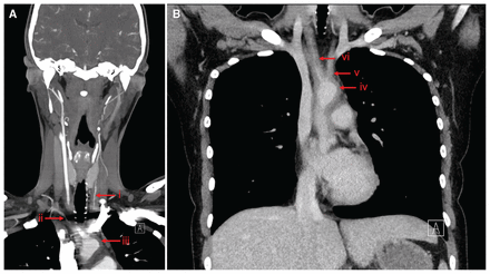
(A) Computed tomography angiogram of a 29-year-old woman with recurrent syncope, showing critical narrowing of left common carotid (i) and right subclavian (ii) arteries, and mural thickening along the aortic arch (iii). (B) Computed tomography infusion scan of the patient’s chest showed diffuse mural enhancement and thickening of aortic arch (iv), with thickening and vessel narrowing of the brachiocephalic (v) and left common carotid (vi) arteries.
Upper and lower extremity blood pressures obtained after imaging were 60/40 mm Hg and 140/70 mm Hg, respectively; this key finding may have led to an earlier diagnosis. Upper extremity pulses that were initially present were now absent. The patient’s erythrocyte sedimentation rate (ESR) and C-reactive protein (CRP) levels were normal. We started her on prednisone (1 mg/kg/d) and azathioprine (50 mg/d, increased to 125 mg/d) but she ultimately required arterial bypass with grafts connecting the descending aorta to the left carotid and the left subclavian artery.
Takayasu arteritis is an uncommon large-vessel vasculitis that predominantly affects young women.1 Insidious symptom onset and rarity makes recognition challenging; the diagnosis is often delayed until after ischemic manifestations occur.1 The ESR and CRP levels are unreliable markers of active disease, with a reported sensitivity and specificity of 75%; therefore, imaging is necessary for diagnosis.2 Treatment involves high-dose glucocorticoids and nonglucocorticoid immunosuppressive agents, with surgery reserved for critical stenosis.3
Diagnosis of Takayasu arteritis requires a high index of suspicion. Clinicians should undertake a thorough pulse examination and measure blood pressure in different limbs in patients with unexplained syncope to investigate for vasculitic causes.
Clinical images are chosen because they are particularly intriguing, classic or dramatic. Submissions of clear, appropriately labelled high-resolution images must be accompanied by a figure caption. A brief explanation (300 words maximum) of the educational importance of the images with minimal references is required. The patient’s written consent for publication must be obtained before submission.
Source: MJA
