Rabies is an ancient, unpredictable and potentially fatal disease − two rabies researchers explain how to protect yourself


Rabies virus (red) has an incubation period that can last from days to months.

Rodney E. Rohde has received funding from the American Society of Clinical Pathologists, American Society for Clinical Laboratory Science, U.S. Department of Labor (OSHA), and other public and private entities/foundations. Rohde is affiliated with ASCP, ASCLS, ASM, and serves on several scientific advisory boards.

Charles Rupprecht is a global biomedical consultant for academia, government, industry and NGOs. He is affiliated with LYSSA LLC.

A feral kitten in Omaha, Nebraska, tested positive for rabies in November 2023. It died of the raccoon variant of the virus, which is typically found only in the Appalachian Mountains. Detecting this variant hundreds of miles away in the Midwest raised concerns about a potential outbreak and launched a public health task force to vaccinate all raccoons in the area.

While the case was likely contained, a better understanding of how rabies is transmitted can help prevent future outbreaks. Researchers Rodney Rohde and Charles Rupprecht explain how rabies vaccination works and how to protect yourself from infection.

What causes rabies?

Rabies is an ancient viral disease that has been around for thousands of years. Considered a neglected tropical disease, rabies typically occurs in poorer communities without the infrastructure for adequate surveillance, prevention and control.

Rabies is unpredictable. Stages of infection include an incubation period that ranges from days to months, early flu-like symptoms, a period of severe neurological effects, coma and then death. Common early-stage symptoms in people, such as fatigue, fever and nausea, are often nonspecific. Neurological symptoms can involve aggression, confusion, difficulty swallowing and paralysis.

The pathogens that cause rabies belong to a genus of viruses called Lyssavirus that target warm-blooded vertebrates. Although researchers believe that all mammals are susceptible to infection, only certain animals are reservoirs: environments, habitats or populations where a pathogen can live, multiply and transmit. In the U.S., the highest-risk animal reservoirs for rabies are skunks, bats, foxes, coyotes and raccoons. A kitten found in Omaha infected with rabies spurred a push to vaccinate local wildlife.

Most people who become infected with rabies get it from an animal bite. Less common routes include contact with open wounds or the mucous membranes of the eyes, nose or mouth. Once the virus enters the body, it can begin replicating in muscle tissue or after traveling directly to the brain. Once it spreads to other organs, patients typically die of brain inflammation.

Are rabies cases increasing?

Measuring the global burden of rabies is difficult because surveillance is often inadequate.

While human incidence of rabies are rare in the U.S., averaging one to three cases per year, this disease causes tens of thousands of deaths worldwide annually.

Rabies rates in animals vary each year. During 2021, 54 U.S. jurisdictions reported 3,663 rabid animals, an 18.2% decrease from the previous year. Lower- and middle-income countries, especially since the COVID-19 pandemic, have experienced disruptions to animal vaccination for rabies due to vaccine production and access issues and increased feral animal populations.

Human rabies cases have risen in several countries because of multiple ecological and socioeconomic factors. For example, in China, rabies cases are associated with proximity to urban populations and transportation hubs. The closer a susceptible animal is to a community experiencing an outbreak, the greater the likelihood for spread.

Increased temperatures due to climate change are also linked to increased rabies transmission because of changes in animal ranges. For example, as regions warm, the relative distribution and abundance of certain reservoirs, such as tropical species like vampire bats, may increase. Rising Arctic temperatures may increase how often red and Arctic foxes interact and lead to outbreaks.

Higher rates of interactions between humans and animals, as well as lower levels of rabies education and prevention measures, are also linked to an increased risk of infection.

Has controlling rabies in wildlife been successful?

Prior attempts to control rabies include animal culling and vaccination. Culling animal populations did not lead to reduced infection. Rather, it raised significant economic, ecological and ethical concerns. Besides killing likely healthy animals, culling also isn’t cost-effective.

Animal vaccination, on the other hand, can protect both animals and humans with minimal risk and reduced costs. Oral rabies vaccination of wildlife began during the 1970s with the distribution of vaccine-laden baits in the local environment. Officials saw success in rabies control among coyote, fox and raccoon populations in Europe and North America.

Two stray dogs playing with each other on a street
Vaccination of strays can reduce the spread of rabies. CDC/Nicholas S. Tenorio, Health Communication Specialist

We both participated in the inaugural oral rabies vaccination campaign in Texas. This effort eventually led to the elimination of canine rabies in the state.

Oral vaccines are also being considered for the prevention and control of rabies in animals in other countries, such as dogs in India and Thailand.

How do rabies vaccines work?

There is no proven treatment for rabies, so prevention through education and vaccination is critical. Rabies can be prevented by avoiding exposure or receiving vaccination before or after an exposure.

Preexposure prophylaxis, or PrEP, involves exposing the immune system to a harmless version of the virus to prevent a future infection. For people who work in high-risk occupations, such as wildlife biologists, veterinarians and animal control personnel, two doses of a rabies vaccine can offer significant protection.

The Centers for Disease Control and Prevention currently recommends a booster dose for people at elevated risk of exposure. People traveling to areas with a high prevalence of rabies may also want to consider vaccination.

Postexposure prophylaxis, or PEP, means taking a vaccine or medications to prevent an infection as soon as possible after exposure. In most cases, this involves a dose of human rabies immune globulin, or HRIG, as well as a rabies vaccine dose on the day of exposure.

Also known as passive immunization, HRIG gives your body the antibodies to neutralize rabies virus until your immune system can produce its own antibodies. Three more doses of the rabies vaccine are given three to 14 days after exposure. People who are immunocompromised may receive a fifth dose as well.

Microscopy image of clusters of dark circles called Negri bodies against a background of pink brain tissue
Negri bodies (purple) are proteins that collect in neurons containing rabies virus. CDC/Dr. Daniel P. Perl

Regular and appropriate pet and livestock vaccinations are also important to help curb rabies virus exposures. Animals typically receive a yearly rabies booster.

There are several new rabies vaccines for animals and people in development to improve their safety, cost and efficacy. Researchers are also developing treatments to control rabies when the infection reaches the central nervous system.

How do you protect yourself from rabies?

Some people may not realize they were bitten by an animal if the bites are small. Because of the long incubation period of the virus, they also may not recall a previous interaction with an infected animal.

Laboratory tests can confirm whether an animal has rabies, as well as which rabies virus variant is present. A physician may begin vaccination even without laboratory confirmation based on the risk factors of a case. For example, since most recent fatal rabies cases in the U.S. have been from unknown bat bites, rabies vaccination is recommended after any suspected bat exposure.

There are many practical ways to protect yourself from rabies such as:

  • Vaccinating and supervising your pets.
  • Never handling wild animals that seem to be acting strangely.
  • Not touching sick, injured or dead animals.
  • Never attempting to feed wildlife.
  • Treating animals respectfully. Do not tease an animal, disturb its sleep or handle its offspring.
  • Always reporting a bite to an animal control officer, game warden or health care professional.

WHO Guidelines for Post-exposure prophylaxis of Rabies


https://speciality.medicaldialogues.in/who-guidelines-for-post-exposure-prophylaxis-of-rabies/

Guide for post-exposure prophylaxis


The recommendations given here are intended as a general guide. It is recognized that, in certain situations, modifications of the procedures laid down may be warranted. Such situations include exposure of infants or mentally disabled persons and other circumstances where a reliable history cannot be obtained, particularly in areas where rabies is enzootic, even though the animal is considered to be healthy at the time of exposure. Such cases may be treated as category II or III.

Post-exposure treatment, which consists of local treatment of the wound, followed by vaccine therapy (with or without rabies immunoglobulin) should be initiated immediately with contacts of categories II and III. Treatment may be discontinued if the animal involved (dog or cat) remains healthy throughout an observation period of 10 days; or if the animal is killed humanely and found to be negative for rabies by laboratory examination. Any biting animal suspected of being rabid should be immediately killed humanely and tissues examined using appropriate laboratory technique(s). Modification of the recommended procedures would be indicated in a rabies-free area where animal bites are encountered. In areas where canine or wildlife rabies is epizootic, adequate laboratory and field experience, indicating that there is no infection in the species involved, may justify local health authorities in not recommending specific anti-rabies treatment.

The indication for post-exposure vaccination with or without rabies immune globulin depends on the type of contact with the rabid animal.

Types of contact are:
  • category I – touching or feeding animals, licks on the skin
  • category II – nibbling of uncovered skin, minor scratches or abrasions without bleeding, licks on broken skin
  • category III – single or multiple transdermal bites or scratches, contamination of mucous membrane with saliva from licks; exposure to bat bites or scratches

For category I no treatment is required, whereas for category II immediate vaccination and for category III immediate vaccination and administration of rabies immune globulin are recommended in addition to immediate washing and flushing of all bite wounds and scratches. Depending on vaccine type, the post-exposure schedule prescribes intramuscular doses of 1 ml or 0.5 ml given as four to five doses over four weeks. For rabies-exposed patients who have previously undergone complete pre-exposure vaccination or post-exposure treatment with cell-derived rabies vaccines, two intramuscular doses of a cell-derived vaccine separated by three days are sufficient. Rabies immune globulin treatment is not necessary in such cases. The same rules apply to persons vaccinated against rabies who have demonstrated neutralizing antibody titres of at least 0.5 IU/ml.

In order to reduce the cost of post-exposure treatment, intradermal multi-site regimens using a fraction of the intramuscular volume per intradermal inoculation site have been developed. Purified Vero cell vaccine has been given intradermally to more than 70 000 recipients in Thailand, where it has been in routine use for several years. Intradermal rabies vaccination is also recommended by the ministries of health of Sri Lanka (since 1995) and the Philippines (since 1997). In each of these countries the introduction of this route for post-exposure treatment has permitted the discontinuation of the local production of vaccines prepared on brain tissue. Only the cell-derived vaccines that meet the WHO requirements regarding safety, potency and efficacy for this application may be considered for intradermal use. Although rabies vaccines are usually administered under qualified medical supervision, field experience from routine infant immunization programmes with other intradermally injected vaccines highlights the potential difficulties in assuring proper delivery. This emphasizes the need for appropriate staff training to ensure correct storage, reconstitution and injection. Provided that a correct sterile technique is used, the remaining doses may be kept in the vial at 2–8°C and used for another patient within six hours after reconstitution.

Tissue-culture or purified duck-embryo vaccines of potency at least 2.5 IU per single intramuscular immunizing dose should be applied according to the following schedules.

Intramuscular schedules

One dose of the vaccine should be administered on days 0, 3, 7, 14 and 30. All intramuscular injections must be given into the deltoid region or, in small children, into the anterolateral area of the thigh muscle. Vaccine should never be administered in the gluteal region.

Abbreviated multisite schedule

In the abbreviated multisite schedule, the 2-1-1 regimen, one dose is given in the right arm and one dose in the left arm at day 0, and one dose applied in the deltoid muscle on days 7 and 21. The 2-1-1 schedule induces an early antibody response and may be particularly effective when post-exposure treatment does not include administration of rabies immunoglobulin.

Intradermal schedule

WHO recommended the following intradermal regimen and vaccines for use by the intradermal route:

  • 2-site intradermal method (2-2-2-0-1-1) for use with PVRV (Verorab TM, Imovax TM, Rabies vero TM, TRC Verorab TM) and PCECV (Rabipur TM)
For 2-site intradermal method (2-2-2-0-1-1)

The volume per intradermal site is:

  • 0.1 ml for PVRV (Verorab TM, Imovax TM, Rabies vero TM, TRC Verorab TM)
  • 0.1 ml for PCECV (Rabipur TM)

Brain-tissue vaccines

The use of brain-tissue vaccines should be discontinued. WHO does not recommend any schedule using brain-tissue vaccine. National authorities should recommend a schedule of immunization that has been shown to induce an adequate level of protection when brain tissue vaccines are available in that country.

Combined immunoglobulin-vaccine

Combined immunoglobulin-vaccine treatment was considered in the eighth report of the WHO Expert Committee as the best specific systemic treatment available at that time for the post-exposure prophylaxis of rabies in humans, although experience indicated that vaccine alone was sufficient for minor exposures (category II). Immunoglobulin should be given in a single dose of 20 IU per kg of body weight for human anti-rabies immunoglobulin, and 40 IU per kg of body weight for heterologous (equine) immunoglobulin; the first dose of vaccine should be inoculated at the same time as the immunoglobulin, but in a different part of the body. Sensitivity to heterologous immunoglobulin must be determined before it is administered. The physician should be prepared to deal with anaphylactic shock reactions. Administration of rabies immunoglobulin (RIG) should be infiltrated into the depth of the wound and around the wound as much as anatomically feasible. Any remainder should be injected at an intramuscular site distant from that of vaccine inoculation e.g. into the anterior thigh.

Treatment should be started as early as possible after exposure, but in no case should it be denied to exposed persons whatever time interval has elapsed.

Local treatment of wounds

Local treatment of wounds involving possible exposure to rabies – recommended in all exposures.

How rabies attacks the brain, seen in Israel for the first time .


Israeli scientists watch as virus hijacks neuron ‘train’ and speeds to central nervous system
An illustration of a rabies virus in the nervous system. (photo credi: Rabies image

The virus has long been known to travel along neurons, the cells that transmit the electrical and chemical signals enabling movement, feeling, and thought. Until now, though, nobody had been able to figure out how.
Using powerful live cell imaging, the scientists found that the virus hijacks the “train” that transports cell components along a neuron, and drives it full throttle into the spinal cord. From there, the virus likely takes other trains to the brain and then throughout the peripheral nervous system, they say – shutting down the body as it goes along.
A microscope image of a sensory neuron, with an inset of the rabies virus (green) binding to the p75 receptor (red). (photo credit: Courtesy)

“The rabies virus is transported through railway-like machinery in the neurons,” said Shani Gluska, a doctoral student at Tel Aviv University, who led the study along with Prof. Eran Perlson, a physiologist at the university. “With very high-end microscopy, we saw for ourselves how the virus not only hijacks the transport machinery, but also makes it go faster.”

The scientists say their findings, published in the journal PLOS Pathogens in August, could one day enable scientists to take control of the neuron train system to treat rabies, as well as other neurodegenerative diseases.

Seeing is believing
Rabies is infamous for its dramatic symptoms, like aggression, psychosis, wild movements, and “foaming at the mouth.” Without treatment in time by vaccination, rabies severely inflames the brain, ultimately leading to paralysis of the heart and lungs and, with a few recent exceptions, to death. More than 55,000 people die every year from rabies, mostly in Africa and Asia, according to the World Health Organization.

To see how the rabies virus travels through the nervous system, the Israeli scientists grew mouse sensory neurons in the lab and infected them with the virus. They tagged the virus with a fluorescent marker, then watched and recorded its movements in real time with a high-power microscope.

‘If we can learn how rabies manipulates the system, we can maybe try to manipulate it ourselves’
The scientists saw that the virus takes a route normally reserved for nerve growth factors, proteins that are responsible for development and health of neurons. The virus enters a neuron in the peripheral nervous system by binding to a nerve growth factor receptor called p75.

Once inside, the virus boards a “train car,” a bubble-like vesicle, and departs from the cell membrane. “Engines” – nano-sized motor proteins that typically chug up and down neurons to keep them alive – then hitch themselves to the car and pull it along “tracks,” microtubules. The ride continues through the neuron’s tail-like axon, which can stretch up to a meter in length, and on to its cell body, which is located in the spinal cord.
Doctoral student Shani Gluska working in Dr. Eran Perlson’s lab at Tel Aviv University. (photo credit: Courtesy)

In the spinal cord, the scientists believe the virus catches the first available train to the brain, where it wreaks havoc before embarking on a tour of the body – though the study did not include the kinds of neurons that run directly to or from the brain. Compared to the growth factors that take the same p75 route, the virus travels much more quickly.

The scientists say the virus may speed up the train by pushing the engines harder, by dumping more “fuel,” or ATP, into the engines, or by demanding more engines or a better track. A minority of the rabies viruses in the study took other, slower routes along the neurons.

Putting the brakes on nerve disease
The results reveal what is likely a major mechanism the rabies virus uses to enter the peripheral nervous system, usually from the muscles, and to travel rapidly to the central nervous system, the scientists say.

Based on previous research, they say the virus probably travels in a similar way elsewhere in the nervous system: along motor neurons to the spinal cord, along interneurons in the spinal cord to the brain, and along motor neurons from the brain back to the peripheral nervous system.

Improved understanding of how the neuron train works could lead to new disease treatments, they say.

“If we can learn how rabies manipulates the system, we can maybe try to manipulate it ourselves,” said Perlson, who oversaw the study in his lab, which focuses on neuron signaling and transport. When it comes to rabies, interfering with the virus’ travel plans could extend the window of time for treatment, the scientists say.

The rabies vaccine is only effective until the virus reaches the central nervous system and begins causing symptoms, which usually takes one to three months. On the other hand, disruptions of the neuron train system contribute to neurodegenerative diseases, like Alzheimer’s disease, Parkinson’s disease, and amyotrophic lateral sclerosis (ALS).

The scientists say getting the train back on track could treat and possibly even cure such diseases.


Rabies Vaccine Dropped from the Sky


Story at-a-glance

  • There are only 2-4 human rabies cases in the US each year, but annual prevention costs are more than $300 million
  • The Texas Department of Health is using helicopters to spread 100,000 rabies vaccines for skunks in the wilderness; other states have also conducted similar vaccination efforts
  • No one knows if such programs are effective or if the indiscriminate spreading of a pharmaceutical product into the environment is going to have any unforeseen consequences to wildlife or the surrounding ecosystem.
  • rabies

In 2009, there were just four human cases of rabies in the US. In 2010, there were two1… yet each year, the US spends more than $300 million for rabies prevention,2 which includes the vaccination of companion animals, animal control programs, maintenance of rabies laboratories and medical costs.

Even at the turn of the century, rabies-related human deaths only numbered around 100 annually, and by the 1990s, this had dropped to one or two.  While rabies is a serious, potentially deadly, illness, it is most often transmitted through the bite of a rabid wild animal – a risk factor that is negligible for many in the US.

Texas Department of Health Is Dropping Experimental Rabies Vaccines from the Sky

About 92 percent of the reported rabies cases in 2010 were in wild animals, including raccoons, skunks, bats, foxes, rodents and others. This poses a theoretical risk not only to humans but also to family pets, which could then transmit rabies to their owners.

Nonetheless, human rabies cases remain extremely rare… but efforts are still underway to knock out the rabies virus in wild skunk populations in Texas.

The Texas Department of Health is actually using helicopters to spread 100,000 rabies vaccines in two counties. The vaccines, which are contained in plastic cases coated with fishmeal to entice wildlife to eat them, are part of a pilot program to help reduce the number of rabid skunks in the area.

No one knows yet if the program is going to work – skunks will need to be caught and tested for rabies 30-60 days after the vaccines are dropped – or if the indiscriminate spreading of a pharmaceutical product into the environment is going to have any unforeseen consequences to wildlife or the surrounding ecosystem.

Should Wildlife Be Vaccinated Against a Disease That Infects 2-4 People a Year?

It’s also unclear why Texas is taking such aggressive measures against rabies. There has so far been only one reported case of human rabies in Texas in 2013, and the man was exposed in Guatemala, Mexico — not in Texas. The last case of human rabies in Texas prior to that was in 2009 and prior to that in 2004 – for a total of just 6 human cases in the last decade.3

For comparison, there were 2,390 cases of campylobacteriosis in Texas in 2012 alone… an illness largely spread by contaminated poultry raised on concentrated animal feeding operations (CAFOs). This illness, too, can be deadly if it infects a person with a compromised immune system, yet we’re not hearing about widespread efforts to curb its transmission…

Even if you factor in data from the US Centers for Disease Control and Prevention (CDC), which states there were 6,153 reported cases of rabies in animals in 2010, that’s for animals in the entire US, and not only skunks but also raccoons, foxes, bats and others. Texas isn’t the only state to opt for preventative rabies vaccination of wildlife, either. According to the Human Society of the United States (HSUS):4

“Federal and state wildlife officials have been vaccinating wildlife in many regions over the past 15 years. They distribute vaccine-laden baits that the target animals eat and thereby vaccinate themselves. Right now, oral rabies vaccination of wildlife focuses on halting the spread of specific types of rabies in targeted carrier species. Next, it’s hoped that this tool can shrink the diseases’ range.”

The end question remains the same, not only for Texas but for the entire US: is it really necessary to spend $300 million a year on rabies prevention… and what are the potential consequences of vaccinating wildlife?

What Exactly Is Rabies?

Rabies is a viral disease that most often enters your body through a bite or wound contaminated by the saliva from an infected animal. If it manages to infect the central nervous system, it can lead to early symptoms that include fever, headache, weakness and discomfort. As the disease progresses, it can lead to insomnia, anxiety, confusion, paralysis, hallucinations, difficulty swallowing, fear of water and death.

If you have been bitten by a wild animal (or a dog with unknown rabies status), wash the wound thoroughly with soap and water, as this will help to decrease your risk of infection.

Next, talk to a doctor about your next steps. He or she will probably contact the local or state health department and, if it’s deemed that the animal was rabid or at high risk of being rabid, you may need to start postexposure prophylaxis (PEP), which consists of a series of vaccines that can protect you from developing rabies. But remember, though rabies is serious, and frightening, it’s extremely rare. HSUS puts it into perspective:

Given all the media attention that rabies regularly receives, it may be somewhat surprising to learn that very few people die from rabies nationwide each year. Over the past 10 years, rabies has killed only a total of 28 people in the U.S. This amounts to fewer than 3 fatalities a year nationwide.

People who contracted rabies in the United States were mostly infected by a bat. Most didn’t even know they were bitten. Some may have been sleeping when bitten. Others handled a bat bare-handed without realizing they’d been potentially exposed to rabies. But don’t panic over every bat sighting. Less than one-half of one percent of all bats in North America carries rabies. Although raccoons suffer from rabies more than any other mammal in the United States (about 35 percent of all animal rabies cases), only one human death from the raccoon strain of rabies has been recorded in the United States.”

Transmission of Rabies Virus from an Organ Donor to Four Transplant Recipients.


Rabies is an acute encephalitis caused by viruses in the genus Lyssavirus, family Rhabdoviridae, that is nearly uniformly fatal in unvaccinated hosts. Although the virus is present in animal reservoirs, infection in humans is rare in the United States, with only two cases reported in 20031,2 and no more than six cases reported in any year in the past decade.3 The primary mode of transmission is through the bite of an infected animal, most commonly a bat in the United States.4 Although transmission of rabies virus from corneal transplants has previously been described,5 to our knowledge, no cases ascribed to organ or vascular-tissue transplants have been reported.

In May 2004, physicians at a hospital in Texas diagnosed encephalitis in three recipients of a liver and two kidneys from a common organ donor. It was later discovered that encephalitis also developed in a fourth patient, who had received a vascular graft from the same donor during liver transplantation. All four patients became progressively obtunded, lapsed into coma, and died within 50 days after transplantation. The initial diagnostic evaluation revealed no cause of the encephalitis, and assistance was sought from the Centers for Disease Control and Prevention (CDC) and the Texas Department of State Health Services. We report the results of this investigation.

CASE REPORTS

Transplant Recipients

In May 2004, encephalitis was diagnosed in three recipients of a liver and two kidneys (Patients 2, 3, and 4 in Figure 1FIGURE 1The Clinical Course of Four Recipients of Rabies-Infected Tissue or Organs.) from a common organ donor. In all three patients, signs and symptoms of altered mental status and progressively worsening encephalitis developed within 30 days after transplantation. Major clinical events and immunosuppressive medications are summarized in Figure 1. All patients had rapid neurologic deterioration characterized by agitated delirium and seizures. Respiratory failure requiring intubation developed within 48 hours after the onset of neurologic symptoms. Examination of cerebrospinal fluid from the three patients showed pleocytosis, with an average of 18 cells per cubic millimeter (range, 7 to 35), and elevated protein levels (mean, 135 mg per deciliter; range, 17 to 331). Neurologic imaging in the week after the onset of symptoms showed no evidence of an acute cerebral process. Magnetic resonance imaging (MRI) performed later in the course of illness demonstrated diffuse signal abnormalities, most often in the temporal lobes, basal ganglia, brain stem, and hippocampi on T2-weighted and fluid-attenuated inversion recovery images (Figure 2FIGURE 2Axial Fluid-Attenuated Inversion Recovery MRI Scan Showing Profound Signal Abnormalities within the Bilateral Frontal and Temporal Lobes, Hippocampi, Basal Ganglia, and Medulla in Patient 2.). There was minimal enhancement after the administration of gadolinium. The patients died an average of 13 days after the onset of neurologic symptoms (range, 7 to 23).

Organ Donor

Four days before death, the organ donor was seen twice at an emergency department for nausea, vomiting, and difficulty swallowing. He was subsequently admitted to another hospital with altered mental status requiring intubation. Physical examination revealed a temperature of 38.1°C (100.5°F) and fluctuating blood pressures, including systolic measurements of more than 200 mm Hg. On admission, a urine toxicology screen was positive for cocaine and marijuana, and computed tomography of the brain demonstrated a subarachnoid hemorrhage. The hemorrhage progressed, and the neurologic symptoms, including seizures and coma, worsened. The patient was declared brain-dead within four days after presentation. Donor-eligibility screening and testing performed by an organ-procurement organization, including a review of premortem blood, urine, and sputum bacterial cultures, did not detect any signs or symptoms of infection precluding solid-organ donation. The patient’s kidneys, lungs, and liver were removed for transplantation; in addition, iliac arteries were harvested for potential use in vascular reconstruction during the liver transplantation. In part because of the positive toxicology result, nonorgan tissues (e.g., tendons) were not removed. During contact investigations conducted after the rabies diagnoses were made, friends of the donor indicated he had reported being bitten by a bat.

METHODS

Clinical and Epidemiologic Review

Medical records of the donor and infected transplant recipients were reviewed to characterize clinical courses and diagnostic evaluations. After the laboratory diagnosis of rabies infection in the three organ recipients, case finding was performed to search for other possible cases. Hospital autopsy records on patients with encephalitis were reviewed for pathological findings consistent with the presence of rabies. Also, charts of patients who had been on the same floor as a patient with rabies and who had also had a lumbar puncture or neurology consultation for altered mental status were examined for documented clinical findings consistent with the presence of rabies. Procedures for organ recovery and handling were also reviewed.

Laboratory Methods

Formalin-fixed, paraffin-embedded tissue specimens, obtained at autopsy, were stained with hematoxylin and eosin and various immunohistochemical stains according to a method described previously.6 For immunohistochemical assays, 3-μm tissue sections were deparaffinized, rehydrated, and digested in proteinase K. Tissue sections were incubated for 60 minutes at room temperature with a hyperimmune rabbit antiserum or mouse ascitic fluid with reactivity to rabies virus. After sequential application of the appropriate biotinylated linked antibody, avidin–alkaline phosphatase complex, and naphthol fast-red substrate, sections were counterstained in Meyer’s hematoxylin and mounted with the use of aqueous mounting medium. Serologic analyses, detection of viral antigen in tissue by means of fluorescence microscopy, and identification of rabies virus variants were performed as described previously.7,8 Controls included serum specimens from noninfected animals, tissues from humans with nonrabies encephalitides, and rabies-infected human tissues. Immunohistochemical assays for various other viral, rickettsial, and protozoan agents of encephalitis were also performed on tissues from recipients.

Vero E6 cells were inoculated with CSF and 10 percent tissue suspensions from three of the four rabies-infected recipients (Patients 2, 3, and 4). Suckling mice were inoculated intracranially and intraperitoneally with cerebrospinal fluid and 10 percent clarified homogenates of brain tissue, spinal cord, and kidney suspensions. Tissue cultures and suckling mice were observed daily for cytopathic effects and signs of illness, respectively. Tissues obtained from suckling mice that developed neurologic signs or died were fixed in 10 percent neutral buffered formalin or 2.5 percent buffered gluteraldehyde or were frozen for further evaluation. At 14 days, the Vero E6 cells were suspended in saline, fixed on glass slides, and tested for the presence of rabies virus antigen by means of a direct fluorescence antibody assay according to a previously described method.9 Immunohistochemical studies were performed as described above, and formalin-fixed tissues were embedded for examination by electron microscopy.

RESULTS

Review of Transplantation Records

All organs obtained from the donor were transplanted; the lung recipient died of intraoperative complications. Iliac arteries from the donor were not used during the liver transplantation in Patient 2 and were placed in a sterile container and stored for potential use in subsequent transplantation procedures. One day after the organs were transplanted, the iliac-artery segment was retrieved and used to construct a vascular graft for another liver-transplant procedure (in Patient 1).

Rabies Case Finding

In addition to the three initial cases noted by physicians, autopsy review identified a fourth patient (Patient 1 in Figure 1) in whom progressive, fatal encephalitis had developed after liver transplantation. This patient had received the vascular segment from the rabies-infected donor. A review of the medical records of patients who had been on the same floor as a patient with rabies and who had had a lumbar puncture or neurology consultation for altered mental status revealed no further cases of encephalitis consistent with the presence of rabies.

Pathological Findings

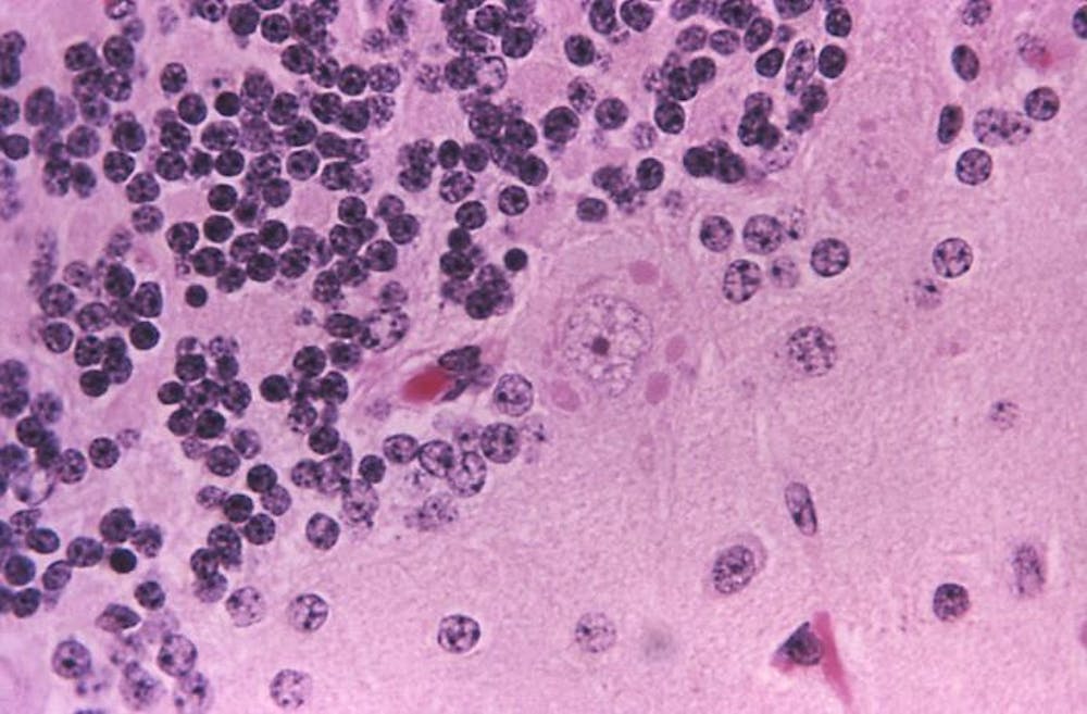
Histopathological evaluation of tissues from all four rabies-infected transplant recipients demonstrated diffuse, predominantly lymphohistiocytic, infiltrates and microglial nodules involving the cerebrum, brain stem, cerebellum, and spinal cord. Cytoplasmic inclusions consistent with Negri bodies were identified throughout the central nervous system (CNS), particularly in the Purkinje cells of the cerebellum and in neurons of the frontal cortex, thalamus, hippocampus, midbrain, and pons (Figure 3AFIGURE 3Histopathological Findings in Patient 4.). Lymphohistiocytic infiltrates involving the peripheral nerves, heart, and kidneys were also noted in some patients. Electron microscopy of the midbrain of Patient 4 demonstrated abundant rhabdovirus particles (Figure 3B). Intracytoplasmic rabies virus antigens were detected on immunohistochemical staining in neurons from multiple areas of the CNS (Figure 3C); in peripheral nerves of the transplanted kidneys, liver, and arterial graft (Figure 4FIGURE 4Immunohistochemical Staining (Red) of Rabies Virus Antigens in Peripheral Nerves of the Liver (Panels A and B), Kidney (Panel C), and Arterial-Graft Transplants (Panel D).); and in renal tubular epithelium, smooth muscle, histiocytes, and vascular endothelium. No tissues were positive for enteroviruses, human herpesviruses 1 and 2, West Nile and other flaviviruses, eastern equine encephalomyelitis virus, lymphocytic choriomeningitis virus, Cache Valley virus, henipaviruses, measles virus, spotted fever and typhus group rickettsiae, Toxoplasma gondii, or Trypanosoma cruzion immunohistochemical analysis. Direct fluorescence antibody staining also demonstrated rabies virus antigens in CNS tissues from all recipients.

Serologic Analyses and Viral Identification

Antibodies (IgM and IgG) reactive to rabies virus were present in the donor’s serum at the time of death. Antibodies were also present in three of the four recipients in samples obtained on postoperative days 35 and 36; both IgM and IgG antibodies were present in one kidney recipient (Patient 3) and the recipient of the donor’s liver (Patient 2), whereas only IgG antibodies were present in the patient who received the arterial segment (Patient 1). Antigenic typing revealed a previously characterized rabies virus variant associated with bats.

Cell Culture and Mouse Inoculations

All suckling mice had neurologic abnormalities or had died seven to eight days after inoculation. Thin-section electron microscopy of CNS tissue demonstrated rhabdovirus particles, and IHC testing detected rabies virus antigens in mouse CNS tissues. Cultures of Vero E6 cells inoculated with brain, spinal cord, and kidney from a kidney recipient demonstrated rabies virus antigen on staining with DFA.

DISCUSSION

This report describes the transmission of rabies virus through the transplantation of solid organs and vascular material. Four patients who received transplants — three organs and one vascular segment — from a donor with unrecognized rabies infection subsequently died of rabies. The transmission of rabies from corneal transplants has been described previously.5

Rabies is seldom included in the differential diagnosis of encephalitis in the absence of a documented exposure or suggestive history.8,10 The symptoms in the cases reported here, including fever, changes in mental status, and autonomic instability, were, in retrospect, consistent with a diagnosis of rabies. However, the diagnosis was complicated by the absence of a history of exposure at presentation and by the number of other potential causes of illness in these immunosuppressed patients. A history of a bat bite in the donor was discovered during contact interviews only after rabies had been diagnosed, and the investigation initiated. The diagnosis in the donor was further complicated by the presence of a subarachnoid hemorrhage in the setting of hypertension and a positive toxicology screen for cocaine. It is not known whether rabies infection was the cause of the subarachnoid hemorrhage, since this finding has not been noted in previous reports.11-13

Signs of rabies developed in all four transplant recipients within 30 days after infection. According to previous reports, symptoms developed within 30 days after an animal bite in only 25 percent of patients.10 It is unknown whether the shorter incubation period in these patients was due to the immunosuppression, the route of transmission, or both. The effect of immunosuppression on rabies infection is currently not well understood. In reports of rabies transmission from corneal transplants in patients who were not immunosuppressed and did not receive postexposure prophylaxis, symptoms developed an average of 26 days after transplantation,14-17 suggesting that implantation of material from infected donors may lead to a shorter incubation period. Three of our patients presented with commonly described symptoms of tremors and changes in mental status, whereas the fourth presented with abdominal and flank pain, which may have been neuropathic, and changes in mental status occurred about 48 hours later. The rapidly progressive encephalitis, with death occurring an average of 13 days after the onset of symptoms, is consistent with the course in other reports.4

There is only one reported case of recovery from clinical rabies by a patient who had not received preexposure or postexposure prophylaxis against rabies.18 However, administration of postexposure prophylaxis with rabies immune globulin and vaccine is highly effective in preventing infection after exposure. In a previous report, administration of postexposure prophylaxis probably prevented infection in a patient who had received a cornea from a donor with rabies.19

This report and another, describing the transmission of West Nile virus through solid-organ transplantation,20 underscore the potential for transmission of unexpected infectious diseases through organ transplantation. Recognition and prevention of transplant-transmitted infections may be improved in various ways, including enhanced donor screening and testing, the development of standardized procedures related to storage and use of donor vascular segments, as well as methods to track their use or nonuse, and enhanced means of detection and diagnosis of illnesses in recipients.

To minimize the risk of transmitting infections during organ transplantation, the Organ Procurement and Transplantation Network (OPTN) has established standards that require organ-procurement organizations to assess the risks of infectious diseases through screening questions and blood testing for selected bloodborne viral pathogens and syphilis.21 Questions about potential exposure to rabies are generally not included, and laboratory testing for rabies infection is not performed. Organs can be procured from donors who are febrile, provided that the medical director of the organ-procurement organization and the transplantation physicians agree that the cause of the fever does not pose an unacceptable risk to the recipient. Given the growing importance of emerging and reemerging infectious diseases, the ability of general improvements in the donor-screening process, rather than disease-specific measures, to increase organ safety should be evaluated. A proposed revision of OPTN policies would expand the list of potentially transmittable diseases and conditions that clinicians should consider in determining a donor’s eligibility.22 The revision emphasizes that when any of these conditions is known or suspected in a donor, this information should be conveyed immediately to the organ-procurement organization as well as to all transplantation centers that received organs from the donor.

The successful use of donor arterial conduits has been reported in liver transplantation23-26 and in the management of vascular complications in recipients of both hepatic transplants27,28 and renal transplants.29 As with organs, these vessel segments have the potential to transmit infection. A careful accounting of and an ability to track donated material, such as vessel conduits, are essential in efforts to link unexplained illnesses or deaths to a common organ donor and will increase the probability of quickly identifying all recipients who may be at risk from donor infections. Proposed revisions of the policies of both OPTN22 and the Joint Commission on Accreditation of Healthcare Organizations30 may help address the storage of vessel conduits and documentation of their use or nonuse.

Our investigation underscores the challenge in detecting and diagnosing infections that occur in recipients of organs or tissues from a common donor. The potential for disease transmission from a donor as a cause of illness or death may not be considered in the evaluation of an individual recipient. In this investigation, and in the previous report of the transmission of West Nile virus through transplantation, the ability to connect illnesses to a common organ donor was facilitated by the fact that multiple recipients were hospitalized at the same facility. Improved national detection of unexpected or serious outcomes among transplant recipients may facilitate the discovery of transplant-related transmission of emerging and unusual pathogens by allowing connections to common donors to be made. The ability to make retrospective diagnoses of infections in organ donors when unexplained deaths or illnesses occur in recipients is hampered by the limited availability of donor samples, particularly tissue; currently, only serum samples from organ donors are retained for any length of time. Investigations into possible transplantation-associated infections would be facilitated by the availability of selected, archived tissue samples from the donor and by autopsy reports and materials. An improved diagnostic ability may have important implications for other patients who received material from the donors and for contacts of the patients and donors.

As organ and tissue transplantation becomes more common, the potential risks of disease transmission may increase. Cases of transplantation-associated infections provide important opportunities to review practices in an attempt to enhance the safety of transplantation without affecting the organ supply. The Department of Health and Human Services, including the CDC, is working with other partners in the organ- and tissue-transplantation community to review donor-screening practices, the use of retained vascular segments, and surveillance of recipients for illness. Clinicians who care for organ-transplant recipients should continue to be aware of the potential for disease transmission through transplantation and the challenges in recognizing atypical presentations of infections in this immunosuppressed population. Clinicians should report unexpected outcomes or unexplained illnesses in transplant recipients to their local organ- and tissue-procurement organization.

We are indebted to the state health departments in Oklahoma and Alabama, to the Southwest Transplant Alliance, and to the staff of the Baylor University Medical Center for their assistance with this investigation.

Source: NEJM


Patient Caught Rabies Through Organ Transplant, CDC Says.


A patient who recently died of rabies in Maryland contracted the illness from a kidney transplant received over a year ago, the CDC reported on Friday. The lengthy incubation period, while longer than the typical 1 to 3 months, is unusual but not unprecedented.

Tests on tissue samples from the patient and donor confirmed that they were both infected with raccoon-type rabies. The three other patients who received organs from the donor have been identified and are receiving anti-rabies shots.

The CDC notes: “If rabies is not clinically suspected [in a potential donor], laboratory testing for rabies is not routinely performed, as it is difficult for doctors to confirm results in the short window of time they have to keep the organs viable for the recipient.”

Source: CDC