FDA Approves First Oral Appliance for Sleep Apnea Therapy


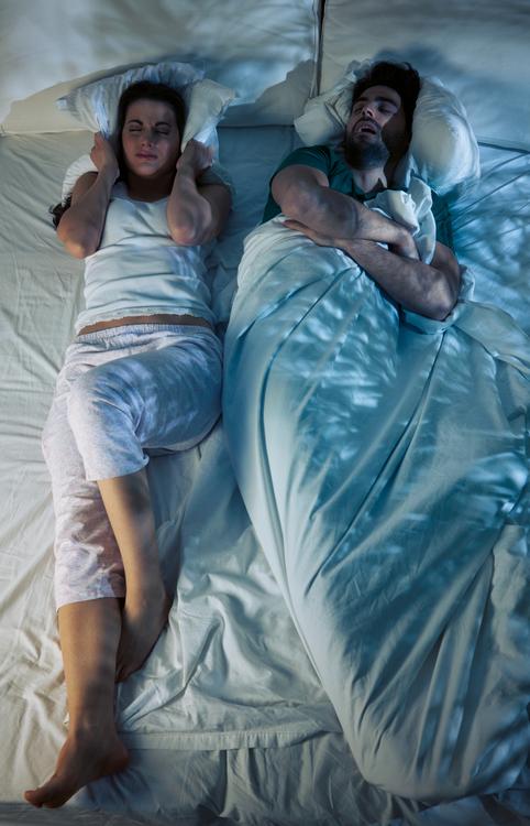
For the first time, the Food and Drug Administration (FDA) recently approved oral devices that treat severe obstructive sleep apnea (OSA). These devices offer an alternative to more common sleep apnea treatments, like continuous positive airway pressure (CPAP) therapy and surgery.

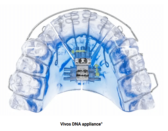
Vivos Therapeutics, Inc. manufactures the oral devices, called Complete Airway Repositioning and/or Expansion (CARE) appliances. They are dental appliances that look similar to retainers. Trusted Source National Library of Medicine, Biotech Information The National Center for Biotechnology Information advances science and health by providing access to biomedical and genomic information. View Source Some appliances are worn only during sleep, while others are worn day and night.

With FDA clearance, the manufacturer can market CARE appliances in the U.S. The approval is significant because it may allow more people with sleep apnea who can’t use CPAP therapy or get surgery to receive treatment. Without treatment, a person with sleep apnea faces a higher risk of severe health problems.  

Data the company submitted to the FDA on 73 people with severe OSA treated with CARE appliances show that about 80% experienced at least some improvement in sleep apnea symptoms. Another study by a team of researchers that included some members of the medical advisory board for Vivos Therapeutics, found that about 64% of participants had less severe OSA following treatment with a CARE appliance. About 26% recovered from OSA completely.

Although approved by the FDA, CARE appliances may only be appropriate for some with OSA. A dentist must evaluate patients and custom-fit CARE appliances to each individual’s teeth and mouth. And people should check with their insurance company about covering the costs.

An advantage of the CARE appliances is that, unlike CPAP therapy, they may not have to be used for a person’s lifetime. Instead, they are worn daily for at least six months and, at most, a year and a half. After that, the benefits should remain for many people without any further need for the devices.

Some people develop OSA because they have naturally narrow mouths and throats. Muscles in their upper airway relax while they’re asleep, narrowing or blocking their airway and causing repeated pauses in breathing during the night. Trusted Source National Heart, Lung, and Blood Institute (NHLBI) The NHLBI is the nation’s leader in the prevention and treatment of heart, lung, blood and sleep disorders. View Source CARE appliances used to treat OSA gradually expand the roof of a person’s mouth, called the palate, by pushing their teeth outward. Dentists must periodically adjust the appliances to keep expanding the palate. Some CARE appliances also move the lower jaw forward. Both of these changes help create space in the airway, making it less likely to become blocked during sleep.

Up to 83% of people with sleep apnea who are prescribed CPAP therapy do not use it as often as directed. Also, surgery can be expensive, risky, and potentially less effective than other treatments. For these reasons, CARE appliances may become an attractive treatment option. 

Studies of CARE appliances suggest they are not likely to cause major side effects. In some instances, they might create space between teeth.

Nasal spray treatment may help patients with obstructive sleep apnea


A drug administered through a nasal spray may be an alternative treatment option for patients with obstructive sleep apnea by improving upper airway collapsibility, according to a study published in CHEST.

This is the first time the drug, a novel, potent, TWIK-related acid sensitive K+ (TASK) 1/3 channel antagonist, was studied in humans.

woman using nasal spray
Additionally, researchers found that all applications and doses of BAY2586116 that they tested lowered and improved the critical closing pressure among patients.

“Although a small study, our findings represent the first detailed investigation of this new treatment in people with OSA, with promising results,” Amal M. Osman, PhD, of Flinders University’s sleep lab FHMRI: Sleep Health, said in a press release. “The drug we tested is designed to target specific receptors that are expressed on the surface of the upper airways, triggering them more easily to activate the surrounding muscles to keep the airway open during sleep. While there’s still a long way to go in terms of clinical testing and development, our study shows targeting these receptors may be a promising avenue for future treatments.”

In a double-blind, randomized, crossover study, Osman and colleagues studied 12 patients (50% women; median age, 57 years) with obstructive sleep apnea to find out if BAY2586116 (Bayer), a TASK 1/3 K+ channel antagonist, could help airways stay open during sleep.

For two overnight physiology studies, researchers randomly gave patients either 160 µg of BAY2586116 or a placebo nasal spray. After these two nights, the participants could also take part in three more overnight studies in which they all received BAY2586116 but through a different application method each night, including BAY2586116 nasal drops (n = 10), half-dose nasal spray (n = 10) or direct application using an endoscope (n = 8).

Various tools, including polysomnography equipment, an epiglottic pressure catheter, pneumotachograph and a nasal mask, were used to track patients’ sleeping and breathing patterns over the course of receiving the drug.

Measuring for upper airway critical closing pressure during sleep, researchers found a mean value of 2.1 ± 1.8 cmH2O (range, –0.16 to 5 cmH2O) among patients who received the placebo nasal spray. However, the BAY2586116 spray lowered the upper airway critical closing pressure to 0.1 ± 4.4 cmH2O (range, –5.7 to 9.7 cmH2O).

Additionally, researchers found that all applications and doses of BAY2586116 that they tested lowered and improved the critical closing pressure among patients, with nasal drops at 0.9 ± 2.6 cmH2O, the half-dose spray at 0 ± 2.2 cmH2O and endoscope application at 0.4 ± 3.3 cmH2O.

Although approximately 60% of patients had a greater than 2 cmH20 improvement with the nasal spray method compared with only 20% of patients using the nasal drop method, results of a mixed model showed no systemic differences between the different modes of application and dose.

When CPAP had transient reduction to atmospheric pressure, 82% of 11 patients had no flow for breaths 3 to 5 during treatment with placebo, but the average peak inspiratory flow responses increased when BAY2586116 high-dose spray and endoscope were used.

“At the moment, there are no approved drug treatments for OSA,” Danny J. Eckert, PhD, director of the Flinders’ sleep lab FHMRI: Sleep Health, said in the release. “However, with advances in our understanding of the different reasons people get OSA, the potential for effective new medications is growing stronger each year.”

More severe obstructive sleep apnea may increase risk for incident venous thromboembolism


Patients with more severe obstructive sleep apnea, measured by the apnea-hypopnea index, were more likely to have incident venous thromboembolism, researchers reported in Chest.

“We found that VTE was more common in patients with OSA, as we expected. However, much of this risk seems to be driven by obesity, which may be the common risk factor

Older woman sleeping
Source: Adobe Stock.

that predisposes patients to both OSA and VTE,” Michael V. Genuardi, MD, MS, FACC, assistant professor of clinical medicine at the Center for Sleep and Cardiovascular Outcomes Research at the University of Pittsburgh School of Medicine and the division of cardiology at the Perelman School of Medicine at the University of Pennsylvania, told Healio. “Separate from obesity, patients who have low oxygen saturation levels overnight are also at increased risk of developing blood clots.”

Researchers evaluated clinical outcomes of 31,309 patients (mean age, 50.4 years; 50.1% women) who were undergoing overnight polysomnography in six sleep laboratories at the University of Pittsburgh Medical Center from July 2004 to December 2018. The researchers assessed associations between OSA severity and incident VTE.

Michael V. Genuardi, MD, MS, FACC

During a mean follow-up period of 5.3 years, 1,791 incident VTE events, the primary outcome, occurred.

Every 10-event per hour increase in the apnea-hypopnea index was associated with a 4% increase in risk for incident VTE in age- and sex-adjusted analyses (HR = 1.04; 95% CI, 1.02-1.06). However, when the researchers adjusted for BMI, the association disappeared (HR = 1.01; 95% CI, 0.99-1.03).

The researchers reported an independent association with nocturnal hypoxemia and incident VTE. Patients with more than 50% sleep time spent with oxyhemoglobin saturation of less than 90% had a 48% increased risk for VTE compared with patients without nocturnal hypoxemia (HR = 1.48; 95% CI, 1.16-1.69).

According to the researchers, the association between elevated apnea-hypopnea index and risk for VTE may be largely explained by differences in patient adiposity.

“We found that patients who have low oxygen saturation levels overnight are at higher risk of VTE,” Genuardi said. “More research is needed to understand this risk factor, since it may broadly apply to patients with multiple underlying medication conditions, including obstructive and central sleep apnea, chronic lung disease, and congestive heart failure.”

Obstructive sleep apnea is associated with low GABA and high glutamate in the insular cortex


Summary

The insular cortex is injured in obstructive sleep apnea (OSA) and responds inappropriately to autonomic challenges, suggesting neural reorganization. The objective of this study was to assess whether the neural changes might result from γ-aminobutyric acid (GABA) and glutamate alterations. We studied 14 OSA patients [mean age ± standard deviation (SD): 47.5 ± 10.5 years; nine male; apnea–hypopnea index (AHI): 29.5 ± 15.6 events h−1] and 22 healthy participants (47.5 ± 10.1 years; 11 male), using magnetic resonance spectroscopy to detect GABA and glutamate levels in insular cortices. We localized the cortices with anatomical scans, and measured neurochemical levels from anterior to mid-regions. Left and right anterior insular cortices showed lower GABA and higher glutamate in OSA versus healthy subjects [GABA left: OSA n = 6: 0.36 ± 0.10 (mean ± SD), healthy n = 5: 0.62 ± 0.18; < 0.05), right: OSA n = 11: 0.27 ± 0.09, healthy n = 14: 0.45 ± 0.16; < 0.05; glutamate left: OSA n = 6: 1.61 ± 0.32, healthy n = 8: 0.94 ± 0.34; < 0.05, right: OSA n = 14: 1.26 ± 0.28, healthy n = 19: 1.02 ± 0.28; < 0.05]. GABA and glutamate levels were correlated only within the healthy group in the left insula (r: −0.9, < 0.05). The altered anterior insular levels of GABA and glutamate may modify integration and projections to autonomic areas, contributing to the impaired cardiovascular regulation in OSA.

Introduction

The insular cortex in obstructive sleep apnea (OSA) shows structural and functional alterations, which likely contribute to adverse autonomic and affective symptoms characteristic of the syndrome (Harperet al., 2003; Kumar et al., 2012; Macey et al., 2008). The altered functions include distorted insular functional magnetic resonance (fMRI) signals accompanying sympathetic nervous system challenges, and probably underlie the high sympathetic outflow in OSA, presumably contributing to the profuse sweating, cardiac arrhythmia and hypertension in the disorder. The insular injury in OSA also probably facilitates the development of depression and anxiety in the condition (Asghari et al., 2012; Kurthet al., 2010; Marin et al., 2012). Mechanisms underlying insular dysfunction in OSA are unclear, but understanding the nature of the brain alterations could provide insights into symptoms that persist even with treatment (Marin et al., 2012), as well as processes underlying partial or complete resolution with continuous positive airway pressure (CPAP) of other symptoms, such as high sympathetic tone (Fatoulehet al., 2015).

Structural brain alterations in OSA could arise from multiple pathologies. Reduced mean diffusivity, apparent in many OSA brain regions (Kumar et al., 2012), reflects an increase in barriers to intercellular water movement, which could arise from cell swelling due to inflammation or increased cell size due to glial activation. fMRI differences appear in the insular cortex during autonomic challenges (Harper et al., 2003). The processes underlining those differences could be elucidated by determining the levels of the neurotransmitters glutamate and γ-aminobutyric acid (GABA), which can now be measured non-invasively. Levels of these chemicals are sensitive to neural reorganization in response to injury, and can reflect different functional states (Arckens et al., 2000). High levels of glutamate, in particular, are associated with excitotoxicity, a probable mechanism of injury in some brain areas in OSA.

The objective of this study was to assess GABA and glutamate in the anterior insular cortex in OSA patients, relative to healthy subjects. The anterior mid-insula was selected, as that subregion preferentially serves autonomic roles (Macey et al., 2012). We hypothesized that, relative to healthy people, OSA patients would show elevated glutamate, based on the potential for hypoxia-induced excitotoxic conditions and altered GABA levels, based on impaired insular action.

Methods

We studied 14 OSA patients [mean age: 47.5 ± 10.5 years; nine male; mean apnea–hypopnea index (AHI): 29.5 ± 15.6 events h−1; SAO2 min: 83 ± 9%] and 22 healthy subjects (mean age: 47.5 ± 10.1 years; 11 male). Patients were diagnosed at the UCLA Sleep Disorders Center. No participants had a history of head trauma or disease, current mental illness, use of psychoactive medications, cancer or cardiac disease. Patients were untreated, apart from two who had a prior history of CPAP use. Healthy subjects were screened to exclude sleep disorders. Procedures were approved by the UCLA Institutional Review Board, and participants provided written informed consent.

Neurotransmitters were measured with a magnetic resonance spectroscopy (MRS) method based on ‘two-dimensional’ MRS (Sarma et al., 2014; see Supporting information). Neurochemicals changes were measured using compressed sensing (CS)-based four-dimensional (4D) echo-planar J-resolved spectroscopic imaging (EP-JRESI). High-resolution T1-weighted scans were acquired for localization of insular cortex. Group differences in GABA and glutamate concentrations (ratios with respect to creatine) in the anterior to mid-insular cortex and adjacent tissue were assessed with t-tests. The relative concentrations of ‘Glx’, a combination of glutamate and glutamine indicative of glutamate levels, were also assessed, as Glx is a more robust MRS measure than glutamate.

Results

Neurotransmitter concentrations were recorded successfully from subsets of the 22 healthy and 14 OSA subjects (Table 1; example spectra Fig. 1). Differences between OSA and healthy subjects emerged for GABA (Fig. 2a) and glutamate (Fig. 2b) in the right and left insulae. The left insula showed poorer data quality in more subjects than the right (right successful measures: 69% GABA, 92% glutamate; left successful measures: 31% GABA, 39% glutamate). Success rates were similar for the OSA and healthy groups. When the two OSA patients with a prior history of CPAP were excluded the mean differences were similar, and the significant differences between OSA and healthy subjects remained, with the exception of glutamate in the right insula. However, Glx showed significantly higher levels in OSA versus healthy subjects in the right and left insulae, with and without excluding the two previously treated subjects (Table 1). Correlations between GABA and glutamate were significant only in healthy subjects in the left insula (r: −0.90, P: 0.04; Table 2; Fig. 2c,d).

Table 1. Levels of GABA and glutamate in subsets of 14 OSA (12 never treated) and 22 healthy subjects
Left insula Right insula
OSA Healthy P OSA Healthy P
n Mean ± SD n Mean ± SD n Mean ± SD n Mean ± SD
  1. P, t-test for group differences; Glx, glutamate + glutamine; GABA, γ-aminobutyric acid; OSA, obstructive sleep apnea; SD, standard deviation.
GABA 6 0.36 ± 0.10 5 0.62 ± 0.18 0.03 11 0.27 ± 0.09 14 0.45 ± 0.16 < 0.01
Glutamate 6 1.61 ± 0.32 8 0.94 ± 0.34 < 0.01 14 1.26 ± 0.28 19 1.02 ± 0.28 0.02
Glx 6 1.92 ± 0.38 6 1.22 ± 0.44 0.01 14 1.75 ± 0.42 19 1.34 ± 0.42 0.01
Never treated OSA Never treated OSA
GABA 5 0.35 ± 0.10 (as above) 0.03 9 0.27 ± 0.10 (as above) < 0.01
Glutamate 5 1.50 ± 0.21 (as above) < 0.01 12 1.20 ± 0.25 (as above) 0.08
Glx 5 1.79 ± 0.18 (as above) 0.01 12 1.69 ± 0.43 (as above) 0.04

Left: example insula voxel is shown in red (15 mm3) overlaid an individual anatomical scan. Right: example two-dimensional magnetic resonance spectroscopy (2D-MRS) spectra from right insulae in obstructive sleep apnea (OSA) (56-year-old female) and control (53-year-old male) subjects, with resonances of various neurochemicals indicated (Cho, choline; Cr, creatine; mI, myo-inisotol; MM, other macromolecules; NAA, N-acetylaspartate; Tau, taurine).

Insular (a) gamma aminobutyric acid (GABA) and (b) glutamate levels in subsets of 14 obstructive sleep apnea (OSA) and 22 healthy subjects [mean ± standard error of the mean (SEM)]. Scatterplots of glutamate versus GABA for subjects with both measures in the left (c) and right (d) insular cortices.

Table 2. Correlation table in subjects with GABA and glutamate measures
Left insula Right insula
OSA Healthy OSA Healthy
n r P n r P n r P n r P
  1. P, correlation between γ-aminobutyric acid (GABA) and glutamate; OSA, obstructive sleep apnea.
Glutamate versus GABA 6 0.16 0.76 5 −0.90 0.04 11 −0.47 0.14 14 −0.04 0.90

Discussion

The anterior insular cortex in OSA shows low GABA and high glutamate, conditions which would modify neural patterns within the structure. GABA normally acts as an inhibitory neurotransmitter, while glutamate is normally excitatory to neural processes. The influences of the insular cortices are complex, with both inhibitory and excitatory influences on hypothalamic, cingulate and frontal cortical structures, among others. On a simplistic level, the combination of a reduction in the inhibitory GABA and increase in the excitatory glutamate could reflect a more active state overall; however, the complexity of the insular projections to multiple structures precludes such an exclusive generalization (Kurth et al., 2010). For excitatory projections, enhanced glutamate could enhance insular influences, and the normal inhibition over hypothalamic structures could be reduced by the lower GABA levels, leading potentially to the exaggerated sympathetic tone in the condition. The altered neurotransmitter levels could also modify cingulate and hippocampal projections influencing mood, such as depression and anxiety, both of which are common in OSA. Low GABA in the insula has been associated with high levels of anxiety (Rosso et al., 2014), and levels appear to be associated with processing of interoceptive stimuli, a function linked with depressive mechanisms (Wiebking et al., 2014). The structures involved in anxiety and depression include the anterior cingulate, recipient of fibres from the cingulum and insular cortices. High glutamate has been found in these limbic areas in bipolar disorder (Soeiro-De-Souza et al., 2015). The hypoxia exposure during apneic events probably leads to excessive excitation of the insular connecting fibres, and such excitotoxic processes are facilitated by glutamate. It is unknown whether the altered neurotransmitter levels revert to normal with CPAP treatment, but such knowledge would assist the selection of interventions and the determination of whether regional GABA and glutamate levels could be useful biomarkers of local neural tissue state in OSA.

The findings of reduced GABA and higher glutamate build upon an earlier demonstration of alterations in the anterior insular cortex in OSA, including changes in structure, function and metabolite levels. The insula shows impaired responses to autonomic challenges in OSA, especially during periods of sympathetic activity increases (Harper et al., 2003). Resting state functional patterns also differ from heathy subjects when those patterns are correlated with other regions (Zhang et al., 2015). Changes in water diffusivity reflect structural changes arising probably from glial activation or inflammation due to hypoxia, and damage to neurones or axons and myelin (Kumar et al., 2012; Macey et al., 2008). Increased myo-inositol in the left insula of OSA patients is also consistent with glial alterations (Yadavet al., 2014). Increased glutamate levels may arise directly from astrocyte activation, as astrocytic glutamate is an energy source for neurones (Pellerin and Magistretti, 1994); if metabolism is altered by OSA in the insular cortex, glutamate from astrocytes may increase to compensate. The combined chemical, functional and structural findings confirm injury and reorganization of the insula in OSA, which probably contribute to clinical symptoms in the condition (Asghari et al., 2012; Kurth et al., 2010; Macey et al., 2013; Marin et al., 2012).

The high glutamate may reflect damaging excitotoxic processes arising from intermittent hypoxia (Jagadapillai et al., 2014). Only 5 h of intermittent hypoxia simulating OSA effects can lead to cellular death (Pae et al., 2005); thus, similar processes are probably operating in people with OSA. However, the high glutamate measured here was in whole tissue, and does not necessarily correspond to concentrations in extracellular fluid, where the neurochemical is key for excitotoxicity.

Interventions complementary to CPAP that influence neurotransmitter levels may help to normalize brain function in OSA. Both GABA and glutamate can be manipulated directly with pharmacological approaches and indirectly with behavioural changes. The measurements presented here could serve as a biomarker of any intervention targeting these brain chemicals.

Measurements were successful in 58% of instances, with the remainder not meeting quality criteria, especially in the left insula. Because OSA and healthy measurements were affected similarly, the low data quality is due probably to technical rather than biological reasons (e.g. poor shimming). The resolution of the MRS voxels was limited (example in Fig. 1), so the measures did not cover the entire anterior to mid-portion of the structure, and included adjacent tissue; the subregion studied will have varied slightly between subjects.

In conclusion, the anterior to mid-insular cortex shows lower GABA and higher glutamate levels in OSA compared with healthy subjects. These alterations probably reflect a functional neural reorganization that contributes to the autonomic and psychological symptoms in OSA, such as high sympathetic tone and refractory hypertension, and anxiety, depression and cognitive difficulties. High glutamate would facilitate excitotoxic processes. The altered neurotransmitter levels provide a biomarker of regional functional status of the OSA brain, and will allow insights into the neurological effects of treatment with CPAP or other interventions.

New, Low-Cost Screening Process for Obstructive Sleep Apnea


Measuring the oxygen desaturation index (ODI) with overnight pulse oximetry is an effective, low-cost way to screen for obstructive sleep apnea (OSA), a new study published in the Journal of Clinical Sleep Medicine suggests.
OSA is often underrecognized in hospitalized patients and associated with significant morbidity and mortality, according to researchers from Thomas Jefferson University and Hospitals, in Philadelphia.
“Sleep disordered breathing is associated with cardiovascular complications and sudden death,” said lead study author Sunil Sharma, MD, FAASM, associate professor of pulmonary medicine in the Sidney Kimmel Medical College at Thomas Jefferson University, in a press release.
The researchers conducted a prospective study with 754 patients to test the efficacy of this screening for identifying OSA in patients admitted to a tertiary care hospital. The patients were admitted between February 2013 and February 2014.
Obese patients (body mass index >30 kg/m2) were automatically screened for OSA—being overweight is a risk factor for OSA—using the STOP (snoring, tiredness during daytime, observed apnea, high blood pressure) questionnaire. Patients with positive results were advised to have a follow-up evaluation, which included an overnight polysomnography.
The optimal screening cutoff for OSA was ODI greater than or equal to 10 events per hour (Matthews correlation coefficient, 0.36; 95% CI, 0.24-0.47). The researchers found that 129 of the 149 patients who underwent polysomnography were shown to have OSA.
“The results showed that our screening process identified sleep disordered breathing in 87% of patients who followed up with a polysomnography,” said Dr. Sharma. “We confirmed the high prevalence of undetected sleep-disordered breathing among hospitalized patients and also validated a low-cost protocol to detect it.”
The authors noted a significant increase in the number of hospitalized patients recommended to undergo polysomnography after implementing this screening process.
“This study should be of great interest to hospitals looking at ways to reduce complications,” said Dr. Sharma. “Recent data suggests that for inpatients with cardiovascular disorders, early diagnosis and intervention for sleep apnea may lead to reduced readmission rates.”

Sleep Apnea Linked to Depression in Men


Obstructive sleep apnea (OSA) is associated with an increased risk of depression in men, a large Australian study found.

Among 1,875 men ages 35 to 83 who were assessed for depression at two time points about 5 years apart in the Men Androgen Inflammation Lifestyle Environment & Stress Study (MAILES), previously undiagnosed severe OSA was associated with depression (OR 2.1, 95% CI 1.1-4, P<0.05), reported Carol J. Lang, PhD, of the University of Adelaide, at theannual meeting of the American Thoracic Society here.

This statistical significance remained even after adjustment for age, waist circumference, smoking, relationship status, financial difficulties, erectile dysfunction, and nocturia, she noted.

“Depression is highly prevalent in OSA, reaching 39% in clinic studies. However, few population-based studies have been done and results have been mixed,” Lang said.

In one longitudinal study that included 1,400 men and women, a dose-dependent association between a sleep-related breathing disorder and depression was seen, with a 2.6-fold increased risk of depression and a moderate or severe sleep-related breathing disorder.

A cardinal symptom of OSA is excessive daytime sleepiness, although not all affected patients report this problem. It’s unclear whether daytime sleepiness is associated with depression in OSA, and in the longitudinal study, sleepiness was not found to be an explanatory factor for the observed relationship between the sleep-related breathing disorder and depression.

Further complicating this relationship was the finding in another study that residual sleepiness persisting after continuous positive airway pressure treatment was linked with refractory depression.

In an attempt to clarify the association of OSA and depression, in 2010 Lang and colleagues conducted telephone interviews asking men if they had ever been diagnosed with sleep apnea with a sleep study, and those answering in the negative were invited to participate. A total of 857 men then underwent at-home polysomnography testing.

Depression was assessed using the Center for Epidemiologic Studies Depression Scale/Beck’s Depression Inventory, and daytime sleepiness was evaluated according to the Epworth Sleepiness Scale.

OSA was defined as an apnea-hypopnea index higher than 10. Mild-to-moderate OSA was an index score of 10 to 29, and severe was 30 or higher.

Logistic regression analysis determined that, along with severe OSA, daytime sleepiness was associated with depression (OR 1.1, 95% CI 1-1.2, P<0.05).

Then, in a model that included both previously undiagnosed OSA and excessive daytime sleepiness, individuals with both had 4.2 times greater odds of depression than those with neither, and 3.5 times greater likelihood of depression than those with either alone.

“The message is that clinicians need to be aware of these risks and assess for the other if one is present,” Lang said.

The precise mechanisms underlying the link between these conditions are uncertain, but may involve low oxygen levels, arterial inflammatory responses, and neurologic changes in the brain, she said.

The press conference moderator, Mihaela Teodorescu, MD, of the department of pulmonary and critical care at the University of Wisconsin in Madison, agreed that this is an important clinical issue.

“Sleep apnea leads to more refractory depression and patients get more treatment, including with benzodiazepines, which can aggravate and further contribute to depression,” she said.

Urgent Adenotonsillectomy in Kids Results in Good Outcome


Patients who require urgent tonsillectomy without or with adenoidectomy (TA) tend to be younger, have elevated obstructive apnea-hypopnea indices, and more persistent desaturations below 80% than children who receive timely TA. Their desaturations also tend to be unresponsive to supplemental oxygen. After surgery, approximately half of children who receive urgent TA will no longer require supplemental oxygen because of a dramatic improvement in gas exchange.

Conan Liang, BA, from the University of Colorado School of Medicine in Aurora, and colleagues published the results of their retrospective medical record review online January 2 in JAMA Otolaryngology–Head & Neck Surgery. They report that at their institution, pediatric patients who receive urgent TA (n = 35) resemble those who meet hospital admission criteria for bronchiolitis. In particular, the decision to perform urgent TA was typically (85% of the time) based on persistent desaturation below 80% despite receipt at least 0.5 L of oxygen.

Patients receiving urgent TA had an average age of 3.8±2.1 years compared to the 6.6±4.2 years seen in the control group. Patients in the urgent TA group had average apnea-hypopnea indices of 39.4 events per hour.

TA is considered a relatively safe procedure with a low incidence of postoperative complications. Results from the current study are consistent with this, and the authors report that 94% of patients had a favorable postoperative course. The authors acknowledge, however, that that their hospital had a low (26%) follow-up rate for postoperative polysomnography.

The authors did not find an increased number of pulmonary comorbidities in the urgent TA cohort compared with the control cohort. This finding led the authors to suggest that children in the urgent TA group may have had subclinical findings that predisposed them to developing hypoxemia.

Urgent vs Timely TA
As more polysomnographies are performed, an increasing number of children are likely to be diagnosed with severe obstructive sleep apnea (OSA) with gas exchange abnormalities. The severity of OSA is correlated with adenotonsillar hypertrophy, and so, not surprisingly, TA is commonly performed in response to sleep-related breathing disorders.

OSA is now understood, however, to be a chronic as opposed to an acute condition, suggesting that it may not require urgent treatment. The study underscores that there are no clearly defined indications for urgent TA in the pediatric population.

7 Ways To Stop Your Snoring


Before you resort to sleeping in separate beds, try these at-home remedies for snoring. 

7 Ways To Stop Your Snoring

Forget about the monster under your bed. If you have a partner who snores, you’re dealing with a monster in your bed — and it’s often a near-nightly showdown. In a new National Sleep Foundation survey, 40 percent of Americans admitted to snoring a few nights per week (or more).

And the ones who are suffering aren’t usually the folks sawing logs. “The most common side effect of snoring is waking up other people, whether in the same bed or the next bedroom, depending on how loud it is,” says Eric Kezirian, MD, a professor of otolaryngology at the University of Southern California, who specializes in the treatment of snoring.

In fact, people with a snoring significant other tend to lose an hour of sleep per night, according to Craig Schwimmer, MD, founder of The Snoring Center. Perhaps as a result, “couples in snoring relationships report lower marital satisfaction scores, they have less sex, and they often resort to sleeping apart,” he tells Yahoo Health.

That’s why snoring is considered a social issue more so than a medical one, although in some cases, it does indicate a more serious problem: obstructive sleep apnea. “When we go to sleep at night, the muscles in the throat relax, and as we breathe in and out, this relaxed tissue tends to vibrate,” explains Schwimmer. If that tissue simply vibrates — and nothing more — you’ll probably just bother your bedmate. But if that tissue closes as it vibrates, blocking your airway, you may have obstructive sleep apnea. “Snoring and sleep apnea are really just different points on a continuum,” says Schwimmer.

Obstructive sleep apnea, of course, requires serious medical intervention. But simple snoring can often be treated with these at-home remedies:

Adjust your position

If you’re a chronic snorer, back isn’t best. “Most people snore more on the back than they do on the side, and more on the side than they do on the stomach,” says Schwimmer. It can be tough to switch your preferred sleeping position, so sleep doctors often suggest this trick to encourage people to stay on their side: Sew a pocket on the back of a T-shirt between the shoulder blades, and slip two tennis balls inside.

“When people sleep on their side, their shoulder can get sore. So they roll on their back,” says Kezirian. “The tennis balls aren’t very comfortable, so they end up rolling to their other side.”

Not extreme enough? Try the Night Shift Sleep Positioner, a device you wear around your neck that vibrates when you roll onto your back, increasing in intensity until you shift to your side. “I wore it one night, and it drove me crazy,” says W. Christopher Winter, MD, director of Charlottesville Neurology and Sleep Medicine. “But it worked. After a few days of that, you would not be sleeping on your back.”

Play with pillows

For some people, the tennis ball trick works — but only because it keeps them up all night. If you simply can’t sleep with sports equipment attached to your PJs, try resting a body pillow between your legs, which helps align your spine and makes side sleeping more comfortable. Or wedge a C-shaped pregnancy pillow behind your back, suggests Winter.

If you still can’t adjust to lying on your side, lie on your back, but prop up your head and shoulders. “You want to make a little incline — a wedge — with a couple pillows,” says Kezirian. “It’s not just lifting up your head.” Try placing one underneath your shoulders to elevate your chest, then another two under your head. That may help keep the back of your throat open.

Avoid alcohol before bed

It’s not just your inhibitions that loosen up when you’re drinking. “Alcohol preferentially relaxes the muscles in the throat, so everybody’s snoring is worse after a couple drinks,” says Schwimmer. Plus, since you’re more sedated after drinking, your snoring is less likely to stir you awake, leaving your bedmate to suffer longer. “Most wives will tell you, ‘When Walter goes out drinking with his buddies, he’s going to snore like crazy. I don’t even sleep in the bed with him that night,’” says Winter. The simple fix: Stop your imbibing within four hours of bedtime.

Open your nose

Sometimes, snoring isn’t due to flapping muscles in your throat — it may simply be a problem of clogged or narrow nasal passages. If they’re consistently congested, a saltwater nasal spray may be the only fix you need. “When you brush your teeth in the morning and at night, put a spray or two in either side of your nose,” says Kezirian. Not only will that keep your nostrils clear, it will also maintain the moisture in your nose, preventing the dryness and irritation that can promote snoring.

Another way to keep your nasal passages open: Breathe Right strips. “If your airway is collapsing in the back of your throat, putting a sticker across your nose is not really going to help,” says Winter. But if narrow nasal passages are the problem (or if they’re chronically clogged due to allergies), the sticky strips could make a big difference.

Try this test to see if these strips might be the right remedy for you: While looking into a mirror, inhale deeply through your nose, and see if the sides of your nose collapse. If your nostrils cave in, you probably have narrow nasal passages, so the strips could do the trick, says Kezirian.

Control your acid reflux

What’s happening in your esophagus may not seem relevant to the noises you make at night, but acid reflux can actually play a major role in snoring. When stomach acid coats your throat, it creates inflammation, says Schwimmer. “The tissue is swollen, so that narrows the airway,” he says. “Swollen tissue is more vibratory.” To tame your reflux, stop eating two to three hours before bedtime, and if that doesn’t work, try taking Tums or Rolaids before bed.

Be a mouth breather

People who snore often sound like a choo-choo train while they snooze. “They’re puffing up their cheeks, and exhaling against a closed mouth,” which can lead to snoring, says Winter. ProVent stickers turn your nostrils into a one-way valve, allowing you to breath in, but not out, through your nose. “That creates extra pressure in the back of your airway and holds it open,” he explains. In other words, the stickers force you to exhale through your mouth. “They’re really for sleep apnea, but I have patients who say that they help with their snoring,” says Winter.

Belt it out

Here’s motivation to turn your morning commute into a concert: In a 2013 British study, people who did singing exercises — a series of simple, repetitive noises put to music — for 20 minutes a day showed a significant reduction in snoring after three months. But you don’t necessarily have to do the specific exercises in the study — just belting it out may have a benefit. “There are a lot of muscles in your upper airway that don’t get used a tremendous amount,” says Winter. By singing, you may strengthen and tone those muscles, which could potentially reduce your snoring, he says.

SLEEP APNEA IN CHILDREN


Sleep apnea is defined as pause/brief cessation in breathing while sleeping.It is not uncommon in children.A good comfortable sleep is necessary for proper growth and functioning of brain.The most common cause of sleep apnea in children is obstructive sleep apnea(OSA) caused by adenotonsillar hypertrophy in which adenoid and tonsils are unduely enlarged causing obstruction in the airway more while sleeping leading to sleep apnea. Obesity is a predisposing factor.According to Philips survey 2009 ,14% Indians are suffering from this disorder.
The chief symptom of OSA is snoring which is seen in 15-20 % of children without having this disease ,so in majority of cases it goes unrecognised.If your child have any of these symptoms,look for OSA
Restless night sleep
Excessive day time sleepiness
Hyperactivity with /without aggressive or violent behaviour
Poor attention span
Subnormal academic performance
Low stamina at sports
Poor growth
Abnormal behaviour
The diagnostic test for this disorder is POLYSOMNOGRAPHY(PSG),which is available at very few centres in India.Sleep medicine is a new branch in India.In PSG the child has to be admiited in hospital and by a device his/her ,breathing,heart rate , rhythm,and brain activities are recorded while the child is sleeping.It costs nearly 15000 INR in Indian private set up.
In most of the cases ,weight reduction and operative removal of adenoid and tonsils are curative.

Unaddressed OSA may lead to early development of these but not limited to these:
Hypertension
Stroke
Diabetes Mallitus

Respiratory disorders in children by DR DK JHA's photo.

Respiratory disorders in children by DR DK JHA's photo.

Suspect OSA in patients with unexplained daytime sleepiness


Adult patients who are excessively sleepy during the day for no apparent reason should be targeted for assessment of obstructive sleep apnea (OSA) using a sleep study, a new guideline from the American College of Physicians (ACP) recommends.

Sleep study records patients’ brain activity, eye movements, heart rate, and blood pressure during sleep to diagnose OSA and determine disease severity.

In patients whom OSA is suspected, the guideline recommends full-night, in-laboratory polysomnography (PSG) to establish the diagnosis. This test requires specialized facilities, is expensive and demands that patients spend the night under observation in a foreign environment, but yields the most accurate diagnostic information. [Ann Intern Med 2014;161:210-220]

Home-based portable sleep monitors may be used as an alternative in patients without comorbid conditions or when PSG is not available, although these can yield substantially different scores on the apnea-hypopnea index (AHI), which accounts for the number of pauses in breathing per hour of sleep.

Daytime sleepiness (hypersomnia) is a clinically relevant symptom of OSA, but clinicians should also include in their assessment other symptoms such as fatigue, insomnia and snoring, and risk factors such as obesity, said the guideline authors.

Clinicians are not advised to assess OSA in the absence of daytime sleepiness or treat patients with low AHI scores as evidence suggests neither improves clinical outcomes. Of note, portable monitoring in patients with chronic lung disease, congestive heart failure, neurologic disorders and other major comorbidities is also not recommended as very few studies included these patients. There is also little evidence for pre-operative screening for OSA and its effect on surgical outcomes.

The guideline, which is based on reviews of peer-reviewed studies published from 1996 to May 2013, is developed to provide clinicians with guidance on diagnosing OSA. For guidance on treatment, clinicians could refer to the ACP guideline on the management of OSA.

“Obstructive sleep apnea is a serious health condition that is associated with cardiovascular disease, hypertension, cognitive impairment, and type 2 diabetes,” said ACP president Dr. David Fleming. “It is important to diagnose individuals with unexplained daytime sleepiness so that they can get the proper treatment.”