Study finds that microglia could regulate sleep via the modulation of norepinephrine transmission


Study finds that microglia could regulate sleep via the modulation of norepinephrine transmission
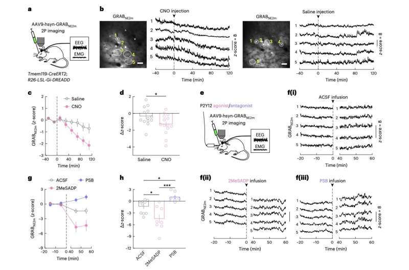
Activation of microglia Gi signaling suppresses NE transmission. a, Schematic of GRABNE2m imaging in the prefrontal cortex. b, Example imaging session with chemogenetic Gi activation in microglia. Left, field of view (scale bar, 50 µm); five ROIs are outlined, whose NE traces are shown on the right. Dashed line, time of CNO or saline injection. c, Effect of chemogenetic Gi activation in microglia on NE signals averaged across 13 (saline) or 14 (CNO) sessions from five mice: three female and two male. A total of 8–12 ROIs were assessed for each session. The dashed line indicates the time of injection. d, Difference in NE before and after (20–120 min) saline or CNO injection. Each circle represents data from one session (saline, n = 13 sessions; CNO, n = 14 sessions; from five mice). Data are presented as the mean ± s.e.m.; *P .

Sleep is known to play a key role in facilitating various physiological processes, while also contributing to the healthy functioning of the brain. Lack of sleep and poor sleep quality have been linked to various chronic health and mental health issues, including high blood pressure, depression, stroke, obesity, and heart disease.

Sleep disturbances have also been implicated in the development of neurodegenerative diseases. Interestingly, neurodegenerative diseases have also been associated with the dysfunction of microglia, the primary mammalian immune cells, yet the link between microglia and sleep has not been thoroughly studied yet.

Researchers at University of California Berkeley (UC Berkeley), Huazhong University of Science and Technology and other institutes in China recently carried out a study exploring the potential role of microglia in regulating sleep. Their findings, published in Nature Neuroscience, suggest that microglia regulate sleep by modulating the transmission of the neurotransmitter norepinephrine, which is known to contribute to arousal, attention and stress reactions.

“We show in mice that microglia can regulate sleep through a mechanism involving Gi -coupled GPCRs, intracellular Ca2+ signaling and suppression of norepinephrine transmission,” Chenyan Ma, Bing Li and their colleagues wrote in their paper.

To investigate the role of microglia in sleep regulation, the researchers carried out a series of experiments on mice, using chemogenetic techniques to manipulate and image microglia signaling in their brain. Specifically, they selectively activated or blocked P2Y12, a Gi -protein-coupled ATP/ADP receptor that has been found to be central to the function of microglia.

The team observed the neural mechanisms that followed the experimental activation/blocking of P2Y12–Gi signaling and recorded the sleeping behavior of the mice. This allowed them to gather new insight about how microglia may regulate sleep in mice, and potentially also in humans and other mammals.

“Chemogenetic activation of microglia Gi signaling strongly promoted sleep, whereas pharmacological blockade of Gi -coupled P2Y12 receptors decreased sleep,” the researchers wrote.

“Two-photon imaging in the cortex showed that P2Y12–Gi activation elevated microglia intracellular Ca2+, and blockade of this Ca2+ elevation largely abolished the Gi -induced sleep increase. Microglia Ca2+ level also increased at natural wake-to-sleep transitions, caused partly by reduced norepinephrine levels. Furthermore, imaging of norepinephrine with its biosensor in the cortex showed that microglia P2Y12–Gi activation significantly reduced norepinephrine levels, partly by increasing the adenosine concentration.”

Overall, the results collected by Ma, Li and their colleagues suggest that microglia play a key role in the regulation of sleep. Specifically, these immune cells appear to regulate sleep via reciprocal interactions with the transmission of norepinephrine.

These initial observations could soon pave the way for further studies investigating the role of microglia in sleep regulation, focusing on norepinephrine transmission.

As microglia dysfunction and sleep disturbances have been linked to Alzheimer’s and other neurodegenerative diseases, this work may also broaden neuroscientists’ understanding of these diseases, potentially aiding the future development of new therapeutic strategies.

Study finds that microglia could regulate sleep via the modulation of norepinephrine transmission


Study finds that microglia could regulate sleep via the modulation of norepinephrine transmission
Activation of microglia Gi signaling suppresses NE transmission. he prefrontal cortex. b, Example imaging session with chemogenetic Gi activation in microglia. Left, field of view (scale bar, 50 µm); five ROIs are outlined, whose NE traces are shown on the right. Dashed line, time of CNO or saline injection. c, Effect of chemogenetic Gi activation in microglia on NE signals averaged across 13 (saline) or 14 (CNO) sessions from five mice: three female and two male. A total of 8–12 ROIs were assessed for each session. The dashed line indicates the time of injection. d, Difference in NE before and after (20–120 min) saline or CNO injection. Each circle represents data from one session (saline, n = 13 sessions; CNO, n = 14 sessions; from five mice). Data are presented as the mean ± s.e.m.; *P . Credit: Ma et al.

Sleep is known to play a key role in facilitating various physiological processes, while also contributing to the healthy functioning of the brain. Lack of sleep and poor sleep quality have been linked to various chronic health and mental health issues, including high blood pressure, depression, stroke, obesity, and heart disease.

Sleep disturbances have also been implicated in the development of . Interestingly, neurodegenerative diseases have also been associated with the dysfunction of microglia, the primary mammalian immune cells, yet the link between microglia and sleep has not been thoroughly studied yet.

Researchers at University of California Berkeley (UC Berkeley), Huazhong University of Science and Technology and other institutes in China recently carried out a study exploring the potential role of microglia in regulating sleep. Their findings, published in Nature Neuroscience, suggest that microglia regulate sleep by modulating the transmission of the neurotransmitter norepinephrine, which is known to contribute to arousal, attention and stress reactions.

“We show in mice that microglia can regulate sleep through a mechanism involving Gi -coupled GPCRs, intracellular Ca2+ signaling and suppression of norepinephrine transmission,” Chenyan Ma, Bing Li and their colleagues wrote in their paper.

To investigate the role of microglia in sleep regulation, the researchers carried out a series of experiments on mice, using chemogenetic techniques to manipulate and image microglia signaling in their brain. Specifically, they selectively activated or blocked P2Y12, a Gi -protein-coupled ATP/ADP receptor that has been found to be central to the function of microglia.

The team observed the neural mechanisms that followed the experimental activation/blocking of P2Y12–Gi signaling and recorded the sleeping behavior of the mice. This allowed them to gather new insight about how microglia may regulate sleep in mice, and potentially also in humans and other mammals.

“Chemogenetic activation of microglia Gi signaling strongly promoted sleep, whereas pharmacological blockade of Gi -coupled P2Y12 receptors decreased sleep,” the researchers wrote.

“Two-photon imaging in the cortex showed that P2Y12–Gi activation elevated microglia intracellular Ca2+, and blockade of this Ca2+ elevation largely abolished the Gi -induced sleep increase. Microglia Ca2+ level also increased at natural wake-to-sleep transitions, caused partly by reduced norepinephrine levels. Furthermore, imaging of norepinephrine with its biosensor in the cortex showed that microglia P2Y12–Gi activation significantly reduced norepinephrine levels, partly by increasing the adenosine concentration.”

Overall, the results collected by Ma, Li and their colleagues suggest that microglia play a key role in the regulation of sleep. Specifically, these  appear to regulate sleep via reciprocal interactions with the transmission of norepinephrine.

These initial observations could soon pave the way for further studies investigating the role of microglia in sleep regulation, focusing on norepinephrine transmission.

As  dysfunction and  have been linked to Alzheimer’s and other neurodegenerative diseases, this work may also broaden neuroscientists’ understanding of these diseases, potentially aiding the future development of new therapeutic strategies.

HOW STRESS HORMONES PROMOTE BRAIN’S BUILDING OF NEGATIVE MEMORIES.


When a person experiences a devastating loss or tragic event, why does every detail seem burned into memory; whereas, a host of positive experiences simply fade away?

It’s a bit more complicated than scientists originally thought, according to a study recently published in the journal Neuroscience by Arizona State University researcher Sabrina Segal.

When people experience a traumatic event, the body releases two major stress hormones: norepinephrine and cortisol. Norepinephrine boosts heart rate and controls the fight-or-flight response, commonly rising when individuals feel threatened or experience highly emotional reactions. It is chemically similar to the hormone epinephrine – better known as adrenaline.

In the brain, norepinephrine in turn functions as a powerful neurotransmitter or chemical messenger that can enhance memory.

Research on cortisol has demonstrated that this hormone can also have a powerful effect on strengthening memories. However, studies in humans up until now have been inconclusive – with cortisol sometimes enhancing memory while at other times having no effect.

A key factor in whether cortisol has an effect on strengthening certain memories may rely on activation of norepinephrine during learning, a finding previously reported in studies with rats.

In her study, Segal, an assistant research professor at the Institute for Interdisciplinary Salivary Bioscience Research (IISBR) at ASU, and her colleagues at the University of California– Irvine showed that human memory enhancement functions in a similar.

140723131247-large

Conducted in the laboratory of Larry Cahill at U.C. Irvine, Segal’s study included 39 women who viewed 144 images from the International Affective Picture Set. This set is a standardized picture set used by researchers to elicit a range of responses, from neutral to strong emotional reactions, upon view.

Segal and her colleagues gave each of the study’s subjects either a dose of hydrocortisone – to simulate stress – or a placebo just prior to viewing the picture set. Each woman then rated her feelings at thetime she was viewing the image, in addition to giving saliva samples before and after. One week later, a surprise recall test was administered.

What Segal’s team found was that “negative experiences are more readily remembered when an event is traumatic enough to release cortisol after the event, and only if norepinephrine is released during or shortly after the event.”

“This study provides a key component to better understanding how traumatic memories may be strengthened in women,” Segal added. “because it suggests that if we can lower norepinephrine levels immediately following a traumatic event, we may be able to prevent this memory enhancing mechanism from occurring, regardless of how much cortisol is released following a traumatic event.”

Further studies are needed to explore to what extent the relationship between these two stress hormones differ depending on whether you are male or female, particularly because women are twice as likely to develop disorders from stress and trauma that affect memory, such as in Posttraumatic Stress Disorder (PTSD). In the meantime, the team’s findings are a first step toward a better understanding of neurobiological mechanisms that underlie traumatic disorders, such as PTSD.

Septic Shock? Reach for Norepinephrine After Fluid Resuscitation.


A meta-analysis shows that dopamine is associated with increased risk for death and arrhythmic events compared with norepinephrine.

Although current guidelines recommend either dopamine or norepinephrine as the vasopressors of choice for septic shock, a recent meta-analysis of six interventional studies suggested that norepinephrine is the superior agent (JW Emerg Med Apr 22 2011). Investigators conducted a meta-analysis of the same six interventional studies — excluding patients with nonseptic shock — and five observational studies; the analysis involved a total of 2768 adult patients.

Observational studies showed significant heterogeneity in results overall and no difference in mortality between patients treated with dopamine and those treated with norepinephrine. After exclusion of one trial that accounted for the heterogeneity, dopamine was associated with an increased risk for death at 28 days over norepinephrine (relative risk, 1.23). Interventional trials were homogeneous and likewise showed a significantly increased risk for death with dopamine use (RR, 1.12). Two interventional studies that reported arrhythmic events showed a significant increase in these events in the dopamine groups (RR, 2.34).

An editorialist suggests that dopamine, a more powerful β-agonist than norepinephrine, could still be considered in patients with septic shock, hypotension (systolic blood pressure <90 mm Hg), and either a low cardiac index (<2.5 L/minute/m2) or low heart rate (<90 beats per minute).

Comment: This study supports norepinephrine as the vasopressor of choice for adult patients with septic shock. Dopamine is relegated to a secondary role, perhaps to be used when cardiac output is insufficient despite optimal use of norepinephrine.

Source: Journal Watch Emergency Medicine


Norepinephrine Outperforms Dopamine in Adults with Septic Shock.


Use of norepinephrine was associated with a 9% reduction in mortality compared with dopamine.

According to the Surviving Sepsis Campaign guidelines, norepinephrine or its precursor, dopamine, are both recommended as first-line treatments to improve organ perfusion in patients with septic shock. To determine which vasopressor is better, researchers conducted a meta-analysis of six randomized trials that compared the two agents in patients with septic shock and that reported in-hospital or 28-day mortality.

The trials included a total of 995 patients randomized to norepinephrine and 1048 randomized to dopamine. Overall, mortality was significantly lower in the norepinephrine group than in the dopamine group (48% vs. 53%). Arrhythmias were significantly less common with norepinephrine than with dopamine (relative risk, 0.43).

Comment: This study suggests that norepinephrine is superior to dopamine for adult patients with refractory septic shock. The finding that dopamine is associated with more arrhythmias might explain the higher mortality, as arrhythmias can impair cardiac function, thereby leading to worse outcomes.

Source: Journal Watch Emergency Medicine.