Cases of Rare, Fatal Cancer Linked to Celiac Disease Increasing


VANCOUVER, B.C.—The incidence of enteropathy-associated T-cell lymphoma—a rare and aggressive T-cell, non-Hodgkin lymphoma—is rising, according to data presented at the 2023 annual meeting of the American College of Gastroenterology.

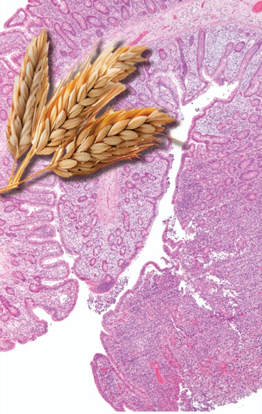
The growing number of EATL cases could be connected to the increase in celiac disease, given the strong association between the two conditions, researchers said.

“Although [EATL is] rare, most of the cases that we see develop in patients with celiac disease,” said lead investigator Isabel Hujoel, MD, the clinic director of the Celiac Disease Center at UW Medical Center, in Seattle. “We don’t know why the incidence of EATL has steadily risen over the past two decades, but we suspect that it’s because celiac disease continues to increase in prevalence.”

The study, which used the Surveillance, Epidemiology, and End Results (SEER) program database, found 463 cases of EATL between 2000 and 2020, with an age-adjusted incidence rate of 0.014 per 100,000 people (abstract P1249). However, the incidence of EATL increased by 2.58% annually over this 20-year period.

image

Poor Survival Outcomes

Findings from the study, which won Presidential Poster and Outstanding Research Awards, also showed that EATL was more common in men, and that the median age at diagnosis was 65 years. Most cases (42%) were treated with both surgery and chemotherapy.

Older age at the time of diagnosis was associated with a higher risk for death, while factors such as sex, race, year of diagnosis and time to treatment initiation showed no significant impact on survival.

Notably, despite medical advances over the past two decades, the data showed no change in survival over the study period. “Unfortunately, survival outcomes of patients with EATL have not improved over time,” Dr. Hujoel said. “Mean survival for this disease remains approximately six months.”

A separate, retrospective cohort analysis of 259 patients in the SEER database found that the majority of patients underwent surgical resection (69.9%), followed by chemotherapy (47.5%) (abstract P1594).

Investigator Sophia Dar, MD, a gastroenterology fellow at Southern Illinois University School of Medicine, in Springfield, told Gastroenterology & Endoscopy News that treatment with these methods was associated with improved survival rates compared with no treatment. Chemotherapy alone also decreased the hazard ratio when compared with no treatment.

“Patients in the chemotherapy group marginally outperformed their surgical counterparts, indicating perhaps chemotherapy as a more viable treatment option in certain scenarios,” Dr. Dar said. “Unfortunately, irrespective of treatment, 83.7% of patients died in the five-year follow-up period.”

Researchers from both studies emphasized the need for further study, especially considering the strong association between EATL and celiac disease.

“Better understanding of the factors contributing to the high mortality rate could help medical practitioners develop more efficient treatment plans for EATL,” Dr. Dar concluded.

Screen Only for Refractory Disease

Debra Silberg, MD, PhD, the chief scientific officer of the nonprofit Beyond Celiac, based in Ambler, Pa., told Gastroenterology & Endoscopy News that the research does not address whether the increase in EATL is accounted for by patients with celiac disease, but “since there has also been an annual increase in celiac disease diagnosis, it follows that there would be an increase in EATL. Therefore, the overall percentage of EATL in patients with celiac disease may not have changed—which is around 0.22 to 1.9 per 100,000—and is still extremely rare,” she added.

Regarding screening, Dr. Silberg referred to the American Gastroenterological Association guidelines for refractory celiac disease (Gastroenterology 2022;163[5]:1461-1469). If a patient is diagnosed with type 2 refractory celiac disease, which, she said, “is associated with clonal T-cell expansion, then small bowel imaging is recommended to exclude EATL and ulcerative jejunoileitis. Since EATL is still rare even with this increased incidence,” she added, “only patients with refractory celiac disease or a suspicion of a complication of celiac disease should be screened.”

R-CHOP Treatment for Non-Hodgkin Lymphoma


MedMaven
Research demonstrates that some patients with NHL can be treated with 4 instead of 6 cycles of R-CHOP.

Rituxan® (rituximab) combined with cyclophosphamide, doxorubicin, Oncovin® and prednisone (R-CHOP) is the standard of care for many patients with Non-Hodgkin’s lymphoma (NHL). Although R-CHOP is the standard researchers continue to try to improve R-CHOP treatment either by adding additional drugs to the regimen in NHL patients at high risk of recurrence or be reducing the total treatment in low risk individuals. Most recently doctors from Germany have reported that 4 cycles of R-CHOP treatment is just is good as 6 cycles in certain low risk individuals. (1) All NHL patients should understand the role of R-CHOP and the research being conducted to improve its effectiveness.

Frequently Asked Questions About R-CHOP

What is R-CHOP Chemotherapy?

CHOP (cyclophosphamide, doxorubicin, vincristine, and prednisone) is a common treatment used for NHL; when combined with Rituxan it is referred to as R-CHOP and is a standard treatment option for NHL.

R-CHOP consists of the following drugs:

  • Rituximab (Rituxan) Rituxan is a monoclonal antibody that has been designed to recognize and bind to B-cells. The binding of Rituxan stimulates the immune system to attack the B-cells and may also be involved in the direct killing or disabling of the B-cell.
  • Cyclophosphamide (Cytoxan) is a chemotherapy drug tht targets the DNA of cancer cells and signals them to stop dividing.
  • Doxorubicin hydrochloride (Adriamycin) is a chemotherapy drug that blocks an enzyme cancer cells need to grow and reproduce.
  • Vincristine (Oncovin, Vincasar, Vincrex) Vincristine is an alkaloid chemotherapy drug and is a vesicant, meaning it can damage tissues that it comes in contact with.

· Prednisolone a corticosteroid oral medication that works with your immune system to help reduce inflammation.

How is R-CHOP administered?

R CHOP is given in “cycles.” Each cycle begins with intravenous doxorubicin, vincristine and cyclophosphamide and is followed by oral prednisolone. Depending on the type of NHL patients will receive 4-8 cycles of R-CHOP. Cycles are typically administered every 21 days unless patients are receiving “dose dense” R-CHOP. Dose-dense R-CHOP is given every 14 days and is supported with a white blood cell growth factor to prevent low white blood counts, infection and fever.

Before each treatment a blood test is performed to check the white blood cell count and determine if the liver and kidneys are functioning well enough. If they’re not, the doctor may need to adjust the treatment.

What are the side effects of R-CHOP chemotherapy?

Chemotherapy is directed at killing or eradicating cancer cells. Unfortunately, cancer treatments may also damage normal, healthy cells that are not affected by the cancer. The result of this damage is a complication, or side effect, of treatment. Side effects occur because most cancer treatments cannot distinguish between cancer cells and normal, healthy cells. The main side effects of R-CHOP include the following. The most important side effect to understand is neutropenia or a low white blood count.

Caring for Yourself During Chemotherapy

Common Side Effects Include:

  • irritation around the intravenous or port site
  • red or pink urine for a few days due to doxorubicin
  • appetite changes
  • weight changes
  • indigestion
  • nausea
  • vomiting
  • fatigue
  • sleeping difficulties
  • low blood counts
  • allergic reactions

What Does the Research Show?

Four Cycles of R-CHOP can be Used to Treat Favorable Prognosis DLBCL

Standard therapy for young patients with favorable-prognosis diffuse large B-cell lymphoma (DLBCL) is six cycles of R-CHOP (rituximab, cyclophosphamide, doxorubicin, vincristine and prednisolone) given every 21 days. (1)

New data from the FLYER clinical trial suggests that some patients can be spared two cycles of CHOP, with Rituxan still given over six cycles, and have excellent clinical outcomes, similar to those seen after the standard six cycles of R-CHOP.

In the FLYER clinical trial doctors from Germany treated 592 patients aged 18 to 60 years with “low risk” stage I/II DLBCL, with either six or four cycles of CHOP chemotherapy every 21 days plus the standard six cycles of Rituxan. “Low risk” was defined as an age-adjusted IPI of 0, age less than 60 and maximum disease diameter < 7.5 cm.

The 3-year progression-free survival with four cycles of CHOP was 96% compared to 94% seen with six cycles of CHOP, and overall survival was 99% and 98% respectively.

The authors concluded that low risk DLBCL patients can be spared two cycles of chemotherapy which reduces the average time on treatment from 126 to 84 days.

It’s important to understand that the results do not apply to the majority of patients with DLBCL and the results should not be applied to individuals over the age of 60.

Dose-Dense R-CHOP for Non-Hodgkin Lymphoma

Among patients with NHL supportive treatment with Neulasta® (pegfilgrastim) allows for the safe administration of the dose-dense treatment regimen R-CHOP-14 given every 14 days.

Dose-dense chemotherapy is a treatment approach where chemotherapy is administered as frequently as possible with the goal of delivering the greatest amount of chemotherapy over the shortest period of time, thereby delivering the maximum amount of chemotherapy drug to the cancer. Increasing the dose and frequency of chemotherapy administration appears to increase survival in some patients with non-Hodgkin’s lymphoma because dose-dense regimens may kill more cancer cells, but they also allow less time for bone marrow recovery and are associated with anemia (low red blood cell level) and neutropenia (low white blood cell level).

Neutropenia increases susceptibility to infection and can become a serious condition for several reasons: Many patients who develop neutropenia will require a delay in treatment or a dose reduction (both events can prevent them from receiving full benefits of treatment); patients who develop neutropenia may require hospitalization; and even minor infections can become life-threatening. Neulasta is a drug that is used to stimulate the production of immune cells in order to reduce or prevent neutropenia.

In order to evaluate the R-CHOP-14 researchers in Italy conducted a phase II clinical trial among 50 patients with DLBCL. Neulasta was given on the third day of each treatment cycle.

  • 92% of treatment cycles were delivered on time.
  • Severe (grade 4) neutropenia developed in 19% of treatment cycles and fever developed in 4% of treatment cycles.
  • 74% of patients experienced a complete remission following treatment.
  • After two years, overall survival was 68%.

The researchers conclude that a single dose of Neulasta in each treatment cycle allowed most patients with diffuse large B-cell lymphoma to receive their chemotherapy on time, with few cases of febrile neutropenia.

Six Cycles of R-CHOP-14 Remains Standard of Care for Elderly with Diffuse Large B-cell Lymphoma

Among elderly patients with diffuse large B-cell lymphoma, treatment with six cycles of Rituxan® and CHOP-14 produces better outcomes than CHOP-14 alone and may also produce better outcomes than eight cycles of Rituxan and CHOP-14.

To compare different approaches of administering CHOP with or without Rituxan in the treatment of elderly patients with diffuse large B-cell lymphoma, researchers in Germany conducted a study among more than 1,000 patients between the ages of 61 and 80 years.

Study participants were assigned to receive either six or eight cycles of CHOP administered every two weeks (CHOP-14). Half the patients also received Rituxan.

  • The addition of Rituxan improved outcomes compared to CHOP-14 alone.
  • Patients who received six cycles of Rituxan and CHOP-14 appeared to have the best overall survival.

The researchers concluded that six cycles of Rituxan and CHOP-14 appeared to produce the best outcomes in the treatment of elderly patients with diffuse large B-cell lymphoma. (3)

References:

  1. American Society of Hematology (ASH) 2018. Presented December 2, 2018. Abstract 781.
  2. Pfreundschuh M, Kloess M, Zeynalova S, et al. Six vs. eight cycles of bi-weekly CHOP-14 with or without rituximab for elderly patients with diffuse large B-cell lymphoma (DLBCL): Results of the completed RICOVER-60 trial of the German High-Grade Non-Hodgkin’s Lymphoma Study Group (DSHNHL).Blood. 2006;108;64a. Abstract 205.
  3. Brusamolino E, Rusconi C, Montalbetti et al. Dose-Dense R-CHOP-14 Supported by Pegfilgrastim in Patients with Diffuse Large B-Cell Lymphoma: A Phase II Study of Feasibility and Toxicity. Haematologica. 2006;91:496-502.

Diet soda’s Aspartame now associated with cardiovascular issues in addition to brain tumors, non-Hodgkin lymphoma and kidney function decrease


Are you still thirsty? Or just addicted to this toxic chemical? Well, here’s another reason why you ought to refuse to pour this poison cocktail down your esophagus.

 Image: Diet soda’s Aspartame now associated with cardiovascular issues in addition to brain tumors, non-Hodgkin lymphoma and kidney function decrease

According to an article in Collective Evolution, researchers at the University of Iowa have been taking another look at aspartame, although I really don’t know why they need any more proof of it’s toxicity.  It was kept off the market until 1981, thanks to the consumer advocate and lawyer James Turner.

60,000 women took part in the research and here’s what they found:

“… Women who consumed two or more diet drinks a day are 30 percent more likely to experience a cardiovascular event, and 50 percent more likely to die from a related disease.”

Of course the folks who created this study have merely called for more research:

“‘It’s too soon to tell people to change their behaviour based on this study; however, based on these and other findings we have a responsibility to do more research to see what is going on and further define the relationship, if one truly exists,’ says Dr. Ankur Vyas, because ‘This could have major public health implications.’”

Hmmm. The major health implication of these neurotoxins were pointed out over four decades ago. Since it was ole Donald Rumsfeld who commandeered this poison into the food supply, a quote from the liar himself might be appropriate.

Watch the video discussion. URL:https://youtu.be/M7vV0XpK3Pw

Nutritional factors and non-Hodgkin lymphoma survival in an ethnically diverse population: the Multiethnic Cohort


Background/Objectives:

To understand the possible effect of modifiable health behaviors on the prognosis of the increasing number of non-Hodgkin lymphoma (NHL) survivors, we examined the pre-diagnostic intake of major food groups with all-cause and NHL-specific survival in the Multiethnic Cohort (MEC).

Subjects/Methods:

This analysis included 2339 participants free of NHL at cohort entry and diagnosed with NHL as identified by cancer registries during follow-up. Deaths were ascertained through routine linkages to state and national death registries. Cox proportional hazards regression was applied to estimate hazard ratios (HRs) and 95% confidence intervals (CIs) for overall and NHL-specific mortality according to pre-diagnostic intake of vegetables, fruits, red meat, processed meat, fish, legumes, dietary fiber, dairy products and soy foods assessed by food frequency questionnaire.

Results:

The mean age at diagnosis was 71.8±8.5 years. During 4.5±4.1 years of follow-up, 1348 deaths, including 903 NHL-specific deaths, occurred. In multivariable models, dairy intake was associated with higher all-cause mortality (highest vs lowest tertile: HR=1.14, 95% CI 1.00–1.31, Ptrend=0.03) and NHL-specific (HR=1.16, 95% CI 0.98–1.37) mortality. Legume intake above the lowest tertile was related to significant 13–16% lower all-cause and NHL-specific mortality, whereas red meat and fish intake in the intermediate tertiles was associated with lower NHL-specific mortality. No association with survival was detected for the other food groups.

Conclusions:

These data suggest that pre-diagnostic dietary intake may not appreciably contribute to NHL survival, although the higher mortality for dairy products and the better prognosis associated with legumes agree with known biologic effects of these foods.

Targeting BTK with Ibrutinib in Relapsed or Refractory Mantle-Cell Lymphoma.


BACKGROUND

Bruton’s tyrosine kinase (BTK) is a mediator of the B-cell–receptor signaling pathway implicated in the pathogenesis of B-cell cancers. In a phase 1 study, ibrutinib, a BTK inhibitor, showed antitumor activity in several types of non-Hodgkin’s lymphoma, including mantle-cell lymphoma.

METHODS

In this phase 2 study, we investigated oral ibrutinib, at a daily dose of 560 mg, in 111 patients with relapsed or refractory mantle-cell lymphoma. Patients were enrolled into two groups: those who had previously received at least 2 cycles of bortezomib therapy and those who had received less than 2 complete cycles of bortezomib or had received no prior bortezomib therapy. The primary end point was the overall response rate. Secondary end points were duration of response, progression-free survival, overall survival, and safety.

RESULTS

The median age was 68 years, and 86% of patients had intermediate-risk or high-risk mantle-cell lymphoma according to clinical prognostic factors. Patients had received a median of three prior therapies. The most common treatment-related adverse events were mild or moderate diarrhea, fatigue, and nausea. Grade 3 or higher hematologic events were infrequent and included neutropenia (in 16% of patients), thrombocytopenia (in 11%), and anemia (in 10%). A response rate of 68% (75 patients) was observed, with a complete response rate of 21% and a partial response rate of 47%; prior treatment with bortezomib had no effect on the response rate. With an estimated median follow-up of 15.3 months, the estimated median response duration was 17.5 months (95% confidence interval [CI], 15.8 to not reached), the estimated median progression-free survival was 13.9 months (95% CI, 7.0 to not reached), and the median overall survival was not reached. The estimated rate of overall survival was 58% at 18 months.

CONCLUSIONS

Ibrutinib shows durable single-agent efficacy in relapsed or refractory mantle-cell lymphoma.

Source: NEJM


If Your Doctor Insists That Vaccines Are Safe, Then Have Them Sign This Form.


vac

The average person that consents to a vaccine injection, either for themselves or for their children, genuinely believes it is for the betterment of health. What they are not aware of is that even their doctor is likely unfamiliar with the toxic ingredients contained in vaccines which can immediately begin to degrade both short- and long-term health. If your doctor insists that vaccines are safe, then they should have absolutely no problem in signing this form so that you may archive it for your own records on the event of an adverse reaction.


The reality of vaccines is that they are a far greater risk to human health than benefit and always have been. In fact, two centuries of official death statistics show conclusively and scientifically that modern medicine is not responsible for and played little part in substantially improving life expectancy and survival from diseases in developed nations.

In North America, Europe, and the South Pacific, major declines in life-threatening infectious diseases occurred historically either without, or far in advance vaccination efforts for specific diseases.

Whenever I personally inform medical doctors of these realities, many of them are quite shocked with the data. That’s not surprising considering the fact that medical students are still brainwashed that vaccines immunize which is a myth in itself, since natural or “real” immunity can never be artificially induced by a vaccine.

Other misinformed educators also still rely on the myth of herd immunity which is nothing short of medical fraud. It is a shame and embarrassment that brilliant students are deceptively led down the path of ignorance every single year at prestigious medical institutions in the hopes of obtaining an education. These students then become the physicians of a good percentage of the population.

One of the problems we have in a society filled with misinformation about health, is that people sit on the fence. They want to conform to the societal norms ingrained in our minds about conventional medicine, but they also want to stand up for their beliefs and conscience. These fence sitters are made up of those who understand that current vaccination practices are unsafe, yet somehow also believe you can make vaccines safer or more effective. That is where we have to shift the opinions of those who are on the fence and have them fall off on the side of natural health rather than conventional medicine. See my article When It Comes to Vaccines, Don’t Sit On The Fence!

I have previously written that if your doctor cannot answer these 4 questions, don’t vaccinate. Well, if your doctor does make an attempt to answer these questions and a verbal response and statement is not satisfactory for your own peace of mind, then your doctor should be at least willing to provide you with his or her personal declaration of the safety and efficacy of the vaccines he or she (or attending physician or nurse) is about to inject in your or your child’s body. Effectively, this becomes your doctor’s warranty that the risk factors he or she has identified justify the recommended vaccinations with the benefits exceeding the risks.


Physician’s Warranty of Vaccine Safety Form

The following form was adapted from Ken Anderson’s original. Perhaps you can find a physician that will sign it because I have no record of that ever happening:

PHYSICIAN’S WARRANTY OF VACCINE SAFETYI (Physician’s name, degree)_______________, _____ am a physician licensed to practice medicine in the State/Province of _________. My State/Provincial license number is ___________ , and my DEA number is ____________. My medical specialty is _______________I have a thorough understanding of the risks and benefits of all the medications that I prescribe for or administer to my patients. In the case of (Patient’s name) ______________ , age _____ , whom I have examined, I find that certain risk factors exist that justify the recommended vaccinations. The following is a list of said risk factors and the vaccinations that will protect against them:
Risk Factor __________________________
Vaccination __________________________
Risk Factor __________________________
Vaccination __________________________
Risk Factor __________________________
Vaccination __________________________I am aware that vaccines may contain many of the following chemicals, excipients, preservatives and fillers:

* aluminum hydroxide
* aluminum phosphate
* ammonium sulfate
* amphotericin B
* animal tissues: pig blood, horse blood, rabbit brain,
* arginine hydrochloride
* dog kidney, monkey kidney,
* dibasic potassium phosphate
* chick embryo, chicken egg, duck egg
* calf (bovine) serum
* betapropiolactone
* fetal bovine serum
* formaldehyde
* formalin
* gelatin
* gentamicin sulfate
* glycerol
* human diploid cells (originating from human aborted fetal tissue)
* hydrocortisone
* hydrolized gelatin
* mercury thimerosol (thimerosal, Merthiolate(r))
* monosodium glutamate (MSG)
* monobasic potassium phosphate
* neomycin
* neomycin sulfate
* nonylphenol ethoxylate
* octylphenol ethoxylate
* octoxynol 10
* phenol red indicator
* phenoxyethanol (antifreeze)
* potassium chloride
* potassium diphosphate
* potassium monophosphate
* polymyxin B
* polysorbate 20
* polysorbate 80
* porcine (pig) pancreatic hydrolysate of casein
* residual MRC5 proteins
* sodium deoxycholate
* sorbitol
* thimerosal
* tri(n)butylphosphate,
* VERO cells, a continuous line of monkey kidney cells, and
* washed sheep red blood

and, hereby, warrant that these ingredients are safe for injection into the body of my patient. I have researched reports to the contrary, such as reports that mercury thimerosal causes severe neurological and immunological damage, and find that they are not credible.

I am aware that some vaccines have been found to have been contaminated with Simian Virus 40 (SV 40) and that SV 40 is causally linked by some researchers to non-Hodgkin’s lymphoma and mesotheliomas in humans as well as in experimental animals. I hereby warrant that the vaccines I employ in my practice do not contain SV 40 or any other live viruses. (Alternately, I hereby warrant that said SV-40 virus or other viruses pose no substantive risk to my patient.)

I hereby warrant that the vaccines I am recommending for the care of (Patient’s name) _______________ do not contain any tissue from aborted human babies (also known as “fetuses”).

In order to protect my patient’s well being, I have taken the following steps to guarantee that the vaccines I will use will contain no damaging contaminants.

STEPS TAKEN: _________________________
_______________________________________
_______________________________________
_______________________________________

I have personally investigated the reports made to the VAERS (Vaccine Adverse Event Reporting System) and state that it is my professional opinion that the vaccines I am recommending are safe for administration to a child under the age of 5 years.

The bases for my opinion are itemized on Exhibit A, attached hereto, — “Physician’s Bases for Professional Opinion of Vaccine Safety.” (Please itemize each recommended vaccine separately along with the bases for arriving at the conclusion that the vaccine is safe for administration to a child under the age of 5 years.)

The professional journal articles I have relied upon in the issuance of this Physician’s Warranty of Vaccine Safety are itemized on Exhibit B , attached hereto, — “Scientific Articles in Support of Physician’s Warranty of Vaccine Safety.”

The professional journal articles that I have read which contain opinions adverse to my opinion are itemized on Exhibit C , attached hereto, — “Scientific Articles Contrary to Physician’s Opinion of Vaccine Safety”

The reasons for my determining that the articles in Exhibit C were invalid are delineated in Attachment D , attached hereto, — “Physician’s Reasons for Determining the Invalidity of Adverse Scientific Opinions.”

Hepatitis B

I understand that 60 percent of patients who are vaccinated for Hepatitis B will lose detectable antibodies to Hepatitis B within 12 years. I understand that in 1996 only 54 cases of Hepatitis B were reported to the CDC in the 0-1 year age group. I understand that in the VAERS, there were 1,080 total reports of adverse reactions from Hepatitis B vaccine in 1996 in the 0-1 year age group, with 47 deaths reported.

I understand that 50 percent of patients who contract Hepatitis B develop no symptoms after exposure. I understand that 30 percent will develop only flu-like symptoms and will have lifetime immunity. I understand that 20 percent will develop the symptoms of the disease, but that 95 percent will fully recover and have lifetime immunity.

I understand that 5 percent of the patients who are exposed to Hepatitis B will become chronic carriers of the disease. I understand that 75 percent of the chronic carriers will live with an asymptomatic infection and that only 25 percent of the chronic carriers will develop chronic liver disease or liver cancer, 10-30 years after the acute infection. The following scientific studies have been performed to demonstrate the safety of the Hepatitis B vaccine in children under the age of 5 years.
____________________________________
____________________________________ _____________________________________

In addition to the recommended vaccinations as protections against the above cited risk factors, I have recommended other non-vaccine measures to protect the health of my patient and have enumerated said non-vaccine measures on Exhibit D , attached hereto, “Non-vaccine Measures to Protect Against Risk Factors” I am issuing this Physician’s Warranty of Vaccine Safety in my professional capacity as the attending physician to (Patient’s name) ________________________________. Regardless of the legal entity under which I normally practice medicine, I am issuing this statement in both my business and individual capacities and hereby waive any statutory, Common Law, Constitutional, UCC, international treaty, and any other legal immunities from liability lawsuits in the instant case. I issue this document of my own free will after consultation with competent legal counsel whose name is _____________________________, an attorney admitted to the Bar in the State of __________________ .
_________________________ (Name of Attending Physician)
______________________ L.S. (Signature of Attending Physician)
Signed on this _______ day of ______________ A.D. ________
Witness: _________________ Date: _____________________
Notary Public: _____________Date: ______________________

Source: http://www.realfarmacy.com
 

Malignant B Cells at the Helm in Follicular Lymphoma.


Lymph nodes in patients with follicular lymphoma contain not just malignant B cells but normal immune cells including T cells, intratumoral macrophages, monocytes, dendritic cells, and natural-killer cells. Recent data have shown that the tumor microenvironment plays an important role in the outcome of patients with non-Hodgkin lymphoma and that the composition of the microenvironment has prognostic significance for patients with follicular lymphoma.1 It is becoming increasingly clear that, although the tumor microenvironment supports the growth and survival of malignant B cells, the malignant B cells also define the constitution of the tumor microenvironment.

Malignant B cells from biopsies of tumors in patients with lymphoma do not play a passive role but instead clearly drive the differentiation and function of intratumoral T cells (Fig 1). Malignant B cells have been found to express multiple ligands responsible for inducing regulatory T cells (Treg cells) and suppressing other intratumoral effector cells. Lymphoma B cells have been found to express CD70, and CD70/CD27 signaling promotes the induction of FoxP3-positive Treg cells.2Malignant cells also express PD-L1, and interactions with PD1 expressed on intratumoral immune cells results in immune suppression.3,4 B cells from lymphoma biopsies have been found to secrete chemokines and cytokines responsible for further immune suppression. CCL22 secreted by lymphoma B cells is involved in chemotaxis and migration of intratumoral Treg cells that express the receptor CCR4, which results in recruitment of Treg cells to the tumor microenvironment.5 Treg cells recruited to sites of lymphoma have been shown to suppress both effector CD8+ T cells and intratumoral CD4+ cells.5,6 Cytokines and chemokines such as interleukin 12 and CXCL10 produced by the malignant B cells also have a role in regulating effector T-cell function.7,8 However, interleukin 12 has been shown to induce T-cell exhaustion, resulting in ineffective T cells that are unable to proliferate or lyse target B cells.8 Overall, malignant B cells skew the T-cell balance within lymph nodes promoting Treg cells and inhibiting effector T cells or TH17 cells.

In the article that accompanies this editorial, Kiaii et al10 contribute to this growing body of literature by showing that follicular lymphoma B cells induce changes in gene expression in intratumoral T cells that results in changes in T-cell function. In addition, they show that T cells exhibiting genetic changes induced by lymphoma cells significantly impact the outcome of patients with follicular lymphoma as well as the risk of transformation to large-cell lymphoma. The authors also investigated the molecular mechanisms whereby tumor-infiltrating T cells are changed within the tumor microenvironment. To do this they used highly purified CD4+ and CD8+ T cells from malignant lymph nodes from patients with follicular lymphoma obtained at the time of diagnosis before therapy. These cells were compared with cells from control tissues, including reactive lymph nodes and peripheral blood. They were able to show that intratumoral CD8+ and CD4+ T lymphocytes had a genetic signature that was different from normal CD4+ and CD8+ T cells. Importantly, they could also show that when healthy allogeneic T cells were cocultured with purified lymphoma B cells, a gene expression pattern similar to that seen in intratumoral T cells, was induced. The changes in gene expression seemed to be caused by both direct contact between T cells and malignant B cells and by soluble factors. This supports the previous findings that both secreted and cell surface ligands are important in defining the tumor microenvironment in follicular B-cell lymphoma. Because ACTN1 was among the most downregulated genes in intratumoral T cells and actin-based motility signaling pathways seemed disrupted, the authors evaluated the motility of sorted CD4+ and CD8+ tumor infiltrating lymphocytes. When compared with normal controls, intratumoral T cells were functionally impaired with a significant reduction in their motility index when compared with normal controls. In a multivariable prognostic model, the authors then showed that the number and location of T cells expressing PMCHNAMPT, and ETV1 had prognostic significance and were associated with both overall survival and the time to transformation to large-cell lymphoma. These data confirm the important role that malignant B cells play in defining the function and differentiation of intratumoral T cells. These results also confirm that the number and location of lymphoma infiltrating lymphocytes is associated with patient outcome.

These findings are particularly important as novel T-cell–mediated therapies are being developed for B-cell malignancies. Whereas T cells can mediate antitumor responses in lymphoma patients, the immune tolerance mechanisms described above often result in deletion or inactivation of tumor-specific T cells. To overcome this problem, genes encoding an antibody against a lymphoma-associated antigen such as CD19 have been linked, in the form of a single-chain variable fragment, to genes that encode T-cell signaling domains. This chimeric antigen receptor (CAR) has then been introduced into T cells and these cells used for adoptive T-cell therapy.11 CAR T cells are able to recognize tumor antigens in a HLA-independent manner and this allows CAR-modified T cells to overcome the tumor’s ability to escape immune detection by downregulation of HLA expression. Initial use of CAR T cells resulted in encouraging clinical responses,12 however the clinical utility of particularly the first-generation CAR T cells was limited by their inability to sufficiently activate and sustain themselves in vivo. The subsequent generations of CAR T cells—with the addition of costimulatory domains to the intracellular portion including CD28, 4-1BB, or OX40—have been engineered to enhance cytokine secretion and effector cell expansion and to prevent activation-induced apoptosis and immune suppression by the tumor. Despite these modifications, the best clinical results have been seen in patients with a low tumor burden and in those who first received cytotoxic chemotherapy before CAR T-cell therapy.13 The chemotherapy administered was likely to have not only depleted malignant cells but also decreased immunosuppressive cells such as Treg cells and suppressive monocytes. Malignant B cells, and the immunosuppressive tumor microenvironment they promote, may therefore remain a barrier to effective adoptive immunotherapy in B-cell lymphoma, particularly in patients with chemotherapy-resistant, bulky disease.

Malignant B cells in follicular lymphoma clearly play a defining role regarding the composition and function of the tumor microenvironment. The data presented confirm that malignant B cells promote a profoundly immunosuppressive microenvironment and thereby protect themselves from being targeted by the immune system. Future therapies in follicular lymphoma, including immunotherapies such as CAR T cells, will need to not only deplete malignant B cells but also inhibit the immunosuppressive mechanisms by which malignant B cells suppress the antitumor immune response. A dual approach that both depletes malignant cells and promotes immune function may subsequently result in a better clinical outcome for patients with follicular lymphoma.

Source: JCO

Reproductive organ involvement in non-Hodgkin lymphoma during pregnancy: a systematic review.


Data for pregnancy-associated non-Hodgkin lymphoma are limited to case reports, making it difficult to define this disorder. We did a systematic search for articles published between 1967 and 2011 with the aim to determine the characteristics, management, and outcome of pregnancy-associated non-Hodgkin lymphoma. We identified 121 patients from 74 papers. Most patients with stage information available presented with stage IV disease (75%, 82 of 108 patients). Patients were classified into three clinical groups; those with indolent lymphomas accounted for 5% (five of 108), aggressive lymphomas (diffuse large B-cell lymphoma and T-cell lymphomas) made up 48% of patients (52 of 108), and highly aggressive lymphomas (Burkitt’s lymphoma, immunoblastic lymphoma, and unspecified highly aggressive lymphomas) accounted for 47% of patients (51 of 108). Reproductive organ involvement (breast, ovary, uterus, placenta) was reported in 49% of 110 patients with information available on extranodal involvement, and prevailed in endemic Burkitt’s lymphoma (100%, 19 of 19), followed by non-endemic Burkitt lymphoma (70%, 14 of 20), immunoblastic lymphoma (67%, two of three), peripheral T-cell lymphoma (46%, six of 13), and indolent (40%, two of five) and diffuse large cell lymphoma (23%, nine of 40). Most patients received antepartum (45%, 55 of 121) or postpartum therapy (45%, 54 of 121), resulting in 6-month survival of 53% for mothers and a livebirth rate of 83%. Pregnancy-associated non-Hodgkin lymphoma has unique clinical characteristics with frequent reproductive organ involvement. Collaborative prospective studies are needed to further characterise pathophysiological and clinical aspects of this complication.

Source: lancet


What is Radioimmunotherapy (RIT) Treatment of NHL?


Radioimmunotherapy (RIT) is a type of targeted therapy that delivers radiation directly to cancer cells. It combines a monoclonal antibody—a type of protein that recognizes and binds to certain parts of cancer cells—with radioactive material. When the monoclonal antibody binds to the cancer cell, the radiation kills the cell.

Currently, RIT is used for the treatment of B-cell non-Hodgkin lymphomas (see examples below). RIT is also being evaluated for the treatment of other types of cancer, including prostate cancer and glioblastoma.

RIT is given on an outpatient basis, is generally completed in 10 days (as opposed to the longer duration of conventional chemotherapy), and avoids many of the side effects of chemotherapy. Because RIT may result in a temporary reduction in blood cell counts, patients typically need to have their blood cell levels monitored after treatment.

Examples of RIT

Zevalin® (ibritumomab tiuxetan): Zevalin therapy combines the monoclonal antibody Rituxan® (rituximab) with Zevalin, which is comprised of an anti-CD20 monoclonal antibody and Yttrium-90, a radioisotope that delivers the radiation. When injected into the body, Zevalin attaches to a protein (CD20) found only on the surface of B-lymphocytes, such as cancerous B-cells found in many forms of non-Hodgkin’s lymphoma. The radioactivity that is spontaneously emitted targets the B-cell and destroys it. This approach protects healthy tissue.  To learn more about Zevalin and view stories from other patients living with follicular lymphoma go to www.Zevalin.com. To locate a Zevalin-experienced oncologist click here.

Bexxar® (tositumomab and iodine I 131 tositumomab): Bexxar also targets B lymphocytes, and is comprised of an anti-CD20 monoclonal antibody and radioactive iodine 131. Bexxar is used for the treatment of certain patients with CD20-positive relapsed or refractory non-Hodgkin lymphoma.

Zevalin and Advanced Follicular Lymphoma

Researchers conducted a study that included 414 patients with CD20-positive stage III or IV follicular lymphoma who achieved a complete or partial response after first-line induction treatment. Patients were randomly assigned to receive Zevalin or no further treatment.

After a median follow-up of 3.5 years, the results indicated that Zevalin significantly prolonged median progression-free survival (PFS) in all patients, regardless of whether they had achieved a partial or complete response. Median PFS in patients treated with Zevalin was 36.5 months, compared to 13.3 months for patients in the control group. For patients who achieved a partial response after induction treatment, those who received Zevalin had a median PFS of 29.3 months compared to 6.2 months for those in the control group. Among patients who achieved complete response after induction, those who received Zevalin had a median PFS of 53.9 months compared to 29.5 months in the control group. What’s more, 77 percent of patients who experienced a partial response after induction converted to a complete response, which resulted in a final complete response rate of 87 percent.

The researchers concluded that Zevalin significantly prolonged PFS and resulted in a high conversion rate from partial to complete response, regardless of the type of first-line induction treatment.

Reference:

Morschhauser F, Radford J, Van Hoof A, et al. Phase III trial of consolidation therapy with yttrium-90-ibritumomab tiuxetan compared with no additional therapy after first remission in advanced follicular lymphoma. Journal of Clinical Oncology. 2008; 26: 5156-5164.


Source: cancerconnect.com

Bendamustine plus rituximab versus CHOP plus rituximab as first-line treatment for patients with indolent and mantle-cell lymphomas: an open-label, multicentre, randomised, phase 3 non-inferiority trial.


Rituximab plus chemotherapy, most often CHOP (cyclophosphamide, doxorubicin, vincristine, and prednisone), is the first-line standard of care for patients with advanced indolent lymphoma, and for elderly patients with mantle-cell lymphoma. Bendamustine plus rituximab is effective for relapsed or refractory disease. We compared bendamustine plus rituximab with CHOP plus rituximab (R-CHOP) as first-line treatment for patients with indolent and mantle-cell lymphomas.
METHODS: We did a prospective, multicentre, randomised, open-label, non-inferiority trial at 81 centres in Germany between Sept 1, 2003, and Aug 31, 2008. Patients aged 18 years or older with a WHO performance status of 2 or less were eligible if they had newly diagnosed stage III or IV indolent or mantle-cell lymphoma. Patients were stratified by histological lymphoma subtype, then randomly assigned according to a prespecified randomisation list to receive either intravenous bendamustine (90 mg/m(2) on days 1 and 2 of a 4-week cycle) or CHOP (cycles every 3 weeks of cyclophosphamide 750 mg/m(2), doxorubicin 50 mg/m(2), and vincristine 1.4 mg/m(2) on day 1, and prednisone 100 mg/day for 5 days) for a maximum of six cycles. Patients in both groups received rituximab 375 mg/m(2) on day 1 of each cycle. Patients and treating physicians were not masked to treatment allocation. The primary endpoint was progression-free survival, with a non-inferiority margin of 10%. Analysis was per protocol. This study is registered with ClinicalTrials.gov, number NCT00991211, and the Federal Institute for Drugs and Medical Devices of Germany, BfArM 4021335.
FINDINGS: 274 patients were assigned to bendamustine plus rituximab (261 assessed) and 275 to R-CHOP (253 assessed). At median follow-up of 45 months (IQR 25-57), median progression-free survival was significantly longer in the bendamustine plus rituximab group than in the R-CHOP group (69.5 months [26.1 to not yet reached] vs 31.2 months [15.2-65.7]; hazard ratio 0.58, 95% CI 0.44-0.74; p<0.0001). Bendamustine plus rituximab was better tolerated than R-CHOP, with lower rates of alopecia (0 patients vs 245 (100%) of 245 patients who recieved >/=3 cycles; p<0.0001), haematological toxicity (77 [30%] vs 173 [68%]; p<0.0001), infections (96 [37%] vs 127 [50%]); p=0.0025), peripheral neuropathy (18 [7%] vs 73 [29%]; p<0.0001), and stomatitis (16 [6%] vs 47 [19%]; p<0.0001). Erythematous skin reactions were more common in patients in the bendamustine plus rituximab group than in those in the R-CHOP group (42 [16%] vs 23 [9%]; p=0.024).
INTERPRETATION: In patients with previously untreated indolent lymphoma, bendamustine plus rituximab can be considered as a preferred first-line treatment approach to R-CHOP because of increased progression-free survival and fewer toxic effects.

Source: Lancet.