Brain-Derived Neurotrophic Factor (BDNF) Role in Cannabinoid-Mediated Neurogenesis


The adult mammalian brain can produce new neurons in a process called adult neurogenesis, which occurs mainly in the subventricular zone (SVZ) and in the hippocampal dentate gyrus (DG). Brain-derived neurotrophic factor (BDNF) signaling and cannabinoid type 1 and 2 receptors (CB1R and CB2R) have been shown to independently modulate neurogenesis, but how they may interact is unknown. We now used SVZ and DG neurosphere cultures from early (P1-3) postnatal rats to study the CB1R and CB2R crosstalk with BDNF in modulating neurogenesis. BDNF promoted an increase in SVZ and DG stemness and cell proliferation, an effect blocked by a CB2R selective antagonist. CB2R selective activation promoted an increase in DG multipotency, which was inhibited by the presence of a BDNF scavenger. CB1R activation induced an increase in SVZ and DG cell proliferation, being both effects dependent on BDNF. Furthermore, SVZ and DG neuronal differentiation was facilitated by CB1R and/or CB2R activation and this effect was blocked by sequestering endogenous BDNF. Conversely, BDNF promoted neuronal differentiation, an effect abrogated in SVZ cells by CB1R or CB2R blockade while in DG cells was inhibited by CB2R blockade. We conclude that endogenous BDNF is crucial for the cannabinoid-mediated effects on SVZ and DG neurogenesis. On the other hand, cannabinoid receptor signaling is also determinant for BDNF actions upon neurogenesis. These findings provide support for an interaction between BDNF and endocannabinoid signaling to control neurogenesis at distinct levels, further contributing to highlight novel mechanisms in the emerging field of brain repair.

Introduction

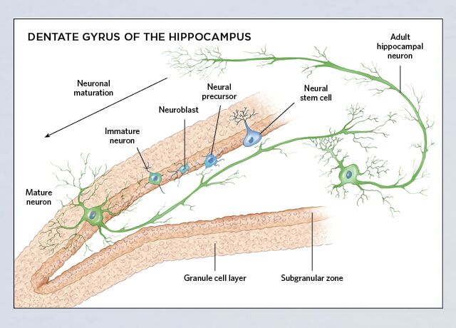
Constitutive neurogenesis occurs in the adult mammalian brain where NSPC are able to differentiate into three neural lineages, neurons, astrocytes and oligodendrocytes (Gage, 2000; Gross, 2000). These multipotent cells exhibit properties of self-renewal and cell proliferation that allow the maintenance of their own pool (Ma et al., 2009). Neurogenesis occurs mainly in two brain areas, the subventricular zone (SVZ) and the subgranular zone (SGZ) within the DG of the hippocampus. These regions are packed with NSPC that originate neuroblasts which migrate toward their final destinations, where they differentiate into mature neurons and are integrated into the neuronal circuitry (Lledo et al., 2006; Zhao et al., 2008; Ming and Song, 2011).

Adult neurogenesis and the neurogenic niches are highly regulated by several factors (intrinsic and extrinsic factors) that control the NSPC rates of proliferation, lineage differentiation, migration, maturation and survival (Ming and Song, 2011). Knowing and understanding the actions of these factors will further contribute to develop new therapeutic strategies useful for brain repair and regeneration. However, there is still a lack of knowledge regarding the key factors that regulate each step of postnatal neurogenesis.

The role of neurotrophins and, in particular, brain-derived neurotrophic factor (BDNF) in adult neurogenesis has been the subject of many studies (Henry et al., 2007; Chan et al., 2008; Vilar and Mira, 2016). BDNF is expressed in both SVZ and SGZ neurogenic niches (Galvão et al., 2008; Li et al., 2008) but its precise role in adult neurogenesis is still not consensual. In fact, some studies suggest that BDNF is important to positively regulate DG cell proliferation and survival (Chan et al., 2008; Li et al., 2008) while others report no BDNF-induced changes in DG neurogenesis (Choi et al., 2009). In SVZ, most studies depict that BDNF does not promote any significant changes in cell proliferation and survival (Henry et al., 2007; Galvão et al., 2008), despite having a role in the migration of SVZ-derived cells (Snapyan et al., 2009; Bagley and Belluscio, 2010). Despite the available contradictory data, BDNF, through TrkB signaling, was shown to have an essential role in the regulation of dendritic complexity as well as synaptic formation, maturation and plasticity of newborn neurons (Chan et al., 2008; Gao et al., 2009; Wang et al., 2015).

Besides expressing BDNF, NSPC present in the neurogenic niches were shown to express all the elements of the endocannabinoid system (Aguado et al., 2005; Arévalo-Martín et al., 2007), including the main cannabinoid receptors type 1 (CB1R) and type 2 (CB2R) receptors (Rodrigues et al., 2017). They are both present in the CNS, although CB2R expression is relatively higher in the immune system (Galve-Roperh et al., 2007). In recent years, the role of cannabinoids in neurogenesis has been of particular interest given their multiplicity of neuromodulatory functions (Mechoulam and Parker, 2013). Cannabinoid receptors modulate adult neurogenesis by acting at distinct neurogenic phases (Prenderville et al., 2015). Importantly, activation of type 1 (Xapelli et al., 2013) or type 2 cannabinoid receptors (Palazuelos et al., 2006) by selective agonists was found to regulate cell proliferation, neuronal differentiation and maturation (Rodrigues et al., 2017).

Several studies have provided molecular and functional evidence for a crosstalk between BDNF and endocannabinoid signaling (Maison et al., 2009; Zhao et al., 2015). Synergism between BDNF and CB1R has been observed both in vitro and in vivo (De Chiara et al., 2010; Galve-Roperh et al., 2013). In particular, BDNF was shown to regulate striatal CB1R actions (De Chiara et al., 2010). Moreover, evidence for BDNF-TrkB signaling interplay with CB1R has been shown to trigger endocannabinoid release at cortical excitatory synapses (Yeh et al., 2017). Importantly, genetic deletion of CB1R was shown to promote a decrease in BDNF expression (Aso et al., 2008) while induction of BDNF expression contributed to the protective effect of CB1R activity against excitotoxicity (Marsicano, 2003; Khaspekov et al., 2004). Moreover, CB1R activity can enhance TrkB signaling partly by activating MAP kinase/ERK kinase pathways (Derkinderen et al., 2003) but also by directly transactivating the TrkB receptors (Berghuis et al., 2005). Δ9-THC, the principal active component of cannabis, was shown to promote upregulation of BDNF expression (Butovsky et al., 2005) whereas increased levels of BDNF were shown to rescue the cognitive deficits promoted by Δ9-THC administration (Segal-Gavish et al., 2017). Interestingly, clinical data suggests that acute and chronic intermittent exposure to Δ9-THC alters BDNF serum levels in humans (D’Souza et al., 2009).

Given the evidence that BDNF and cannabinoid signaling can affect neurogenesis as well as the fact that BDNF may interact with cannabinoid receptors, we hypothesized that cannabinoid receptors could act together with BDNF signaling to fine-tune neurogenesis. We show for the first time that endogenous BDNF is crucial for the cannabinoid-mediated effects on SVZ and DG neurogenesis to happen. Moreover, we demonstrate that CB2R has a preponderant role in regulating some of the BDNF actions on neurogenesis. Taken together, our results suggest an important crosstalk between BDNF and cannabinoid signaling to modulate postnatal neurogenesis.

Discussion

The present work reveals a yet not described interaction between BDNF and cannabinoid receptors (CB1R and CB2R) responsible to modulate several aspects of SVZ and DG postnatal neurogenesis. BDNF was shown to be an important modulator of SVZ and DG postnatal neurogenesis, its actions being under control of cannabinoid receptors. The relevance of each cannabinoid receptor to control the action of BDNF upon neurogenesis is different in the two neurogenic niches. While CB2R has a preponderant role in modulating BDNF actions on DG, BDNF-mediated SVZ postnatal neurogenesis is modulated by both CB1R and CB2R. A constant and clear finding in both neurogenic niches is that BDNF is required for cannabinoid actions to occur. It thus appears that a reciprocal cross-talk between cannabinoids and BDNF exist to modulate postnatal neurogenesis.

BDNF is a neurotrophin important in the regulation of several neuronal processes such as neuronal branching, dendrite formation and synaptic plasticity (Dijkhuizen and Ghosh, 2005; Gómez-Palacio-Schjetnan and Escobar, 2013). In line with this evidence, several studies have shed light on the actions of BDNF in the survival and differentiation of newborn neurons (Benraiss et al., 2001; Henry et al., 2007; Chan et al., 2008; Snapyan et al., 2009). Our findings now demonstrate that BDNF is able to affect early steps of postnatal neurogenesis, such as cell-fate, cell proliferation and neuronal differentiation of SVZ and DG cultures. We observed that BDNF promoted self-renewal of SVZ- and DG-derived cells as observed by an increase in self-renewal divisions, i.e., an increase in the percentage of Sox2+/+ cell-pairs. BDNF-CBR crosstalk has been reported to control several processes at the synaptic level (Zhao and Levine, 2014; Zhong et al., 2015) and we now extended these findings toward very early stages of postnatal neurogenesis. Interestingly, the increase in the SVZ and DG pool of stem/progenitor cells mediated by BDNF was fully abolished in the presence of CB2R antagonist but not CB1R antagonist. An exception is the influence of BDNF upon SVZ cell proliferation, which is not affected by CB1R or CB2R selective antagonism. In what concerns neuronal differentiation, both CB1R and CB2R are required for BDNF actions on SVZ whereas at the DG, only CB2R seem to affect BDNF-promoted neuronal differentiation. Overall, cannabinoid receptor blockade appears to influence more BDNF-induced actions upon early stages of DG neurogenesis in comparison to SVZ, highlighting the fact that cannabinoids distinctly modulate the effects promoted by BDNF in SVZ and DG neurogenesis.

It was previously known that the endocannabinoid system and cannabinoid receptors are important modulators of several stages of neurogenesis (Palazuelos et al., 2012; Xapelli et al., 2013; Prenderville et al., 2015; Rodrigues et al., 2017). In accordance with our previous data, SVZ and DG cells were differently affected by the same cannabinoid pharmacological treatments (Rodrigues et al., 2017). Considering cell fate, we observed that selective activation of CB2R activation promotes self-renewal of DG cells, but not of SVZ cells. This is consistent with several pieces of evidence showing a regulation of cell fate promoted by the activation of several signaling pathways [such as mitogen-activated protein kinase (MAPK) family (ERK, JNK and p38) and the phosphoinositide-3 kinase (PI3K)/AKT pathways] triggered by CBR activation (Molina-Holgado et al., 2007; Gomez et al., 2010; Soltys et al., 2010; Compagnucci et al., 2013).

On the other hand, our results reveal, for the first time, a role of cannabinoid receptors (CB1R and CB2R) in regulating DG cell commitment.

Considering cell proliferation, it is promoted by CB1R but not CB2 at SVZ, while at DG cell proliferation was only induced by co-activation of CB1R and CB2R. These results are in accordance with previous reports that have shown an increase in SVZ cell proliferation promoted by CB1R selective activation (Trazzi et al., 2010; Xapelli et al., 2013) and an increase in DG cell proliferation triggered by CB1R/CB2R non-selective activation (Aguado et al., 2005; Rodrigues et al., 2017). Importantly, while we also detected an effect with the non-selective CB1R/CB2R agonists, none of the selective agonists when applied in the absence of the other agonist were effective to promote cell proliferation in the DG, highlighting the need of caution while interpreting negative results with each of those agonists separately.

Regarding neuronal differentiation, our data indicate that in SVZ and DG neurogenic niches both subtypes of cannabinoid receptors are able to promote neuronal differentiation. These data are in accordance with previous reports in which cannabinoid receptor activation enhanced neuronal differentiation of NSPC by CB1R- (Compagnucci et al., 2013) or CB2R-dependent (Avraham et al., 2014) mechanisms.

The most important finding in the present work is that most of the cannabinoid-induced effects upon cell proliferation and neuronal differentiation depend on the presence of BDNF, suggesting the existence of a BDNF-endocannabinoid feedback loop responsible for regulating these processes. Previous reports have shed light on the existence of a putative interaction between BDNF and cannabinoid receptors (Howlett et al., 2010), but none focused upon neurogenesis. De Chiara et al. (2010) have identified a novel mechanism by which BDNF mediates the regulation of striatal CB1R function. Moreover, others have suggested that BDNF can regulate neuronal sensitivity to endocannabinoids through a positive feedback loop important for the regulation of neuronal survival (Maison et al., 2009). Evidence also shows the involvement of BDNF in the actions mediated by cannabinoids against excitotoxicity (Khaspekov et al., 2004), in synaptic transmission and plasticity (Klug and van den Buuse, 2013; Zhao et al., 2015; Yeh et al., 2017) and in several behavioral outputs (Aso et al., 2008; Bennett et al., 2017). Previous animal studies have shown that acute (Derkinderen et al., 2003) and chronic (Butovsky et al., 2005) Δ9-THC (major psychoactive constituent of cannabis; CB1R and CB2R agonist) administration is associated with an increase in BDNF gene expression. Moreover, it was shown that overexpression of BDNF is able to rescue cognitive deficits promoted by Δ9-THC administration in a mouse model of schizophrenia (Segal-Gavish et al., 2017). In human studies it was found that Δ9-THC increased serum BDNF levels in healthy controls, but not in chronic cannabis users (D’Souza et al., 2009). In fact, cyclic AMP response element-binding protein (CREB) may be the common linking element because it is an important regulator of BDNF-induced gene expression (Finkbeiner et al., 1997), and has been reported to control several steps of the neurogenic process in the adult hippocampus (Nakagawa et al., 2002) and SVZ (Giachino et al., 2005). Consistently, cannabinoids have been shown to induce CREB phosphorylation (Isokawa, 2009) and also to promote changes in BDNF and CREB gene expression (Grigorenko et al., 2002). In addition, the work done by Berghuis et al. (2005) showed that endocannabinoids stimulate TrkB receptor phosphorylation during interneuron morphogenesis. Most importantly, in the same study, the authors observed by co-immunoprecipitation the formation of heteromeric complexes in PC12 cells expressing TrkB receptors and CB1R (Berghuis et al., 2005). Our study brings new and relevant information on the interaction between cannabinoid receptors and BDNF in controlling SVZ and DG neurogenesis, and clearly highlights that this interaction is reciprocal. In fact, neurogenesis promoted by cannabinoid receptor activation depends on the presence of endogenous BDNF, while the effects mediated by BDNF upon neurogenesis are directly regulated by modulation of CB1R or CB2R.

Although our study is based on an in vitro approach, the neurosphere assay, it represents a highly relevant model. In vitro systems of NSPC allow an easier access and better control of experimental variables as well as a thorough analysis of mechanisms happening at cellular and molecular level providing useful information to be further validated in vivo (Singec et al., 2006). Moreover, the heterogeneous composition of the NSPC grown in neurospheres is extremely relevant because it holds some of the features, such as close contact with neighboring cells (newly generated neuroblasts, astrocytes and oligodendrocytes), that resemble those of the physiological niche (Casarosa et al., 2014). These well-established advantages (Aguado et al., 2007; Agasse et al., 2008; Azari et al., 2010) are the reason why we have used this in vitro approach to study the intrinsic properties of NSPC and to understand the interaction between BDNF and cannabinoids in modulating neurogenesis. It is, however, important to mention that the mechanisms governing the regulation of neurosphere dynamics might be different from the ones regulating in vivo adult neurogenesis (Casarosa et al., 2014). Indeed, further in vivo studies will be required to comprehensively understand the role of BDNF in regulating the actions of cannabinoid receptors on postnatal neurogenesis.

Taken together, our data highlight a novel level of complexity for the regulatory mechanisms involved in NSPC dynamics, which involve the interplay of multiple signaling cues, and where BDNF and cannabinoids may play a relevant role. Further in vitro studies are required to detail the molecular mechanisms involved, as well as in vivo studies to determine the functional consequences of the BDNF/cannabinoid crosstalk to control neurogenesis. Nevertheless, our study provides evidence for the need of integrative strategies whenever focusing on NSPC for brain repair.

Yes, you can grow new brain cells. Here’s how.


It’s called neurogenesis.  Your brain’s hippocampus is capable of creating new neurons and certain environmental factors make it speed up and slow down.

The creation of new brain cells, once thought impossible, is related to memory.  Things like eating healthy food and exercising can enhance your body’s ability to create new ones while stress, high levels of sugar and other negatives can slow it down.

Here’s a cross-section; you can see where the new neurons incubate

This is one of those “under the radar” pieces of information that has made its way into the main-stream.  You are constantly regrowing and regenerating your body.

Doctor Sandrine Thuret, neural stem cell researcher at the King’s College of London, has examined the power and potential of the human body to grow new brain cells.  Her research has uncovered a slew of activities that are good for regrowth.  She has also isolated some very dangerous behavior that will slow down the production of new brain cells, including excessive sugar, stress and lack of sleep.


Source:www.minds.com

How Exercise Makes Your Brain Grow


Can exercise help boost your cognitive faculties? Researchers increasingly say the answer is a resounding yes. Recent research reveals that exercise promotes a process now known as neurogenesis, i.e. your brain’s ability to adapt and grow new brain cells, regardless of your age.

Regular Exercise

Story at-a-glance

  • Recent research reveals that exercise promotes a process known as neurogenesis, i.e. your brain’s ability to adapt and grow new brain cells, regardless of your age
  • During exercise, nerve cells release proteins that stimulate the production of brain-derived neurotrophic factor or BDNF, which in turn helps preserve existing brain cells and stimulates the growth of new neurons
  • There’s compelling evidence showing that exercise produces large cognitive gains and helps fight dementia
  • BDNF is also expressed in your neuro-muscular system where it helps protect against age-related muscle atrophy. So BDNF is actively involved in the preservation and rejuvenation of both your muscles and your brain
  • Workouts using nothing but your own body weight are an efficient way to get fit. You can even fulfill the requirements for a high intensity exercise using nothing more than your own body weight, a chair, and a wall

As reported by Forbes Magazine:1

“Not only has research discovered that we can foster new brain cell growth through exercise, but it may eventually be possible to ‘bottle’ that benefit in prescription medication.

The hippocampus, a brain area closely linked to learning and memory, is especially receptive to new neuron growth in response to endurance exercise. Exactly how and why this happens wasn’t well understood until recently.

Research has discovered that exercise stimulates the production of a protein called FNDC5… Over time, FNDC5 stimulates the production of another protein in the brain called Brain Derived Neurotrophic Factor (BDNF), which in turns stimulates the growth of new nerves and synapses… and also preserves the survival of existing brain cells.”

In essence, physical activity produces biochemical changes that strengthen and renew not only your body but also your brain—particularly areas associated with memory and learning.

Researchers Aim to Bottle Exercise Benefits…

Researchers at Harvard Medical School now believe they may be able to recreate the benefits of exercise by putting this protein, FNDC5, into a pill. Bruce Spiegelman, PhD, told Forbes:

 “What is exciting is that a natural substance can be given in the bloodstream that can mimic some of the effects of endurance exercise on the brain.”

They believe such a drug might be useful for those experiencing cognitive decline, including those with early-stage Alzheimer’s and Parkinson’s disease. So far, the hypothesis has only been tested on animals however.

In a recent study published in the journal Cell,2 the researchers successfully increased BDNF in the brains of mice by piggybacking FNDC5 molecules on a virus. According to the authors:

“Perhaps the most exciting result overall is that peripheral delivery of FNDC5 with adenoviral vectors (i.e. a virus) is sufficient to induce central expression of BDNF and other genes with potential neuroprotective functions or those involved in learning and memory.”

Personally, I don’t believe you can fool your body in the long term. It’s important to realize that while a pill may be able to mimic a specific biological effect that exercise produces, such as increasing production of a specific protein or chemical, it will never provide you with ALL the health effects exercise provides, which go far beyond any one specific effect.

Exercise has countless effects on your body — not only on your muscle fibers and brain, but also on your immune system, your ability to fight cancer and much more. To “mimic” all of these benefits, you would literally need handfuls of different pills — and even then they could never reproduce the synergistic benefits that exercise has on your body and mind.

For example, besides boosting memory and learning, regular exercise is also one of the “secret weapons” to overcoming depression. It does this quite effectively by normalizing insulin resistance and boosting natural “feel good” hormones and neurotransmitters associated with mood control in your brain.

Earlier this summer, Princeton University researchers reported3 that physical exercise also helps you combat anxiety by making your brain more resilient during times of stress.

You Cannot Fool Your Body in the Long Run

While actual physical activity can offer you dozens of health benefits, a pill might only be able to recreate one at a time. Besides losing out on the synergistic benefits, taking a pill versus engaging in physical activity will also cost you financially and physically, as there might be unforeseen adverse side effects of the drug to contend with.

All in all, you’re FAR better off just getting physically active. To get the most out of your workouts, I recommend a comprehensive program that includes Peak Fitness high-intensity exercise, strength training, stretching, and core work.

Non-exercise activity and movement is also critical for optimal health, as explained by Dr. Joan Vernikos. Sitting for extended periods of time is actually an independent risk factor for poor health and premature death. Even if you exercise regularly and are fit, uninterruptedly sitting for a great percentage of the time increases your risk of dying prematurely.

Simply standing up, a minimum of 30 times a day, is a powerful antidote to long periods of sitting and is, surprisingly enough, more effective than walking. The good news is that there are virtually unlimited opportunities for movement throughout the day, from doing housework or gardening, to cooking and even just standing up every 10 minutes.

Your Brain Health Is Directly Related to Exercise

That said, let’s look at some of the beneficial effects exercise can have on your brain. According to John J. Ratey, a psychiatrist who wrote the book Spark: The Revolutionary New Science of Exercise and the Brain, there’s overwhelming evidence that exercise produces large cognitive gains and helps fight dementia. Besides triggering the release of BDNF, exercise also protects your brain by:

  • Increasing production of nerve-protecting compounds
  • Improving and increasing blood flow to your brain
  • Improving development and survival of neurons
  • Altering the way damaging proteins reside inside your brain, which appears to slow the development of Alzheimer’s disease. In animal studies, significantly fewer damaging plaques and fewer bits of beta-amyloid peptides, associated with Alzheimer’s, were found in mice that exercised

Ideally, you’d want to make exercise a regular part of your life from as early on as possible. But it’s never too late to start. Even seniors who take up a fitness regimen can improve their cognitive function.

For example, a team at the University of Edinburgh followed more than 600 people, starting at age 70, who kept detailed logs of their daily physical, mental and social habits. Three years later, their brains were imaged for age-related changes, such as brain shrinkage and damage to the white matter, which is considered the “wiring” of your brain’s communication system. Not surprisingly, seniors who engaged in the most physical exercise showed the least amount of brain shrinkage.4

Similarly, Kirk Erickson, PhD of the University of Pittsburgh, found that adults aged 60 to 80 walking moderately (just 30 to 45 minutes, three days per week for one year) increased the volume of their hippocampus by two percent. The hippocampus is a region of your brain important for memory. Erickson told WebMD:5

“Generally in this age range, people are losing one to three percent per year of hippocampal volume. The changes in the size of the hippocampus were correlated with changes in the blood levels of the brain-derived neurotrophic factor (BDNF).”

Erickson also found higher fitness levels associated with a larger prefrontal cortex. He called exercise “one of the most promising nonpharmaceutical treatments to improve brain health.” The most important message from studies like these is that mental decline is NOT inevitable! And exercise is as good for your brain as it is for the rest of your body.

Fasting Can Also Trigger Brain Rejuvenation

Growing evidence indicates that fasting and exercise trigger similar genes and growth factors that recycle and rejuvenate both your brain and muscle tissues. These growth factors include BDNF, as previously mentioned, as well as muscle regulatory factors, or MRFs. These growth factors signal brain stem cells and muscle satellite cells to convert into new neurons and new muscle cells respectively.

Interestingly enough, BDNF also expresses itself in the neuro-muscular system where it protects neuro-motors from degradation. (The neuromotor is the most critical element in your muscle. Without the neuromotor, your muscle is like an engine without ignition. Neuro-motor degradation is part of the process that explains age-related muscle atrophy.)

So BDNF is actively involved in both your muscles and your brain, and this cross-connection, if you will, appears to be a major part of the explanation for why a physical workout can have such a beneficial impact on your brain tissue. It, quite literally, helps prevent, and even reverse, brain decay as much as it prevents and reverses age-related muscle decay.

This also helps explain why exercise while fasting can help keep your brain, neuro-motors, and muscle fibers biologically young. For more information on how to incorporate intermittent fasting into your exercise routine for maximum benefits, please see this previous article. Sugar suppresses BDNF, which also helps explain why a low-sugar diet in combination with regular exercise is so effective for protecting memory and staving off depression.

Almost Anyone Can Improve Their Fitness Without Joining a Gym

In related fitness news, forgoing expensive exercise equipment and focusing on pushing, pulling and lifting your own body weight is becoming a popular alternative that is suitable for most people, regardless of age or fitness level. According to Bret Contreras,6 author of Bodyweight Strength Training Anatomy:

“If more people knew you could get a good physique using your body as a bar bell, they could take matters into their own hands. Find things in the environment: a table to get underneath, hold on to the sides of and then pull the body upward; a rafter for a pull-up. To work your glutes (buttocks muscles), all you need is a couch. It doesn’t have to be intimidating. You could do a 20-minute workout three times a week and have an incredible physique, so long as you push hard and keep challenging yourself.”

Adaptability is a major benefit of body weight exercises: It’s adjustable to almost anyone, from the least fit to the professional athlete. Just learn the basics and try different approaches until you find what works best for you. In the video below, Jill Rodriguez, one of the personal trainers at Mercola.com, demonstrates some basic body weight exercises, and how to add levels of difficulty as you go along.


You can do these exercises just about anywhere… at home, outdoors, or in a gym. You can even fulfill the requirements for a high intensity exercise using nothing more than your own body weight, a chair, and a wall! This program is described in my previous article, “The Scientific 7-Minute Workout.” As the title implies, this science-backed routine only requires a seven minute investment, as the program calls for as little as 10- to 15-seconds of rest between each 30-second exercise, which should be performed in rapid succession.

Need a Portable Fitness Routine? There’s an App for That…

In today’s world, you have plenty of technological allies in fitness. With prices ranging from free to $3.99, a previous article brings you information about six bodyweight apps for your iPad or phone. One helps you work out your own customized workout for your skill and fitness level. Another can keep you body challenged. Other non-bodyweight training apps help you track your progress in jogging or running, keep track of your workouts, or calculate heart rate with a range of tools to keep you on track. These apps let you bring your own personal trainer along on every workout, no matter where you are.

For Total Body-Mind Health, Adopt a Well-Rounded Fitness Program

Ideally, you’ll want to strive for a varied and well-rounded fitness program that incorporates a wide variety of exercises. As a general rule, as soon as an exercise becomes easy to complete, you need to increase the intensity and/or try another exercise to keep challenging your body.

Additionally, as I mentioned earlier, more recent research has really turned the spotlight on the importance of non-exercise movement. Truly, the key to health is to remain as active as you can, all day long, but that doesn’t mean you train like an athlete for hours a day. It simply means, whenever you have a chance to move and stretch your body in the course of going about your day—do it!

And the more frequently, the better. Everything from standing up, to reaching for an item on a tall shelf, to weeding in your garden and walking from one room to another, and even doing dishes count. In short, it’s physical movement, period, that promotes health benefits by the interaction your body gets with gravity. To learn more about this important aspect of health, please see this previous article. That said, I recommend incorporating the following types of exercise into your program:

    1. Interval (Anaerobic) Training: This is when you alternate short bursts of high-intensity exercise with gentle recovery periods.
    2. Strength Training: Rounding out your exercise program with a 1-set strength training routine will ensure that you’re really optimizing the possible health benefits of a regular exercise program. You can also “up” the intensity by slowing it down. For more information about using super slow weight training as a form of high intensity interval exercise, please see my interview with Dr. Doug McGuff.
    3. Stand Up Every 10 Minutes. This is not intuitively obvious but emerging evidence clearly shows that even highly fit people who exceed the expert exercise recommendations are headed for premature death if they sit for long periods of time. My interview with NASA scientist Dr. Joan Vernikos goes into great detail why this is so, and what you can do about it. Personally, I usually set my timer for 10 minutes while sitting, and then stand up and do one legged squats, jump squats or lunges when the timer goes off. The key is that you need to be moving all day long, even in non-exercise activities.
    4. Core Exercises: Your body has 29 core muscles located mostly in your back, abdomen and pelvis. This group of muscles provides the foundation for movement throughout your entire body, and strengthening them can help protect and support your back, make your spine and body less prone to injury and help you gain greater balance and stability.

Foundation Training, created by Dr. Eric Goodman, is an integral first step of a larger program he calls “Modern Moveology,” which consists of a catalog of exercises. Postural exercises such as those taught in Foundation Training are critical not just for properly supporting your frame during daily activities, they also retrain your body so you can safely perform high-intensity exercises without risking injury.

Exercise programs like Pilates and yoga are also great for strengthening your core muscles, as are specific exercises you can learn from a personal trainer.

  1. Stretching: My favorite type of stretching is active isolated stretches developed by Aaron Mattes. With Active Isolated Stretching, you hold each stretch for only two seconds, which works with your body’s natural physiological makeup to improve circulation and increase the elasticity of muscle joints. This technique also allows your body to repair itself and prepare for daily activity. You can also use devices like the Power Plate to help you stretch.

Zika Infects Adult Neural Progenitors Too


A mouse study shows that the virus has tropism for adult proliferative neural progenitor cells and immature neurons.

Zika virus exposure in a mouse model can infect adult neural stem cells in the brain, leading to cell death and reduced proliferation.CELL STEM CELL, H. LI ET AL.Microcephaly and associated birth defects in babies born to mothers infected with the virus during pregnancy is considered the most serious consequence of the ongoing Zika outbreak. However, the increasing incidence of Guillain-Barré syndrome and other neuropathologies linked to the mosquito-borne and sexually transmissible pathogen indicate that Zika virus infection represents a risk to adults, as well.

A number of recent studies have investigated how Zika virus infects fetal brain cells. Working in mice, scientists at Rockefeller University in New York City and their colleagues elsewhere have now examined how Zika virus infection impacts adult brain cells. As it turns out, as it has for fetal neural progenitor cells, Zika virus has tropism for adult proliferative neural progenitor cells and immature neurons. The team’s results were published today (August 18) in Cell Stem Cell.

Zika virus infection can also induce apoptosis of adult neural progenitor cells in the anterior subventricular and subgranular zones of the mouse brain, the researchers reported.

The results of this mouse study show, “for the first time, that [Zika virus] can affect adult neurogenesis by increasing cell death in both adult neurogenic niches,” the anterior subventricular and subgranular zones,”Patricia Garcez, who studies neuoroplasticity at Brazil’s Federal University of Rio de Janeiro and was not involved in the work, wrote in an email to The Scientist. “Humans produce more than 700 neurons a day in adult hippocampus. . . . If the [neural] stem cells are depleted the effect would be long-lasting.”

While the findings “suggest that the virus has the potential to infect and destroy adult neural progenitor cells in . . . the adult mouse brain,” wrote Arnold Kriegstein, director of the University of California, San Francisco, School of Medicine’s developmental and stem cell biology program, they “do not shed light onhow the virus infects the adult cells.” This mechanism has yet to be uncovered.

The researchers worked with six-week-old mice triply deficient in interferon regulatory factor, which were infected by a single strain of Zika virus, and examined only once post-infection. “We limited our results to just this once strain of mice, this one strain of virus, and this one endpoint in order to have robust and quantitative results,” study coauthor Joseph Gleeson of Rockefeller University wrote in an email to The Scientist. “We used one of the strains of Zika that is known to cause human disease,” he added, “so the work is relevant to the current outbreak.”

As to whether the results in mice might translate to humans, Gleeson noted that “future research would require analysis of the stem cell populations as well as neurocognitive outcomes in adults following Zika infection.” Still, he wrote, it stands to reason that “some immunocompromised or even some healthy individuals might have reactions to Zika like we show in mice.”

Overall, said Kriegstein, who was not involved in the work, “this paper highlights the potential risk that Zika virus infection of adults might have unsuspected consequences—in at least some patients—that could affect brain function and behavior.”

Metformin Activates an Atypical PKC-CBP Pathway to Promote Neurogenesis and Enhance Spatial Memory Formation


Highlights

  • aPKCs ζ and ι play distinct roles in the neural precursor to neuron transition
  • Metformin enhances mammalian neurogenesis via the aPKC-CBP pathway
  • Metformin increases adult neurogenesis in vivo and enhances spatial memory

Summary

Although endogenous recruitment of adult neural stem cells has been proposed as a therapeutic strategy, clinical approaches for achieving this are lacking. Here, we show that metformin, a widely used drug, promotes neurogenesis and enhances spatial memory formation. Specifically, we show that an atypical PKC-CBP pathway is essential for the normal genesis of neurons from neural precursors and that metformin activates this pathway to promote rodent and human neurogenesis in culture. Metformin also enhances neurogenesis in the adult mouse brain in a CBP-dependent fashion, and in so doing enhances spatial reversal learning in the water maze. Thus, metformin, by activating an aPKC-CBP pathway, recruits neural stem cells and enhances neural function, thereby providing a candidate pharmacological approach for nervous system therapy.

Neuroprotective and neurorestorative effects of thymosin β4 treatment initiated 6 hours after traumatic brain injury in rats.


Thymosin β4 (Tβ4) is a regenerative multifunctional peptide. The aim of this study was to test the hypothesis that Tβ4 treatment initiated 6 hours postinjury reduces brain damage and improves functional recovery in rats subjected to traumatic brain injury (TBI).

Methods

Traumatic brain injury was induced by controlled cortical impact over the left parietal cortex in young adult male Wistar rats. The rats were randomly divided into the following groups: 1) saline group (n = 7); 2) 6 mg/kg Tβ4 group (n = 8); and 3) 30 mg/kg Tβ4 group (n = 8). Thymosin β4 or saline was administered intraperitoneally starting at 6 hours postinjury and again at 24 and 48 hours. An additional group of 6 animals underwent surgery without TBI (sham-injury group). Sensorimotor function and spatial learning were assessed using the modified Neurological Severity Score and the Morris water maze test, respectively. Animals were euthanized 35 days after injury, and brain sections were processed to assess lesion volume, hippocampal cell loss, cell proliferation, and neurogenesis after Tβ4 treatment.

Results

Compared with saline administration, Tβ4 treatment initiated 6 hours postinjury significantly improved sensorimotor functional recovery and spatial learning, reduced cortical lesion volume and hippocampal cell loss, and enhanced cell proliferation and neurogenesis in the injured hippocampus. The high dose of Tβ4 showed better beneficial effects compared with the low-dose treatment.

Conclusions

Thymosin β4 treatment initiated 6 hours postinjury provides both neuroprotection and neurorestoration after TBI, indicating that Tβ4 has promising therapeutic potential in patients with TBI. These data warrant further investigation of the optimal dose and therapeutic window of Tβ4 treatment for TBI and the associated underlying mechanisms.

Source: Journal of Neurosurgery.