Deadly Crosstalk


Nerve cells found to suppress immune response during lethal lung infections

Neurons that carry nerve signals to and from the lungs suppress immune response during fatal lung infections.

When the body is fighting infection, the immune system kicks into high gear. But emerging evidence hints at the involvement of another, rather surprising, player in this process: the nervous system.

New research from Harvard Medical School, conducted in mice, shows just how the interaction between the nervous and the immune systems occurs in deadly lung infections—a tantalizing clue into a complex interplay between two systems traditionally viewed as disconnected.

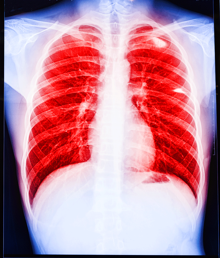
The findings, published March 5 in Nature Medicine, reveal that neurons carrying nerve signals to and from the lungs suppress immune response during infection with Staphylococcus aureus, a bacterium that is growing increasingly impervious to antibiotics and has emerged as a top killer of hospitalized patients, who are often immunocompromised and weakened overall.

The results, the researchers said, suggest that targeting the nervous system could be one way to boost immunity and can set the stage for the development of nonantibiotic approaches to treat recalcitrant bacterial infections.

“With the rapid emergence of drug-resistant organisms, such as methicillin-resistant Staph aureus, nonantibiotic approaches to treating bacterial infections are sorely needed,” said senior study investigator Isaac Chiu, assistant professor in the Department of Microbiology and Immunobiology at Harvard Medical School. “Targeting the nervous system to modulate immunity and treat or prevent these infections could be one such strategy.”

Sensory neurons play a protective role by sensing adverse stimuli and alerting the body that something is awry. In the lungs, the neurons’ projections detect mechanical pressure, inflammation, temperature changes and the presence of chemical irritants, then send an alert to the brain—a notification that can come in the form of pain, airway constriction or a cough that expels harmful agents or particles from the airways.

But the new study reveals that when mouse lungs are invaded by staph bacteria, these guardian neurons interfere with the organ’s ability to cope with infection. Specifically, they reduce the lungs’ ability to summon several types of disease-fighting cells in response to infection. A series of experiments conducted in mice revealed that disabling these neurons promoted immune cell recruitment, increased the lungs’ ability to clear bacteria and boosted survival in staph-infected mice.

The results, the researchers said, suggest that different classes of sensory neurons may be involved in restraining or promoting immune response. Another possibility is that certain pathogens may have evolved to hijack and exploit an immunosuppressive pathway to their benefit—a survival mechanism for some classes of infectious bacteria, said study co-author Stephen Liberles, professor of cell biology at Harvard Medical School.

The team’s interest in the crosstalk between the immune and nervous systems stems from recent work conducted by Chiu and colleagues. Chiu’s earlier research showed that when nerve cells detect bacterial invaders, they produce pain during infection. Other research has revealed nervous system involvement in animal models of allergic asthma.

The team suspected that nerve cells would play a protective role in bacterial infections by boosting immune response to shield the lungs, but the experiments revealed the exact opposite. Much to their surprise, the scientists found that neurons dampened lung immunity and worsened outcomes in mice with bacterial pneumonia.

To determine how nerve cells affect immunity, the scientists genetically or chemically disabled lung neurons and then compared the activity of several types of immune cells involved in infection protection. They also monitored animal survival and took physiological measures such as body temperature and number of bacteria in the lungs.

In an initial set of experiments, researchers injected mice—half with intact neurons and half with chemically disabled neurons—with drug-resistant staph bacteria. Compared with mice with intact nerve receptors, mice with disabled neurons controlled their body temperatures better, harbored 10 times fewer bacteria in their lungs 12 hours after the infection and were markedly more capable of overcoming and surviving the infection. Sixteen of 20 mice with intact neurons succumbed to the infection. By contrast, 17 of 18 mice with disabled neurons survived.

The lungs of mice with genetically or chemically disabled neurons were also better at recruiting neutrophils—the body’s pathogen-fighting troops that provide first responses during infections by devouring disease-causing bacteria. These mice summoned nearly twice as many infection-curbing neutrophils as did mice with intact neurons. But neutrophils in these animals were not simply more numerous. They were also more agile and more efficient in their performance. As a measure of agility, researchers compared how well neutrophils in both groups managed to patrol lung blood capillaries—a key ability that allows these cells to scan for the presence of disease-causing pathogens. Neutrophils in animals with chemically disabled neurons crawled farther, covering greater distances. They were also stickier and thus more capable of adhering to the walls of blood vessels, the site of their pathogen-gobbling action.

“We observed a striking difference in neutrophil presence and behavior between the two groups,” said Pankaj Baral, a research fellow in microbiology and immunobiology at Harvard Medical School and first author on the study. “Neutrophils in mice with disabled neurons were simply better at doing their job.”

Additionally, mice with disabled neurons marshaled more efficiently several types of cytokines, signaling proteins that regulate inflammation, infection and bacterial clearance. In animals with disabled neurons, the levels of these inflammatory cells ramped up and subsided much faster, indicating that these mice were capable of mounting a more rapid immune response in the early stages of infection.

Conversely, mice with intact neurons showed suppressed function in a class of protective immune cells known as gamma delta T cells, a type of protective white blood cell found mostly in barrier tissues that line a variety of organs, including the lungs.

A final set of experiments revealed just how neurons suppressed immunity. The researchers observed that an immune signaling molecule released locally by neurons—a neuropeptide known as CGRP—was markedly increased in mice with intact neuron receptors during infection but absent in mice with disabled neurons. Researchers observed that the release of this molecule interfered with the lungs’ ability to summon immunoprotective neutrophils, cytokines and gamma delta T cells. Experiments in lab dishes revealed that CGRP disrupted immune cells’ ability to kill bacteria. When researchers blocked the production of CGRP in live animals infected with staph, these mice showed an enhanced ability to fight infection.

Taken together, these findings show that lung neurons enable the release of CGRP during lung infections and that blocking the activity of CGRP improves survival in bacterial pneumonia.

“The traditional delineation between nervous and immune systems is getting blurry and our findings underscore the idea that these two systems cross-talk to regulate each other’s function,” Chiu said. “As we move forward, immunologists should think more about the role of the nervous system, and neuroscientists should think more about the immune system.”

Nerve cells actively repress alternative cell fates, researchers find


A neural cell maintains its identity by actively suppressing the expression of genes associated with non-neuronal cell types, including skin, heart, lung, cartilage and liver, according to a study by researchers at the Stanford University School of Medicine.

 It does so with a powerful . “When this protein is missing, neural cells get a little confused,” said Marius Wernig, MD, associate professor of pathology. “They become less efficient at transmitting nerve signals and begin to express genes associated with other cell fates.”

The study marks the first identification of a near-global repressor that works to block many cell fates but one. It also suggests the possibility of a network of as-yet-unidentified master regulators specific to each cell type in the body.

“The concept of an inverse master regulator, one that represses many different developmental programs rather than activating a single program, is a unique way to control neuronal cell identity, and a completely new paradigm as to how cells maintain their throughout an organism’s lifetime,” Wernig said.

Because the protein, Myt1l, has been found to be mutated in people with autism, schizophrenia and major depression, the discovered mode of action may provide new opportunities for therapeutic intervention for these conditions, the researchers said.

Wernig is the senior author of the study, which will be published online April 5 in Nature. Postdoctoral scholars Moritz Mall, PhD, and Michael Kareta, PhD, are the lead authors.

Repressors

Myt1l is not the only protein known to repress certain cell fates. But most other known repressors specifically block only one type of developmental program, rather than many. For example, a well-known repressor called REST is known to block the neuronal pathway, but no others.

“Until now, researchers have focused only on identifying these types of single-lineage repressors,” said Wernig. “The concept of an ‘everything but’ repressor is entirely new.”

In 2010, Wernig showed that it is possible to convert skin into functional neurons over the course of three weeks by exposing them to a combination of just three proteins that are typically expressed in neurons. This “direct reprogramming” bypassed a step called induced pluripotency that many scientists had thought was necessary to transform one cell type into another.

 One of the proteins necessary to accomplish the transformation of skin to neurons was Myt1l. But until this study the researchers were unaware precisely how it functioned.

“Usually we think in terms about what regulatory programs need to be activated to direct a cell to a specific developmental state,” said Wernig. “So we were surprised when we took a closer look and saw that Myt1l was actually suppressing the expression of many genes.”

These genes, the researchers found, encoded proteins important for the development of lung, heart, liver, cartilage and other types of non-neuronal tissue. Furthermore, two of the proteins, Notch and Wnt, are known to actively block neurogenesis in the developing brain.

Blocking Myt1l expression in the brains of embryonic mice reduced the number of mature neurons that developed in the animals. Furthermore, knocking down Myt1l expression in mature neurons caused them to express lower-than-normal levels of neural-specific genes and to fire less readily in response to an electrical pulse.

‘A perfect team’

Wernig and his colleagues contrasted the effect of Myt1l with that of another protein called Ascl1, which is required to directly reprogram skin fibroblasts into neurons. Ascl1 is known to specifically induce the expression of neuronal genes in the fibroblasts.

“Together, these proteins work as a perfect team to funnel a developing cell, or a cell that is being reprogrammed, into the desired cell fate,” said Wernig. “It’s a beautiful scenario that both blocks the fibroblast program and promotes the neuronal program. My gut feeling would be that there are many more master repressors like Myt1l to be found for specific cell types, each of which would block all but one cell fate.”

Source:medicalxpress.com

Eating shuts down nerve cells that counter obesity


Fractions of a second after food hits the mouth, a specialized group of energizing nerve cells in mice shuts down. After the eating stops, the nerve cells spring back into action, scientists report August 18 in Current Biology.  This quick response to eating offers researchers new clues about how the brain drives appetite and may also provide insight into narcolepsy.

orexin in nerve cells

Mouse study offers hints of orexin’s role in weight gain and narcolepsy

These nerve cells have intrigued scientists for years. They produce a molecule called orexin (also known as hypocretin), thought to have a role in appetite. But their bigger claim to fame came when scientists found that these cells were largely missing from the brains of people with narcolepsy.

People with narcolepsy are more likely to be overweight than other people, and this new study may help explain why, says neuroscientist Jerome Siegel of UCLA. These cells may have more subtle roles in regulating food intake in people without narcolepsy, he adds.

Results from earlier studies hinted that orexin-producing nerve cells are appetite stimulators. But the new results suggest the opposite. These cells actually work to keep extra weight off. “Orexin cells are a natural obesity defense mechanism,” says study coauthor Denis Burdakov of the Francis Crick Institute in London. “If they are lost, animals and humans gain weight.”

Mice were allowed to eat normally while researchers eavesdropped on the behavior of their orexin nerve cells.  Within milliseconds of eating, orexin nerve cells shut down and stopped sending signals. This cellular quieting was consistent across foods. Peanut butter, mouse chow, a strawberry milkshake and a calorie-free drink all prompted the same response. “Foods with different flavors and textures had a similar effect, implying that it is to do with the act of eating or drinking, rather than with what is being eaten,” Burdakov says. When the eating ended, the cells once again resumed their activity.

Story continues after graph

Packing it on

Mice in which orexin nerve cells had been killed gained more weight (orange line) than mice that retained those nerve cells (brown line).

J.A. GONZÁLEZ ET AL/CURRENT BIOLOGY 2016

When Burdakov and colleagues used a genetic technique to kill orexin nerve cells, mice ate more food than normal, behavior that led to weight gain, the team found. But a reduced-calorie diet slimmed these mice down.

The results suggest that giving orexin to people who lack it may reduce obesity. But that might not be a good idea. An overactive orexin system has been tied to stress and anxiety, Burdakov says. Orexin’s link to stress raises a different possibility —that anxiety can be reduced by curbing orexin nerve cell activity. “And our study suggests that the act of eating can do just that,” Burdakov says. “This provides a candidate explanation for why people turn to eating at times of anxiety.”

Artificial Skin Sends Touching Signals to Nerve Cells


Sensors transmit pressure changes to neurons and could help prosthetic limbs truly feel

Model robotic hand with artificial mechanoreceptors touching a human hand.
Prosthetic limbs can restore an amputee’s ability to walk or grip objects, but they haven’t yet been able to restore a person’s sense of touch. Researchers at Stanford University have taken a step closer to this type of prosthetic by creating an electronic skin that responds to pressure changes and transmits signals via nerve cells, much as human skin does.

Zhenan Bao and coworkers made the artificial skin by connecting three components: microstructured resistive pressure sensors, flexible printed organic electronic circuits, and nerve cells containing light-activated ion channels (Science 2015, DOI: 10.1126/science.aaa9306).

The pressure sensors are made of a carbon nanotube-elastomer composite shaped into tiny pyramidal structures that are coated onto a surface. The sensor changes conductance in response to applied pressure. Bao previously made similar capacitive sensors, but the new resistive sensors better detect the range of pressures sensed by human skin.

Each sensor is connected to an organic circuit printed with the help of researchers at Xerox’s Palo Alto Research Center (PARC). The circuit converts the pressure signal into a series of electrical pulses and increases pulse frequency in response to increasing pressure. “This circuit is relatively simple to build,” Bao says. “It serves as the perfect electrical readout for our sensors.”

The researchers used the electrical pulses to modulate the frequency of a light-emitting diode. In their proof-of-concept study, they sent light from the LED through an optical fiber to stimulate neurons in mouse brain slices. The nerve cells in these samples were decorated with engineered channelrhodopsins that open in response to light, triggering nerve cells to fire.

The work represents “an important advance in the development of skinlike materials that mimic the functionality of human skin at an unprecedented level,” says Ali Javey, who is developing electronic skin at the University of California, Berkeley. “It could have important implications for the development of smarter prosthetics.”

“This is just the beginning of the path toward building fully integrated artificial skin,” Bao says. Next, she says, her team hopes to mimic other sensing functions of human skin, such as the ability to feel heat, and integrate them into the new platform.

LIVE BROADCAST FROM INSIDE THE NERVE CELL


150123102223

Scientists estimate that our brain consists of about ten to one hundred billions of nerve cells. In order to fulfill their respective tasks as long as possible, these cells have to constantly control their internal proteins with regard to quality and functionality. Otherwise the proteins might clump together and thereby paralyze or even kill the cells. Once the cell recognizes a defect protein, this is marked for degradation and a kind of a molecular shredder, the so-called proteasome, chops it into pieces that are eventually recycled.

For the first time now, researchers have succeeded in visualizing this process in intact nerve cells, which previously could only be investigated in the test tube. Electron cryo-tomography was essential for obtaining the described images. Hereby, cells are cooled down to minus 170°C in a fraction of a second. In a consecutive step, pictures of the interior of the cells are taken from many different angles, which then are merged computationally into a three-dimensional image.

“First time in intact cells”
In the current study, the use of specific technical innovations allowed the researchers to achieve a unprecedented imaging quality, enabling them to distinguish single proteasomes within the cell. “For the first time it is possible to qualitatively and quantitatively describe this important enzyme complex in intact cells”, Asano classifies the results. In the following experiments, the scientists focused on the activity of the proteasomes. For the interpretation of the single particles it is important to know that there are cap-like structures, the so-called regulatory particles, attached to the ends of proteasomes (see picture). They bind proteins that are designated to be degraded and thereby change their shape. The scientists were able to distinguish these states and consequently could deduce how many of the proteasomes were actively degrading proteins.

IN SEARCH OF THE ORIGIN OF OUR BRAIN


141223084134-large


While searching for the origin of our brain, biologists at Heidelberg University have gained new insights into the evolution of the central nervous system (CNS) and its highly developed biological structures. The researchers analysed neurogenesis at the molecular level in the model organism Nematostella vectensis. Using certain genes and signal factors, the team led by Prof. Dr. Thomas Holstein of the Centre for Organismal Studies demonstrated how the origin of nerve cell centralisation can be traced back to the diffuse nerve net of simple and original lower animals like the sea anemone. The results of their research will be published in the journal “Nature Communications”.

Like corals and jellyfish, the sea anemone – Nematostella vectensis – is a member of the Cnidaria family, which is over 700 million years old. It has a simple sack-like body, with no skeleton and just one body orifice. The nervous system of this original multicellular animal is organised in an elementary nerve net that is already capable of simple behaviour patterns. Researchers previously assumed that this net did not evidence centralisation, that is, no local concentration of nerve cells. In the course of their research, however, the scientists discovered that the nerve net of the embryonic sea anemone is formed by a set of neuronal genes and signal factors that are also found in vertebrates.

According to Prof. Holstein, the origin of the first nerve cells depends on the Wnt signal pathway, named for its signal protein, Wnt. It plays a pivotal role in the orderly evolution of different types of animal cells. The Heidelberg researchers also uncovered an initial indication that another signal path is active in the neurogenesis of sea anemones – the BMP pathway, which is instrumental for the centralisation of nerve cells in vertebrates.

Named after the BMP signal protein, this pathway controls the evolution of variouscell types depending on the protein concentration, similar to the Wnt pathway, but in a different direction. The BMP pathway runs at a right angle to the Wnt pathway, thereby creating an asymmetrical pattern of neuronal cell types in the widely diffuse neuronal net of the sea anemone. “This can be considered as the birth of centralisation of the neuronal network on the path to the complex brains of vertebrates,” underscores Prof. Holstein.

While the Wnt signal path triggers the formation of the primary body axis of all animals, from sponges to vertebrates, the BMP signal pathway is also involved in the formation of the secondary body axis (back and abdomen) in advanced vertebrates. “Our research results indicate that the origin of a central nervous system is closely linked to the evolution of the body axes,” explains Prof. Holstein.

Nano air pollutants strike a blow to the brain


Cough. Wheeze. Gasp!

Those sounds echo through the streets of polluted cities. Brown clouds made up of noxious gases, dust, soot and even finer particles hang over buildings and hug the ground. When outside, people can’t help but breathe it all in. And in most parts of the world, windows won’t keep these air pollutants out.

Not all large cities have air pollution like this. But in those urban areas where mountains block the wind from clearing the air, such heavily polluted conditions frequently develop. Mexico City often confronts such pollution. So does Beijing, China. And Los Angeles, Calif. With massive populations, these three cities have huge numbers of cars, buses, trucks and factories spewing pollutants into their air.

Air pollution can lead to serious health problems. Wheezing and gasping occur when people breathe in pollutants for long periods of time. Lungful after lungful of contaminated air gums up the respiratory tract. That’s the branching network of tubes that supplies air to the lungs.

Within the respiratory tract, an inner coating of mucus works like fly paper. This slimy secretion traps large particles, such as pollen grains. Small hair-like structures carry this contaminated mucus up and away from the lungs. Coughing and hacking are the body’s way of clearing the airways. A strong cough — or a good swallow — may remove some pollutants completely.

tractor exhaust

Black soot spews from a truck. People can inhale these fine, black-carbon particles deeply into the lungs, where they can trigger inflammation.

Far smaller particles, called nanoparticles, can sneak past this first line of defense. These airborne particles are measured in the billionths of a meter. (Nano is a prefix meaning a billionth.) These particles can pass all the way into the lungs. Once they settle on lung cells, the pollutants can begin blocking the movement of oxygen into — and carbon dioxide out of — the blood.

The largest nanoparticles are only 100 nanometers across. Scientists are just starting to understand how soot and other nanoparticles interact with the body. Experts already know these pollutants are small enough to slip inside cells. There, they can damage DNA, proteins and other cellular structures. That leads to all kinds of health problems — and not just in the elderly. Kids experience them, too.

Nanoparticles also damage blood vessels. These ultra-small molecules impair the ability to smell. They can even mess with learning and memory. Brains exposed to nanoparticles develop abnormal features similar to those found in people with Alzheimer’s and Parkinson’s diseases. And that has scientists worried. New data have begun showing how nanoparticles can pollute our brains. Especially alarming, some can make a beeline directly through the nose and into our thought centers.

Sneak attack

If inhaled into the lungs, nanoparticles can enter the bloodstream. From there, these materials travel throughout the body. Some share of them will end up polluting the brain.

A layer of tightly packed cells is supposed to control what can — and can’t — move out of the blood and into the brain. This blood-brain barrier attempts to protect delicate nerve cells, called neurons, from substances that might damage them. Most of the time, the barrier does its job. Nanoparticles, however, slip right through.

nanoparticles

Pollutant nanoparticles can inflame cells of the body. That inflammation draws out white blood cells, such as these lymphocytes, a type of immune cell.

Once nanoparticles gain entry to the brain, they damage cells there in a number of ways, notes Caleb Finch. He’s a biologist at the University of Southern California, in Los Angeles.

One type of damage is inflammation. Cells attacked by nanoparticles send out a distress signal. The body responds by dispatchingwhite blood cells to the area. White blood cells are part of the body’s immune response. They help fight infection. Soon, the site becomes swollen with blood. Such inflammation occurs not only in the brain, says Finch, but throughout the body.

Inflammation is supposed to be an acute reaction. That means it should occur briefly, then go away. However, when exposure to air pollution is chronic — persistent over time — inflammation, too, can become persistent. And that’s not healthy.

Particularly worrisome: The inflammation triggered by nanopollutants can lead to hardening of blood vessels. Healthy blood vessels are flexible. This allows large amounts of blood to pass through with each contraction of the heart. But as vessels stiffen, blood must squeeze through narrower spaces. Sometimes blood vessels become completely blocked. Then blood flow stops altogether. When that happens in the brain, a person suffers a stroke.

Nanoparticles also can interfere with a nerve cell’s ability to signal to its neighbors. Nerve cells communicate by releasing chemical messengers. These travel to neighboring nerve cells. There they link up with proteins on the outside of a cell, called receptors. Nerve cells can’t understand what their neighbors are trying to tell them if their receptors aren’t working well. And nanoparticles can damage those receptors. This makes them less sensitive to the chemicals — and messages — they need to detect.

Atherosclerosis diagram

In a healthy artery (top), blood can flow freely. Inflammation triggered by nanoparticles can lead to hardening of blood vessels (bottom). Inflammation can slow — or eventually block — blood flow and foster the build up of fatty plaque.

NHLBI/WIKIMEDIA COMMONS

Damage occurs because many nanoparticles contain what chemists call free radicals. That means some of their molecules contain an atom with an unpaired (missing) outer electron. This makes them unstable. In search of a mate for its lone outer electron, a free radical will swipe an electron from some other molecule. This theft transforms the radical into a stable molecule again. In the process, though, its victim now becomes a free radical. As each victim steals an electron from some neighboring molecule, new free radicals form.

The ongoing chain of electron-theft will damage molecules. It can even kill cells. This happens in the lungs and in the brain. The impact of nanoparticles on the brain, in particular, is severe. That is because the particles meddle with our minds. This makes especially worrisome the recent discovery that nanoparticles can make a beeline for the brain.

Direct connection

Hitching a ride through the bloodstream is a long, roundabout way for pollutants to reach the brain. Unfortunately, if pollutants are small enough, they can take a shortcut, notes Alison Elder. She works at the University of Rochester Medical Center in New York. As a toxicologist, she studies how materials can harm the body.

Working with fellow toxicologist Günter Oberdörster, also at Rochester’s Medical Center, Elder tracked the route that nanoparticles take to the brains of rats. Some travel through the blood and cross the blood-brain barrier, she found. Others, however, enter the brain directly through the nose. To get there, the super-tiny toxic chemicals travel along the olfactory (oal-FAK-tur-ee) — scent-sensing — nerves. These line the inside of the nose.

When rats (or people) inhale through their noses, air passes over the olfactory neurons. Odor molecules link up with receptors on these nerve cells. That causes the cells to signal a brain structure called the olfactory bulb. Different nerve cells in the olfactory bulb, called mitral (MY-trul) cells, relay this incoming information about smells to other parts of the brain.

sense of smell

Nanopollutants can hijack olfactory nerve cells and enter the brain by way of the olfactory bulb. That bulb is highlighted here as a yellow netlike structure coming through the ceiling of the nasal cavity. Earlier work showed the polio virus can use the same pathway.

PATRICK J. LYNCH, MEDICAL ILLUSTRATOR/WIKIMEDIA COMMONS (CC BY 2.5)

But owing to their super-tiny size, nanoparticles can hijack that connection. Scientists had known about this route into the brain since the 1930s (when they realized the polio virus could exploit it). Nanoparticles, at less than one-thousandth the diameter of a human hair, are about the same size as a virus, Elder explains. And just as that small size allows viruses to slip across the blood-brain barrier, it also allows nanoparticles to enter olfactory neurons.

Moving along these sensory neurons, nanoparticles travel straight into the brain by way of mitral cells. Scientists don’t know yet what happens in the nerve cells that allows nanoparticles to travel along them, as if along a highway. Scientists do know, however, what damage the nanoparticles can cause en route. Inside the cells, the particles strip electrons from the atoms that make up cellular structures. This spawns those nasty free radicals.

Nanoparticles also may travel to other parts of the brain. Finch, the biologist at the University of Southern California, exposed mice to air pollution collected near a Los Angeles freeway. Then he examined their brains. Effects of the nanoparticles showed up throughout. Particularly concerning: damage to the hippocampus. This part of the brain is involved in learning and remembering new things.

Sniffing out the problem

Damage to parts of the brain that learn and remember things doesn’t just happen in rodents. Nanopollutants can damage the brains of people, too. Brain injury has shown up even in children. Lilian Calderón-Garcidueñas has seen it. She is a toxicologist at the University of Montana in Missoula. As a pathologist, she also studies the effects of disease on the body’s cells and tissues.

cars on highway

Los Angeles is known for the heavy traffic on its freeways. Caleb Finch, a biologist at the University of Southern California, collected air pollution from near one of LA freeway and later exposed mice to it.

Just a few years of exposure to heavy air pollution can damage cells in the olfactory bulb, Calderón-Garcidueñas finds. And that reduces a person’s ability to smell.

Calderón-Garcidueñas has been testing the ability of children and young adults to smell a variety of odors. Some scents are strong, others weak. Over time, young people living in heavily polluted Mexico City lose some of their ability to smell, she is finding.

That’s bad news, she points out. Loss of smell is one of the first symptoms of Alzheimer’s and Parkinson’s diseases. Indeed, that made her wonder whether damage to the olfactory bulb might be an early warning of more severe problems elsewhere in the brain. And she had good reason to think it might.

Earlier, she had examined the brains of stray dogs in Mexico City. She also had studied the brains of city residents, young and old, who had died in auto accidents or from other types of traumatic events. All showed an accumulation of the same proteins that are also seen in the brains of patients with Alzheimer’s or Parkinson’s disease. Even in families with a history of these diseases, people usually don’t develop those proteins before they turn 40. But in Mexico City, the proteins emerged even in the brains of young children. Their presence might signal harm to thought-processing regions.

It would be impossible to test the impact of these changes on thought or memory in people who had died. So Calderón-Garcidueñas looked for something she could scout for in living folks. And she came up with two things: IQ and computerized scans of the brain.

Calderón-Garcidueñas started by giving an intelligence test to children living in Mexico City. She also tested kids who were the same age but living in a less polluted town. Children in the cleaner town performed better on every aspect of the intelligence test.

Next, she scanned the kids’ brains using an MRI scanner. (MRI is short for magnetic resonance imaging.) The scanner takes a picture of the brain.

Those scans showed areas of damage in the brains of all but one of the Mexico City kids. Children living where the air was cleaner had healthy brains. These findings, says Calderón-Garcidueñas, show that air pollution can start ravaging the brains of even young people.

What you can do

Improving air quality would go a long way toward improving health, Calderón-Garcidueñas concludes. But getting rid of air pollution won’t happen quickly or easily. Luckily, there are things that kids and their families can do to protect themselves even if they can’t escape dirty air.

fruits and veggies

Fruits and vegetables can help to counteract the effects of pollutants on the body, scientists say.

Avoid junk food and cigarettes, suggests Elder, at the University of Rochester. Both can trigger health problems. And sick people generally experience more problems when exposed to nanoparticles, she says.

Snacking on fruits and vegetables also helps. These power foods play an important role in fighting damage by free radicals. That’s because many of those foods contain antioxidants, explains Calderón-Garcidueñas. Antioxidants donate an electron to free radicals without becoming unstable. That stops the destructive chain reaction in its tracks.

If you have to indulge your sweet tooth, choose chocolate over other candies. And not just any chocolate. Dark chocolate is chockablock with antioxidants that can help keep the brain — and the body — up and running. Just don’t overdo it: The sugar in most chocolate just adds unwanted calories.