A National Institutes of Health (NIH)-developed multidimensional MRI method can detect astrogliosis, a neuroinflammatory response that occurs in traumatic brain injury (TBI) and other neurological conditions, according to a study published in Brain. Researchers had previously established that the multidimensional MRI strategy can identify diffuse axonal injury—a microscopic brain injury that, like astrogliosis, cannot be detected by conventional radiological methods.
The two studies, conducted with postmortem human brain tissue, illustrate the potential of using multidimensional MRI with living humans to identify biomarkers for diseases and disorders previously considered radiologically invisible.
The multidimensional MRI method was developed in the laboratory of Peter Basser, PhD, at NIH’s Eunice Kennedy Shriver National Institute of Child Health and Human Development (NICHD). The studies were conducted through a collaboration between NIH, the Center for Neuroscience and Regenerative Medicine (CNRM), and the Henry M. Jackson Foundation for the Advancement of Military Medicine. They were led by Dan Benjamini, PhD, a former NICHD postdoctoral fellow in Dr Basser’s lab and CNRM staff scientist who is now a tenure-track investigator at NIH’s National Institute on Aging.
Conventional MRI methods lack the sensitivity to detect microscopic brain injuries such as axonal injury and astrogliosis. Diffuse axonal injury is a type of TBI that involves tearing of the brain’s white matter fibers when the brain shifts and rotates inside the skull. Astrogliosis is an abnormal increase in the number and size of reactive astrocytes that can occur in TBI, neurodegenerative diseases and disorders such as epilepsy and multiple sclerosis, aging-related diseases such as Alzheimer’s disease, and other neurological conditions.
MRI methods that can detect such microscopic changes would have potential research and clinical applications, including facilitating diagnosis of TBI, a major global public health concern.
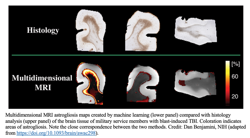
To identify MRI biomarkers for astrogliosis, the researchers compared samples of postmortem brain tissue from seven military service members who had sustained TBI from blast exposure, which results in astrogliosis, and from seven people who had not. The researchers found that astrogliosis induces microscopic changes in the brain tissue that result in a distinct multidimensional MRI pattern. They then developed an approach to compare areas with and without astrogliosis in the same brain, allowing them to specifically map areas of astrogliosis. Their MRI results correlated well with findings from histology images—the current gold standard for detecting astrogliosis in tissue samples.
Their previous work to identify MRI biomarkers of diffuse axonal injury in human brain tissue samples followed a similar approach. Interestingly, they found that astrogliosis and axonal injury have opposite effects on certain parameters of their MRI spectra, suggesting that multidimensional MRI could be used to detect both conditions in the same brain.
If successfully adapted for use in live humans, this multidimensional MRI strategy could be applied to personalized diagnosis and therapy of TBI and other neurological conditions. It also would serve as a useful tool for neuroimaging studies of brain injury, disease, repair, and aging.
Additionally, the method can be used to identify other quantitative MRI biomarkers of injury or tissue damage. “In the past, finding potential MRI biomarkers was a shing expedition,” says NICHD’s Dr Basser. “Now we’re at least fishing with sonar.”
The scientists are now working to adapt their multidimensional MRI method to detect astrogliosis and other neurological diseases and disorders in people. In the future, they aim to conduct a small clinical study to validate the method.
