Dual-phase protocol: a two-for-one deal for brain imaging


A novel nuclear medicine imaging protocol can take the place of two separate imaging scans for the evaluation of brain changes linked to cognitive impairment, cutting costs and radiation exposure for patients as well as providing them greater comfort.

The protocol—known as dual-phase amyloid PET—can assess both amyloid deposition and neurodegeneration with a single tracer injection, assisting physicians as they classify patients on the Alzheimer’s disease continuum. This research was published in the February issue of The Journal of Nuclear Medicine

This graphical Abstract shows the comparability of early-phase amyloid-PET and...

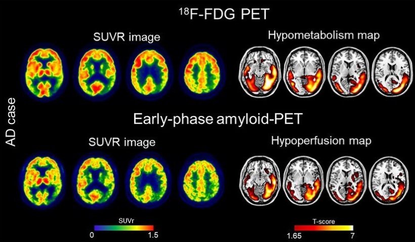
This graphical Abstract shows the comparability of early-phase amyloid-PET and FDG-PET images at the single-subject level.

Image source: SNMMI; Boccalini et al., Journal of Nuclear Medicine 2023

Alzheimer’s disease causes several changes in the brain, including neurodegeneration and beta-amyloid deposition. Currently, 18F-FDG scans are used to measure brain metabolism, which can indicate neurodegeneration. Amyloid PET scans are performed to evaluate beta-amyloid deposition, a key characteristic of Alzheimer’s disease. “During the amyloid PET scans, nuclear medicine physicians wait at least 50 minutes after the radiotracer injection to acquire the image of beta-amyloid deposition. If images are also taken immediately after injection (early-phase), they can provide a measure of brain perfusion. Since brain perfusion and brain metabolism are closely related, the perfusion information could replace an 18F-FDG PET scan,” said Cecilia Boccalini, MSc, PhD candidate at the Laboratory of Neuroimaging and Innovative Molecular Tracers and faculty of medicine at the University of Geneva Neurocenter in Geneva, Switzerland, and faculty of neuroscience at Vita-Salute San Raffaele University in Milan, Italy.

Obtaining both amyloid and perfusion data with a single tracer injection is optimal in terms of patient safety and radiation exposure, comfort, and costsValentina Garibotto

The study aimed to compare early-phase amyloid PET scans and 18F-FDG scans at the individual level, as well as their ability to distinguish patients along the Alzheimer’s disease continuum. A total of 166 patients ranging from cognitively unimpaired to mild cognitive impairment and dementia underwent 18F-FDG and early-phase amyloid PET scans. Brain hypoperfusion maps (from the early-phase amyloid PET) and brain hypometabolism maps (from 18F-FDG PET) were created and compared for each patient. 

Results showed that brain hypoperfusion assessed by early-phase amyloid PET is comparable with brain hypometabolism assessed by 18F-FDG-PET. Hypoperfusion and hypometabolism patterns were also equally able to distinguish patients with neurodegenerative diseases from controls. “This dual-phase amyloid-PET protocol allows physicians to measure brain amyloid and perfusion in a single procedure,” stated Valentina Garibotto, MD, head of the Nuclear Medicine and Molecular Imaging Division at Geneva University Hospitals in Geneva, Switzerland, and associate professor at the University of Geneva. “Obtaining both amyloid and perfusion data with a single tracer injection is optimal in terms of patient safety and radiation exposure, comfort, and costs. Our work supports the routine use of the dual-phase amyloid PET protocol in clinical practice.” 

Source: Society of Nuclear Medicine and Molecular Imaging

Molecular Imaging Flags Risk of AAA Rupture


Uptake of 18F-sodium fluoride (18F-NaF) can point to active vascular calcification associated with high-risk atherosclerotic plaque and may be a marker of high-risk abdominal aortic aneurysms (AAAs), according to a molecular imaging study.

Uptake of the biomarker on positron emission tomography (PET) and CT was significantly higher in the AAA (aortic diameter exceeding 40 mm) than in nonaneurysmal regions of the same aorta in the 20 patients studied. It was also significantly higher than in aortas of 20 controls in the prospective SoFIA3 study from researchers led by Rachael Forsythe, MD, of University of Edinburgh.

In a 72-person longitudinal cohort, the highest tertile of 18F-NaF uptake had aneurysms expand 3.10 mm per year versus 1.24 mm annually for the lowest tertile (P=0.008). The highest tertile also had triple the risk of AAA repair or rupture (15.3% versus 5.6%, log-rank P=0.043).

In this group with a baseline aneurysm diameter of 48.8 mm, 26.4% had their aneurysm repaired and 4.2% had a rupture and died without repair over 1.5 years of follow-up, Forsythe’s group reported in the Feb. 6 issue of the Journal of the American College of Cardiology.

“Fluorine-18-NaF PET-CT is a novel and promising approach to the identification of disease activity in patients with AAA and is an additive predictor of aneurysm growth and future clinical events,” the SoFIA3study authors concluded from their single-center, proof-of-concept study.

“This is the first study to demonstrate that an imaging biomarker of disease activity can add to the risk prediction of AAA and to suggest that this approach might refine clinical decisions regarding the need for surgery and improve patient outcomes,” they said. “We suggest that 18F-NaF uptake again relates to microcalcification and is particular to the most diseased areas associated with tissue disruption and loss of integrity.”

“Importantly, areas of fluoride uptake did not correspond to regions of macrocalcification on CT, suggesting the importance of dynamic calcification process,” noted an accompanying editorial.

In that commentary, Parmanand Singh, MD, of Weill Cornell Medical College, and Jagat Narula, MD, PhD, of Icahn School of Medicine at Mount Sinai, both in New York City, emphasized that “earlier detection of high-risk aneurysms is important to render appropriate care to the highest risk patients.”

“Despite significant advances in aortic imaging, pharmacotherapy and surgical interventions over the past decade, patients with AAA complications continue to have high rates of mortality,” they wrote. “The identification of aortic features linked to aortic vulnerability is crucial, both in guiding selection of patients for preemptive surgical repair and for optimizing timing of intervention to prevent complications. Noninvasive molecular imaging holds promise to identify markers of aortic instability earlier in the course of disease progression, and could offer a major advance in the diagnosis, surveillance and management of AAA.”

Molecular Imaging Tracks Lung Cancer Immunotherapy


Programmed death ligand (PD-L1) expression in tumors may predict response to checkpoint blockade therapy, but tissue samples are not always on hand to guide therapy. Imaging specialists have addressed this issue by developing and evaluating techniques for non-invasive imaging of PD-L1 expression in tumors.

“Non-invasive imaging of therapeutically effective PD-1 [programmed death 1] and PD-L1 antibodies is of high interest for preclinical and potentially also for the clinical development of these drugs, as it provides an elegant opportunity to obtain quantitative and kinetic information on the whole-body biodistribution of these antibodies, including parameters such as tumor accumulation and blood half-life,” explained Gabriele Niedermann, MD, PhD, of the German Cancer Research Center in Heidelburg, and colleagues, writing recently in Theranostics.

Niedermann’s group developed radiotracers, based on therapeutic checkpoint-blocking antibodies, that allowed for high-resolution PET imaging of both PD-1 and PD-L1 in immunocompetent mice. This “immunoPET” of naïve mice showed similar overall expression patterns for PD-1 and PD-L1 in secondary lymphoid organs (spleen and lymph nodes).

The research also found that PD-L1 tracer uptake was reduced in PD-L1 knockout tumors, and that monitoring the expression changes of PD-L1 in response to its main inducer, the effector T cell cytokine IFN-γ, revealed “robust upregulation in the lung.”

“This suggests that T cell responses in the lung, a vital organ continuously exposed to a variety of antigens, are strongly restrained by the PD-1 checkpoint. In turn, this could explain the association of PD-1 checkpoint inhibition with potentially fatal immune-mediated pneumonitis and partially also its efficacy in lung cancer,” Niedermann and colleagues wrote.

In another animal-model study, Samit Chatterjee, PhD, of Johns Hopkins University in Baltimore, and colleagues performed SPECT-CT imaging, biodistribution, and blocking studies in NSG (NOD scid gamma) mice bearing tumors with constitutive PD-L1 expression (CHO-PDL1) and in controls (CHO).

The preclinical evaluation of a humanized radiolabeled anti-PD-L1 antibody showed specific and increased uptake of radioligand in CHO tumors with stable PD-L1 expression compared with control CHO tumors. The results were confirmed in NSCLC xenografts with varying levels of PD-L1 expression, and in triple-negative breast cancer.

“Subcutaneous NSCLC xenografts showed specific uptake in H2444 tumors compared to H1155 tumors,” the researchers wrote.

They explained that the clinical utility of this antibody imaging agent and others would be to use the radiolabeled antibody accumulation in the tumors to guide therapeutic antibody dosing, and correlate that uptake with tumor response.

“This could be used to establish a relationship between tumor PD-L1 status and therapeutic response, which may have prognostic implications.”

Can these results be translated into humans? Yes, according to Jill Fredrickson, PhD, of Genentech in South San Francisco, and colleagues, who utilized FDG-PET-CT imaging to evaluate atezolizumab (Tecentriq) response among chemotherapy-naïve and previously treated stage IIIB/IV non-small cell lung cancer (NSCLC) patients.

Atezolizumab was developed by Roche, the owner of Genentech, and was grantedpriority review by the FDA in April 2016 for the treatment of locally advanced or metastatic NSCLC with PD-L1 expression.

Fredrickson’s group suggested that patients with NSCLC undergoing immunotherapy may benefit from FDG-PET imaging to assess their disease and predict treatment response, presenting the results of their multinational study at the 2016 Society of Nuclear Medicine and Molecular Imaging Annual Meeting.

Atezolizumab inhibits activity between the PD-1 receptor expressed on certain immune cells and PD-L1, leading to enhanced T-cell priming and re-invigoration of suppressed immune cells.

The primary endpoint of the phase II study was objective response rate on the basis of modified immune-related response criteria. Patients underwent single-time-point FDG-PET scanning — 60 minutes of radiotracer uptake prior to imaging — at baseline, at the time of first tumor assessment during week 6 of immunotherapy, and at disease progression.

Fredrickson and colleagues also wanted to determine if the PET modality and its FDG radiotracer could distinguish between radiographic pseudo progression — an increase in apparent tumor burden due to an anti-tumor T-cell response – from true disease progression.

The study enrolled 138 patients at 28 clinical sites in 5 countries, with 103 patients providing evaluable PET scans at baseline and a post-baseline time point. All patients received 1,200 mg of atezolizumab intravenously every 3 weeks. The scans were analyzed using European Organization for Research and Treatment of Cancer (EORTC) criteria.

Patients with metabolic response by EORTC criteria on scans at week 6 had a higher overall response rate (73.9%) than metabolic non-responders did (6.3%). A patient’s whole-body metabolic tumor volume at the FDG-PET baseline scan was found to be a significant negative prognostic maker for overall survival. A further increase of tumor volume at the 6-week scan was also a sign of decreased overall survival.

The researchers noted that response after apparent radiographic progression was seen in only two patients, so the utility of FDG-PET to distinguish pseudo from true progression could not be determined.

“The utility of FDG-PET in patients with NSCLC on immune blockade therapy appeared to be similar to what has been reported with conventional chemotherapeutic treatments, with early metabolic response predicting subsequent benefit,” the team concluded.

“This study is the first to prospectively evaluate FDG-PET imaging in a phase II trial of lung cancer patients receiving the novel immune checkpoint inhibitor atezolizumab,” Fredrickson said in a press statement. “These findings help define the potential role of FDG-PET as a prognostic and predictive biomarker in the treatment of lung cancer with such immunotherapeutics.”

Molecular Imaging of Ovarian Cancer


Ovarian cancer is the most lethal gynecologic malignancy and the fifth leading cause of cancer-related death in women. Over the past decade, medical imaging has played an increasingly valuable role in the diagnosis, staging, and treatment planning of the disease. In this “Focus on Molecular Imaging” review, we seek to provide a brief yet informative survey of the current state of the molecular imaging of ovarian cancer. The article is divided into sections according to modality, covering recent advances in the MR, PET, SPECT, ultrasound, and optical imaging of ovarian cancer. Although primary emphasis is given to clinical studies, preclinical investigations that are particularly innovative and promising are discussed as well. Ultimately, we are hopeful that the combination of technologic innovations, novel imaging probes, and further integration of imaging into clinical protocols will lead to significant improvements in the survival rate for ovarian cancer.

Molecular imaging: Researcher forsees application in myocardial failure.


The landscape of molecular imaging has changed over the past two years, with advances having been made in image-guided drug delivery, gene-based therapy, and cellular therapy. For the latter, cell-labeling methods and image-based magnetic control of cells now enable the monitoring and guidance of therapy. Potential future applications, such as intracellular heating, could place imaging at the very center of therapy delivery.

A panel of experts will show, in a dedicated minicourse today at the European Congress of Radiology (ECR), that molecular imaging can — and soon could — offer improvements in the efficiency of treatment.

2012_02_29_17_52_31_430_p15_Monday_MC23E_Figure8_225 (1)

The recent development of cell-labeling techniques using superparamagnetic nanoparticles has confirmed the role of imaging in monitoring cellular therapies. In particular, researchers have been able to develop high-resolution cellular imaging, using cryoprobes in MR applications. This combination enables high-resolution imaging with a sharp definition of cells in a given organ.

“It’s probably the main discovery we have made over the past two years. By labeling macrophage cells in an ischemic pad of a rat’s foot, we could clearly see the colonized cells and the ischemic territory,” said Professor Olivier Clément, a radiologist working at European Hospital Georges Pompidou in Paris, France.

Projects such as the European Network for Cell Imaging and Tracking (ENCITE), coordinated by the European Institute for Biomedical Imaging and Research and funded by the EU (encite.org), have played a major role in this progress, the researcher underlined.

These techniques have shown no adverse effects on cell proliferation and functionalities while conferring magnetic properties on various cell types. The magnetic labeling of living cells creates opportunities for numerous biomedical applications, such as individual cell manipulation and magnetic control of cell migration.

Clément and his team are currently working on facilitating cell-homing in myocardial infarct cellular therapy, by placing an external magnet over the heart to create a local field gradient which induces magnetic targeting.

“We have been working on a study to improve cell-homing in the heart and succeeded in having more cells when using a magnet. We still haven’t been able to show a therapeutic effect with this technique, but other studies have, and they could show a long-term effect, including scar reduction,” he said.

Intracellular heating will, probably soon, become a field of investigation for many researchers. With this method, one will be able to kill cancerous cells and tumors or liberate drugs contained in a liposome by redirecting the heat, created by the use of magnetic fields, to superparamagnetic nanoparticles.

“Let’s imagine that we have a liposome containing drugs and superparamagnetic nanoparticles. With a magnet, you can drag the liposome exactly where you want it; to a tumor, for instance. Then, you just have to heat your liposome, using external microwaves, after which it will break and allow for chemotherapy. This is still a concept, but it could become a third application in the short term,” Clément said.

The transition of these techniques into clinical practice remains a long-term prospect. Many things are still to be done, including toxicity tests. On the bright side, cellular therapies such as blood transfusion and bone marrow transplant already work without image-based monitoring.

Molecular imaging should, however, prove very useful for tissue transplants and regeneration by showing exactly what happens.

“It is not clear at all whether the injected cells will replace organ function, for instance in myocardial failure. A cell can also trigger signals that help the heart to function better. In this case, imaging helps us understand exactly what happens,” said Clément, who foresees its upcoming clinical application in the heart.

“Imaging can significantly improve the accuracy of treatment,” said Dr. Michal Neeman, a researcher working on the development of reporter genes for MRI at Weizmann Institute in Rehovot, Israel.

Reporter genes encode proteins that can be detected by imaging and serve as a surrogate for the activity of a particular promoter area. For example, reporter genes are used to follow the activation of tumor-associated fibroblasts as they penetrate tumors, and follow the expression of angiogenic and lymphangiogenic growth factors by tumors.

Different reporter genes have been developed to allow detection by different imaging modalities. The most common reporter genes are the fluorescent and bioluminescent proteins that emit light and can be detected using sensitive cameras. Reporter genes are now also available for nuclear imaging and MRI.

“I believe the next steps will be to prove efficacy and safety in specific diseases,” Neeman concluded.

The first success story is the treatment of hemophilia B, as shown by a study recently published in the New England Journal of Medicine by researchers at University College London and St. Jude Children’s Research Hospital in the U.S.

Source: ECR 

Targeted molecular imaging in oncology.


Improvement of scintigraphic tumor imaging is extensively determined by the development of more tumor specific radiopharmaceuticals. Thus, to improve the differential diagnosis, prognosis, planning and monitoring of cancer treatment, several functional pharmaceuticals have been developed. Application of molecular targets for cancer imaging, therapy and prevention using generator-produced isotopes is the major focus of ongoing research projects. Radionuclide imaging modalities (positron emission tomography, PET; single photon emission computed tomography,

SPECT) are diagnostic cross-sectional imaging techniques that map the location and concentration of radionuclide-labeled radiotracers.

99mTc- and 68Galabeled agents using ethylenedicysteine (EC) as a chelator were synthesized and their potential uses to assess tumor targets were evaluated.

99mTc (t1/2 = 6 hr, 140 keV) is used for SPECT and 68Ga (t1/2 = 68 min, 511 keV) for PET. Molecular targets labeled with Tc-99m and Ga-68 can be utilized for prediction of therapeutic response, monitoring tumor response to treatment and differential diagnosis. Molecular targets for oncological research in (1) cell apoptosis, (2) gene and nucleic acid-based approach, (3) angiogenesis (4) tumor hypoxia,and (5) metabolic imaging are discussed. Numerous imaging ligands in these categories have been developed and evaluated in animals and humans. Molecular targets were imaged and their potential to redirect optimal cancer diagnosis and therapeutics were demonstrated.

Source: http://www.jsnm.org