Artificial Intelligence in Medical Imaging—Learning From Past Mistakes in Mammography


Artificial intelligence (AI) in medical imaging shows promise in improving health care efficiency and outcomes. However, automated AI disease detection is not a new concept. Pressure to adopt such emerging technologies is reminiscent of the rapid rise of adjunct computer-aided detection (CAD) tools in mammography 2 decades ago. The CAD cues in mammography mark image features suspicious for breast cancer, while new deep learning AI algorithms similarly mark suspicious image features and provide scores for cancer risk. Despite the intended outcomes of these tools, their use in clinical practice often generates unexpected results. Without a more robust approach to the evaluation and implementation of AI, given the unabated adoption of emergent technology in clinical practice, we have not learned from our past mistakes in the field of mammography.

In 1998, CAD received US Food & Drug Administration (FDA) clearance as an adjunct tool for mammography. Within a few years, the Centers for Medicare & Medicaid Services approved its reimbursement. Nearly a decade after FDA clearance, a seminal article found that CAD did not improve mammography accuracy after dissemination into routine clinical practice.1 Results of initial CAD reader studies were summarized in simple receiver operating characteristic (ROC) curve figures comparing results with CAD vs control. While early reader studies showed improved accuracy with CAD, after FDA clearance and clinical adoption, results were reversed with lower accuracy with CAD.1

By 2016, more than 92% of US imaging facilities used CAD for mammography interpretation despite further research confirming that CAD did not improve radiologist accuracy over 2 decades of use in clinical practice.2,3 The CAD tools are associated with increased false-positive rates, leading to overdiagnosis of ductal carcinoma in situ and unnecessary diagnostic testing. In 2018, Medicare ceased add-on payments for CAD but not before the widespread embrace of CAD had resulted in more than $400 million per year in unnecessary health care expenditures.2 The premature adoption of CAD is consistent with the embrace of emergent technology before its association with patient outcomes is fully understood. As AI algorithms are increasingly receiving FDA clearance and becoming commercially available with ROC curves similar to what we observed prior to CAD clearance and adoption, how can we prevent history from repeating itself?

First, we must remember that there are complex interactions between a computer algorithm output and the interpreting physician. While much research is being done in the development of the AI algorithms and tools, the extent to which physicians may be influenced by the many types and timings of computer cues when interpreting remains unknown. Automation bias, or the tendency of humans to defer to a presumably more accurate computer algorithm, likely affects physician judgment negatively if presented prior to a physician’s independent assessment. In the case of CAD, 2 to 4 markings were shown to radiologists per screening mammogram–as breast cancer is present in about 5 per 1000 screening mammograms, almost all of these markings are false positives. Yet, radiologists do not want to miss cancer, thus leading to higher rates of additional testing, resulting in false-positive results and benign biopsies.4 Before widespread adoption of new AI tools for medical imaging, we need to evaluate the different user interfaces between AI and human interpreters and better understand how and when AI outputs should be presented. Ideally, we need prospective studies incorporating AI into routine clinical workflow.

Second, reimbursement of AI technologies needs to be incumbent on improved patient outcomes, not just improved technical performance in artificial settings. Currently, FDA clearance requires small reader studies and a demonstration of noninferiority to existing technologies (eg, CAD). Newer AI technologies need to demonstrate disease detection that matters. For example, the use of AI in mammography should correspond with increased detection of invasive breast cancers with poor prognostic markers and decreased interval cancer rates. To demonstrate improved patient outcomes, AI technologies need to be evaluated in large population-based, real-world screening settings with longitudinal data collection and linkage to regional cancer registries. If more benefits than harms are identified, then we need to confirm that these results are consistent across diverse populations and settings to ensure health equity. Given the rapid pace of innovation and the many years often needed to adequately study important outcomes, we suggest coverage with evidence development, whereby payment is contingent on evidence generation and outcomes are reviewed on a periodic basis.

Third, we need to embrace revisions to the FDA clearance process for AI algorithms to encourage continued technological improvement. The benefit of deep learning is the ability of machines to continuously improve their algorithms over time. Unfortunately, current FDA review for AI tools in medical imaging is only provided for static unchanging software tools. The consequence is a loss of incentive for continuously improving deep learning algorithms. The FDA is drafting regulatory frameworks for AI-based software as a medical device that pivots from approving static algorithms to oversight over the total product lifecycle including postmarket evaluation.5 The details are yet to be finalized, but will likely require the development of robust data sharing infrastructure necessary for continuous monitoring. One potential avenue for more vigorous, continuous evaluation of AI algorithms is to create prospectively collected imaging data sets that keep up with other temporal trends in medical imaging and are representative of target populations. For instance, the Breast Cancer Surveillance Consortium collects longitudinal data linked to long-term cancer outcomes data permitting prospective image collection, including from the most up-to-date manufacturers and digital breast tomosynthesis (3 dimensional mammograms) technologies. Then, updated algorithms can be independently validated on these representative, population-based imaging examination cohorts continuously.

Fourth, we need to address the impact of AI on medical-legal risk and responsibility in medical imaging interpretation. A great promise of AI is that sophisticated algorithms could eventually interpret images by themselves and free some of physicians’ time to concentrate on more complex tasks. However, truly independent AI is not currently possible because radiologists continue to be the responsible legal parties for accurate imaging interpretation. For example, the recall rate of mammography screening is heavily influenced by medical-legal risk, with missed breast cancer as one of the leading causes of medical malpractice cases in the US.6 Fear of being sued is likely a major reason that American radiologists call back twice as many women after screening mammograms as other countries, even though the cancer detection rate is the same in American and European populations.7,8 Physicians will remain unwilling to bypass inspecting images unless malpractice concerns are addressed. With the Mammography Quality Standards Act now requiring direct patient disclosure of additional risk information (eg, breast density), the appropriate use of supplemental technologies will likely make missed breast cancer even more of a malpractice lawsuit target. One potential solution is to amend the Mammography Quality Standards Act regarding cancer screening liability to better define standards for who can interpret mammograms, how AI can be used for interpretation, and provide guidance on capping limits to AI-related malpractice payouts. Without better guidance on individual party responsibilities for missed cancer, AI creates a new network of who, or what, is legally liable in complex and prolonged, multiparty malpractice lawsuits. Without national legislation addressing the medical and legal aspects of using AI, adoption will slow and we risk opportunities for predatory legal action.

We stand at the precipice of widespread adoption of AI-directed tools in many areas of medicine beyond mammography, and the harms vs benefits hang in the balance for patients and physicians. We need to learn from our past embrace of emerging computer support tools and make conscientious changes to our approaches in reimbursement, regulatory review, malpractice mitigation, and surveillance after FDA clearance. Inaction now risks repeating past mistakes.

Surgeons admit that mammography is outdated and harmful to women


Breast cancer

Every year, millions of women flock to their doctors to get their annual mammograms, a breast cancer screening procedure that involves pressing a woman’s breasts between two metal platforms to scope out tumors. But surgeons everywhere are starting to question the controversial practice, which studies show isn’t even an effective screening tool, and is actually harmful to the bodies of women who receive it.

The public is told that mammograms are the only way to catch breast cancer early, but a review of eight scientific trials evaluating the procedure, found that mammography is neither effective nor safe. After looking at data on more than 600,000 women between the ages of 39 and 74 who underwent the procedure on a routine basis, researchers found that many women are misdiagnosed. Many of these same women are consequently mistreated with chemotherapy, resulting in their rapid demise.

As published in the Cochrane Database of Systematic Reviews, the review concluded that mammography causes more harm than good, because many more women end up being misdiagnosed and mistreated than those actually avoiding the development of terminal breast cancer. Thus, the procedure known as mammography is an outdated scourge that belongs in the history books of failed medical treatments, and not at the forefront of women’s medicine.

“If we assume that screening reduces breast cancer mortality by 15% and that overdiagnosis and overtreatment is at 30%, it means that for every 2000 women invited for screening throughout 10 years, one will avoid dying of breast cancer and 10 healthy women, who would not have been diagnosed if there had not been screening, will be treated unnecessarily,” the authors concluded.

Group of top medical experts admits mammography does more harm than good

One year after this review was published, a second one published in The New England Journal of Medicine (NEJM) came to a similar conclusion. A team of medical professionals that included a medical ethicist, a clinical epidemiologist, a pharmacologist, an oncologic surgeon, a nurse scientist, a lawyer and a health economist, decided that the medical industry’s claims about the benefits of mammography are essentially bunk.

They found that for every 1,000 women screened in the U.S. over a 10-year annual screening period beginning at age 50, one breast cancer death would be prevented, while a shocking 490 to 670 women would have a false positive, while 70 to 100 would undergo an unnecessary biopsy. Between three and 14 of these women, the study found, would also be over-diagnosed for a non-malignant form of cancer that never even would have become “clinically apparent.”

This study out of Switzerland corroborates another out of Canada – the 2014 Canadian National Breast Screening Study – which concluded in lockstep with the others that mammography screenings do not reduce mortality rates from breast cancer any better than a simple physical examination. In other words, the procedure is completely unnecessary, and in many cases exceptionally harmful.

And on and on the list goes, with data out of Norway and elsewhere confirming that mammography isn’t all that it’s cracked up to be. U.S. data spanning the course of nearly 40 years shows that more women are over- or misdiagnosed with breast cancer because of mammograms than are successfully early-diagnosed with breast cancer in such a way as to protect against metastasization. This represents an exceptionally poor track record that calls into question why mammography continues to be used when it clearly doesn’t work.

“I believe that if you did have a tumor, the last thing you would want to do is crush that tumor between two plates, because that would spread it,” says general practitioner Dr. Sarah Mybill, as quoted in the documentary film The Promise.

American Cancer Society Guidelines for Breast Screening with MRI as an Adjunct to Mammography


Abstract

New evidence on breast Magnetic Resonance Imaging (MRI) screening has become available since the American Cancer Society (ACS) last issued guidelines for the early detection of breast cancer in 2003. A guideline panel has reviewed this evidence and developed new recommendations for women at different defined levels of risk. Screening MRI is recommended for women with an approximately 20–25% or greater lifetime risk of breast cancer, including women with a strong family history of breast or ovarian cancer and women who were treated for Hodgkin disease. There are several risk subgroups for which the available data are insufficient to recommend for or against screening, including women with a personal history of breast cancer, carcinoma in situ, atypical hyperplasia, and extremely dense breasts on mammography. Diagnostic uses of MRI were not considered to be within the scope of this review.

INTRODUCTION

Mammography has been proven to detect breast cancer at an early stage and, when followed up with appropriate diagnosis and treatment, to reduce mortality from breast cancer. For women at increased risk of breast cancer, other screening technologies also may contribute to the earlier detection of breast cancer, particularly in women under the age of 40 years for whom mammography is less sensitive. The American Cancer Society (ACS) guideline for the early detection of breast cancer, last updated in 2003, stated that women at increased risk of breast cancer might benefit from additional screening strategies beyond those offered to women at average risk, such as earlier initiation of screening, shorter screening intervals, or the addition of screening modalities (such as breast ultrasound or magnetic resonance imaging [MRI]) other than mammography and physical examination. However, the evidence available at the time was insufficient to justify recommendations for any of these screening approaches. The ACS recommended that decisions about screening options for women at significantly increased risk of breast cancer be based on shared decision making after a review of potential benefits, limitations, and harms of different screening strategies and the degree of uncertainty about each.1

Although there still are limitations in the available evidence, additional published studies have become available since the last update, particularly regarding use of breast MRI. The ACS guideline panel has sought to provide additional guidance to women and their health care providers based on these new data.

GUIDELINE DEVELOPMENT

The ACS convened an expert panel to review the existing early detection guideline for women at increased risk and for MRI screening based on evidence that has accumulated since the last revision in 2002 to 2003. Literature related to breast MRI screening published between September 2002 and July 2006 was identified using MEDLINE (National Library of Medicine), bibliographies of identified articles, and unpublished manuscripts. Expert panel members reviewed and discussed data during a series of conference calls and a working meeting in August, 2006. When evidence was insufficient or lacking, the final recommendations incorporated the expert opinions of the panel members. The ACS Breast Cancer Advisory Group members and the National Board of Directors discussed and voted to approve the recommendations.

SUMMARY OF RECOMMENDATIONS

Table 1 summarizes the ACS recommendations for breast MRI screening.

Table TABLE 1. Recommendations for Breast MRI Screening as an Adjunct to Mammography
  1. * Evidence from nonrandomized screening trials and observational studies.
  2. †Based on evidence of lifetime risk for breast cancer.
  3. †Payment should not be a barrier. Screening decisions should be made on a case-by-case basis, as there may be particular factors to support MRI. More data on these groups is expected to be published soon.
Recommend Annual MRI Screening (Based on Evidence* )
   BRCA mutation
   First-degree relative of BRCA carrier, but untested
   Lifetime risk ∼20–25% or greater, as defined by BRCAPRO or other models that are largely dependent on family history
Recommend Annual MRI Screening (Based on Expert Consensus Opinion†)
   Radiation to chest between age 10 and 30 years
   Li-Fraumeni syndrome and first-degree relatives
   Cowden and Bannayan-Riley-Ruvalcaba syndromes and first-degree relatives
Insufficient Evidence to Recommend for or Against MRI Screening‡
   Lifetime risk 15–20%, as defined by BRCAPRO or other models that are largely dependent on family history
   Lobular carcinoma in situ (LCIS) or atypical lobular hyperplasia (ALH)
   Atypical ductal hyperplasia (ADH)
   Heterogeneously or extremely dense breast on mammography
   Women with a personal history of breast cancer, including ductal carcinoma in situ (DCIS)
Recommend Against MRI Screening (Based on Expert Consensus Opinion)
   Women at <15% lifetime risk

BACKGROUND

MRI

MRI utilizes magnetic fields to produce detailed cross-sectional images of tissue structures, providing very good soft tissue contrast. Contrast between tissues in the breast (fat, glandular tissue, lesions, etc.) depends on the mobility and magnetic environment of the hydrogen atoms in water and fat that contribute to the measured signal that determines the brightness of tissues in the image. In the breast, this results in images showing predominantly parenchyma and fat, and lesions, if they are present. A paramagnetic small molecular gadolinium-based contrast agent is injected intravenously to provide reliable detection of cancers and other lesions. Thus, contrast enhanced MRI has been shown to have a high sensitivity for detecting breast cancer in high-risk asymptomatic and symptomatic women, although reports of specificity have been more variable.2, [3], [4], [5], [6], [7]–8 This high signal from enhancing lesions can be difficult to separate from fat, leading to the use of subtraction images or fat suppression, or both, to assess disease. Because parenchymal tissue also enhances, but generally more slowly than malignant lesions, and also because contrast can wash out rapidly from some tumors, it is important to look at images at an early time point after contrast injection (typically 1 to 3 minutes). MRI examinations may involve examining images at one time point or, more often, will collect a preinjection image with sequential sets of images after contrast injection (dynamic contrast-enhanced [DCE]-MRI). Both the appearance of lesions and, where available, the uptake and washout pattern can be used to identify malignant disease and discriminate it from benign conditions.

These techniques, which have been widely employed for assessing symptomatic disease, have recently been shown to provide good sensitivity as a screening tool for breast cancer in women at increased risk based on family history.9, [10], [11], [12], [13]–14 The approach requires appropriate techniques and equipment, together with experienced staff. Higher quality images are produced by dedicated breast MRI coils, rather than body, chest, or abdominal coils.

IDENTIFICATION OF WOMEN WITH A HIGH RISK OF BREAST CANCER

Three approaches are available for identifying women with a high risk of breast cancer: family history assessment, genetic testing, and review of clinical history. All contribute to identifying women who are candidates for breast MRI screening.

Family History

Although a high proportion of women in the general population have at least one relative with breast cancer, for the majority of these women, this “family history” either does not increase risk at all (ie, the cancer was sporadic) or is associated with, at most, a doubling of lifetime risk (due to either shared environmental risk factors or an inherited gene of low penetrance). Only 1% to 2% of women have a family history suggestive of the inheritance of an autosomal dominant, high-penetrance gene conferring up to an 80% lifetime risk of breast cancer. In some families, there is also a high risk of ovarian cancer. Features of the family history which suggest the cancers may be due to such a high-penetrance gene include 2 or more close (generally first- or second-degree) relatives with breast or ovarian cancer; breast cancer occurring before age 50 years (premenopausal) in a close relative; a family history of both breast and ovarian cancer; one or more relatives with 2 cancers (breast and ovarian cancer or 2 independent breast cancers); and male relatives with breast cancer.15, [16], [17]–18

Two breast/ovarian cancer susceptibility genes, BRCA1 and BRCA2, have been identified.19,20 Inherited mutations in these genes can be found in approximately 50% of families in which an inherited risk is strongly suspected based on the frequency and age of onset of breast cancer cases, and in most families in which there is a much higher than expected incidence of both breast and ovarian cancer.

Several models can assist clinicians to estimate breast cancer risk or the likelihood that a BRCA mutation is present (Online Supplemental Material). The Gail, Claus, and Tyrer-Cusick models estimate breast cancer risk based on family history, sometimes in combination with other risk factors, such as reproductive history or prior breast biopsies.16,21, [22]–23 Although risk prediction is generally similar for the different models, an individual woman’s risk estimate may vary with different models.21,24,25

Two decision models have been developed to estimate the likelihood that a BRCA mutation is present, BRCAPRO18,26 and the Breast and Ovarian Analysis of Disease Incidence and Carrier Estimation Algorithm (BOADICEA)27; the BOADICEA model also provides estimates of breast cancer risk (Online Supplemental Material).

Genetic Testing

The prevalence of BRCA mutations is estimated to be between 1/500 and 1/1,000 in the general population28; however, in women of Jewish ethnicity, the prevalence is 1/50.29,30 Women with cancer-predisposing mutations in either BRCA1 or BRCA2 have an increased risk of both breast and ovarian cancer. From population-based studies, women with BRCA1 mutations are estimated to have a 65% risk by age 70 years for developing breast cancer (95% confidence interval [CI], 44% to 78%); the corresponding risk for BRCA2 mutations is 45% (95% CI, 31% to 56%).31 Risks estimated from cancer-prone families seen in referral centers are higher, with limit of risk in the 85% to 90% range.31 These mutations follow an autosomal dominant pattern of transmission, which means that the sister, mother, or daughter of a woman with a BRCA mutation has a 50% chance of having the same mutation.

The benefits and risks of genetic testing are beyond the scope of this article, but are reviewed in the American Society of Clinical Oncology policy statement update on genetic testing for cancer susceptibility.32 Genetic testing for a BRCA1 or BRCA2 mutation is generally offered to adult members of families with a known BRCA mutation, or to women with at least a 10% likelihood of carrying such a mutation, based on either validated family history criteria or one of the above-mentioned models. If a woman from a family in which a BRCA mutation has been previously identified does not have that mutation, one can generally safely conclude that her breast cancer risk is no higher than it would have been if she did not have a family history of breast cancer. However, in a high-risk family without a known mutation, failure to find a mutation in a particular member does not reduce her risk estimate.

A high risk of breast cancer also occurs with mutations in the TP53 gene (Li-Fraumeni syndrome) and the PTEN gene (Cowden and Bannayan-Riley-Ruvalcaba syndromes).33 Accurate prevalence figures are not available, but these conditions appear to be very rare.34,35

Clinical Indicators of Risk

Some clinical factors are associated with substantial breast cancer risk. Among women with Hodgkin disease, increased breast cancer risk has been consistently and significantly associated with mantle field radiation treatment. In several studies of women treated between 1955 and 1995, risk was inversely related to age at treatment in patients diagnosed between the ages of 10 to 30 years, with only slight or no increased risk when diagnosis was before age 10 years or after age 30 years.36, [37], [38], [39], [40]–41 Risk following treatment with radiation and chemotherapy was half that of treatment with radiation alone in two studies,39,42 which may reflect the effect of chemotherapy on earlier onset of menopause; risk was equivalent in a third study.43 Risk of breast cancer significantly increased 15 to 30 years after radiation therapy.41 More recently, treatment approaches have used lower doses of radiation and limited-field radiotherapy. In one study, which compared patients who received radiation therapy in 1966 to 1974 and 1975 to 1985, treatment in the later timeframe was not related to increased risk of breast cancer after a median follow up of 13 years, whereas patients treated between 1966 and 1974 were at increased risk, suggesting that Hodgkin disease survivors treated with current approaches will not face substantially increased breast cancer risk.44

Lobular carcinoma in situ (LCIS) and atypical lobular hyperplasia (ALH), together described as lobular neoplasia, are associated with substantially increased risk of subsequent breast cancer, with lifetime risk estimates ranging from 10% to 20%.45 This equates to a continuous risk of about 0.5% to 1.0% per year. The invasive cancers may be ipsilateral or contralateral, are usually invasive lobular cancers, and more than 50% of these diagnoses occur more than 15 years after the original diagnosis of LCIS. Similar findings have been reported by Fisher et al,46 describing a 12-year update of 180 women with LCIS who were treated with local excision alone and followed by the National Surgical Adjuvant Breast Project (NSABP), as well as Li et al, who described the risk of invasive breast cancer among 4,490 LCIS patients using Surveillance, Epidemiology, and End Results (SEER) data between 1988 to 2001.47

A typical ductal hyperplasia (ADH) is part of the continuum of ductal proliferative breast diseases ranging from usual ductal hyperplasia to ductal carcinoma in situ (DCIS). The literature review by Arpino et al45 suggests a 4- to 5-fold increased risk of invasive breast cancer (compared with a 6- to 10-fold risk with LCIS) at a median follow up of 17 years, which is doubled if the woman has an associated family history of breast cancer. It is unclear, however, what percentage of the women with this family history and ADH are at this significantly increased risk because they are carriers of a BRCA1 or 2 gene mutation.

Mammographic density has been shown to be a strong independent risk factor for the development of breast cancer.48, [49], [50]–51 In several studies, women with the most breast density were found to have a 4- to 6-fold increased risk of breast cancer, compared with women with the least dense breasts.52, [53], [54], [55]–56 For example, women with 75% or higher mammographic density had a more than five-fold increased risk of breast cancer, compared with women with less than 1% density.57 In addition, it has been shown that malignant tumors of the breast are more likely to arise in the areas of greatest mammographic density, compared with the more fatty areas of the breast.58

The absolute risk of contralateral breast cancer in women with a personal history of breast cancer is estimated to be 0.5% to 1% per year, or 5% to 10% during the 10 years following diagnosis, significantly higher than that of the general population.59 Hormone therapy and/or chemotherapy for the primary cancer is likely to subsequently lower the risk of contralateral breast cancer.

EVIDENCE AND RATIONALE

Evidence of Efficacy from MRI Screening Studies

In the mid to late 1990s, at least 6 prospective, nonrandomized studies were initiated in The Netherlands, the United Kingdom (UK), Canada, Germany, the United States (US), and Italy to determine the benefit of adding annual MRI to (film) mammography for women at increased risk of breast cancer. Some of the studies included ultrasound and/or clinical breast examination, as well. Despite substantial differences in patient population (age, risk, etc.) and MRI technique, all reported significantly higher sensitivity for MRI compared with mammography (or any of the other modalities). All studies that included more than one round of screening reported interval cancer rates below 10%. Participants in each of these 6 studies had either a documented BRCA1 or BRCA2 mutation or a very strong family history of breast cancer. Some of the studies included women with a prior personal history of breast cancer.

Kriege et al screened 1,909 unaffected women aged 25 to 70 years with an estimated 15% or higher lifetime risk of breast cancer (19% proven to have a BRCA mutation) at 6 centers across The Netherlands.9 After a median of 3 rounds of screening, 50 breast cancers (44 invasive) were diagnosed. Eighty percent of the invasive cancers were detected by MRI, compared with 33% by mammography. However, mammography outperformed MRI for detecting DCIS. Of the invasive cancers, 43% were 1 cm or smaller in diameter, and 33% had spread to axillary lymph nodes. The specificity of MRI was 90%, compared with 95% for mammography.

Leach et al screened 649 unaffected women aged 35 to 49 years who had at least a 25% lifetime risk of breast cancer (19% proven to have a BRCA mutation) at 22 centers in the UK.11 After a median of 3 rounds of screening, 35 cancers (29 invasive) were diagnosed. Sensitivity of MRI was 77%, compared with 40% for mammography, with specificities of 81% and 93%, respectively. MRI was most sensitive and mammography least sensitive for women with BRCA1 mutations. Forty-five percent of the cancers were 1 cm or less in size, and 14% had spread to axillary lymph nodes. There were two interval cancers.

Warner et al screened 236 women aged 25 to 65 years with a BRCA mutation at a single center in Toronto for up to 3 years and detected 22 cancers (16 invasive).14 Sensitivity of MRI was 77%, compared with 36% for mammography, with 50% of the cancers 1 cm or smaller, and 13% were node positive. There was one interval cancer. Specificity was 95% for MRI and 99.8% for mammography.

Kuhl et al screened 529 women aged 30 years and older with a lifetime breast cancer risk of at least 20% at a single center in Bonn for a mean of 5 years.10 They detected 43 cancers (34 invasive), with 1 interval cancer. The sensitivity of MRI was 91%, compared with 33% for mammography. The node positive rate was 16%. Specificity of both MRI and mammography was 97%.

The International Breast MRI Consortium screened 390 women aged 25 years and older with more than a 25% lifetime risk of breast cancer at 13 centers (predominantly in the US) on a single occasion.12 Four cancers were found by MRI, and only one of these by mammography. However, because the patients were not followed after screening, the false-negative rate could not be determined. MRI specificity was 95%, compared with 98% for mammography.

In a study in Italy with 9 participating centers, Sardanelli et al screened 278 women aged 25 years and older; 27% carried a BRCA mutation or had a first-degree relative with a BRCA mutation.13 After a median of 1.4 rounds of screening, 18 cancers (14 invasive) were found. MRI sensitivity was 94%, compared with 59% for mammography, 65% for ultrasound, and 50% for clinical breast examination. MRI specificity was 99%.

Overall, studies have found high sensitivity for MRI, ranging from 71% to 100% versus 16% to 40% for mammography in these high-risk populations. Three studies included ultrasound, which had sensitivity similar to mammography. The Canadian, Dutch, and UK studies9,11,14 reported similar sensitivity (71% to 77%) within CIs for MRI, although the single-center study from Germany10 reported a higher sensitivity, which may reflect the concentration of radiological practice and higher patient volume per radiologist at a single center. There is evidence of a learning curve for radiologists conducting MRI breast screening, with the number of lesions investigated falling with experience.60 The three multicenter studies reflect the likely initial effectiveness of this modality in a population context, and it is expected that, with training and advances in technology, sensitivity will increase further.

Table 2 provides a summary of these six screening studies.

Table TABLE 2. Published Breast MRI Screening Study Results
The Netherlands Canada United Kingdom Germany United States Italy
  1. n/a = not applicable.
No. of centers 6 1 22 1 13 9
No. of women 1,909 236 649 529 390 105
Age range 25–70 25–65 35–49 ≥30 ≥25 ≥25
No. of cancers 50 22 35 43 4 8
Sensitivity (%)
   MRI 80 77 77 91 100 100
   Mammogram 33 36 40 33 25 16
   Ultrasound n/a 33 n/a 40 n/a 16
Specificity (%)
   MRI 90 95 81 97 95 99
   Mammogram 95 >99 93 97 98 0
   Ultrasound n/a 96 n/a 91 n/a 0

Most of the available data are based on screening women at high risk due to family history and/or genetic mutations. More recently, smaller studies have provided information on the potential benefit of MRI screening for women with clinical factors that put them at increased risk. Preliminary data were obtained from one retrospective study, in which Port et al61 reviewed the screening results of 252 women with biopsy-confirmed LCIS and 126 women with atypical hyperplasia (either ductal or lobular), of whom half were screened with annual mammography and biennial clinical exams and half were also screened with MRI. The women who were screened with MRI were younger and more likely to have a strong family history. MRI screening offered a small advantage to patients with LCIS, but not atypical hyperplasia, and also resulted in increased biopsies: 6 cancers were detected by MRI in 5 women with LCIS (4% of patients undergoing MRI), and none were detected in women with atypical hyperplasia. Biopsies were recommended for 25% of MRI screened patients; 13% of biopsies had a cancer detected. All of the cancers in women screened with MRI were Stage 0 to I, whereas all of the cancers in women who were not screened with MRI were Stage I to II. Cancer was detected on the first MRI in 4 of 5 patients. The sensitivity of MRI was 75%, the specificity was 92%, and the positive predictive value was 13%.

Technological Limitations and Potential Harms Associated with MRI Screening

Although the efficacy of breast MRI has been demonstrated, it does not achieve perfect sensitivity or specificity in women undergoing screening, and as such, the issue of adverse consequences for women who do, but especially those who do not, have breast cancer is important to address. As with mammography and other screening tests, false negatives after MRI screening can be attributed to inherent technological limitations of MRI, patient characteristics, quality assurance failures, and human error; false positives also can be attributed to these factors, as well as heightened medical-legal concerns over the consequence of missed cancers. A patient’s desire for definitive findings in the presence of a low-suspicion lesion may also contribute to a higher rate of benign biopsies. The consequences of all these factors include missed cancers, with potentially worse prognosis, as well as anxiety and potential harms associated with interventions for benign lesions.

The specificity of MRI is significantly lower than that of mammography in all studies to date, resulting in more recalls and biopsies. Call-back rates for additional imaging ranged from 8% to 17% in the MRI screening studies, and biopsy rates ranged from 3% to 15%.9, [10], [11], [12], [13]–14 However, several researchers have reported that recall rates decreased in subsequent rounds of screening: prevalence screens had the highest false-positive rates, which subsequently dropped to less than 10%.9,62,63 Most call backs can be resolved without biopsy. The call-back and biopsy rates of MRI are higher than for mammography in high-risk populations; while the increased sensitivity of MRI leads to a higher call-back rate, it also leads to a higher number of cancers detected. The proportion of biopsies that are cancerous (positive predictive value) is 20% to 40%.9, [10], [11], [12], [13]–14 Since false-positive results appear to be common, more data are needed on factors associated with lower specificity rates.

Table 3 compares the likelihood of detection and follow-up tests for women who underwent screening MRI and mammography in two screening studies (Dutch and UK). The study populations differed, with the Dutch study having a wider age group and lower risk category, compared with the UK study.9,11 This affected both the prevalence of cancer and the pick-up rate by modality in the two studies. These results, drawn from two trials, demonstrate the relatively high recall rate in the high-risk population, as well as the fact that MRI is a relatively new technique. Despite the high number of recalls, because of the high cancer rate, the rate of benign surgical biopsy in the UK study per cancer detected was similar to that experienced in the population-based national breast screening service. Recalls will inevitably lead to additional investigations, many of which will not demonstrate that cancer is present.

Table TABLE 3. Rates of Detection and Follow-up Tests for Screening MRI Compared with Mammography
MRI Mammography
The Netherlands United Kingdom The Netherlands United Kingdom
Positives 13.7% 19.7% 6.0% 7.2%
Recalls 10.84% 10.7% 5.4% 3.9%
Biopsies 2.93% 3.08% 1.3% 1.33%
Cancers 1.04% 1.44% 0.46% 0.69%
False negatives 0.23% 0.43% 0.81% 1.52%

Given the high rate of cancer combined with the risk of false-positive scans in a high-risk population undergoing MRI-based screening, the psychological health of these women merits study. In a subgroup of 611 women in the UK study, 89% reported that they definitely intended to return for further screening, and only 1% definitely intended not to return. However, 4% found breast MRI “extremely distressing,” and 47% reported still having intrusive thoughts about the examination 6 weeks afterward.64

In a sample of 357 women from the Dutch study, psychological distress remained within normal limits throughout screening for the group as a whole. However, elevated breast cancer-specific distress related to screening was found in excessive (at least once per week) breast self-examiners, risk overestimators, and women closely involved in the breast cancer case of a sister. At least 35% of the total sample belonged to one of these subgroups. It was recommended that patients in one of these vulnerable subgroups be approached for additional psychological support.65

In a small sample of women from the Toronto study followed over a course of 2 years, there was no evidence of any effect on global anxiety, depression, or breast cancer-related anxiety.66 In another sample of 57 women, almost 50% had elevated baseline general and/or breast cancer-specific anxiety, but in 77% of cases this was attributed by the patients to life events, including relatives with cancer. A nonsignificant increase in general anxiety and breast cancer-related anxiety, compared with baseline, was found in the subset of women recalled for further imaging or biopsies.67 Follow-up time is still insufficient to determine whether anxiety scores return to baseline once the work up has been completed.

There is a special responsibility to alert patients to this technology, with its potential strengths and harms, and to be encouraging, while allowing for shared decision making. The interplay between risks, benefits, limitations, and harms is complicated by the fact that individual women likely will weigh these differently depending on their age, values, perception of risk, and their understanding of the issues. Steps should be taken to reduce anxiety associated with screening and the waiting time to diagnosis, and conscientious efforts should be made to inform women about the likelihood of both false-negative and false-positive findings. How information is conveyed to the patient greatly influences the patient’s response: it is important that providers not convey an undue sense of anxiety about a positive MRI finding. While the high rate of biopsies and further investigations is acceptable in women with a high risk of breast cancer, the number of such investigations in women at lower risk will be much higher than would be appropriate, leading to the need to counsel women in lower risk categories that MRI screening is not advisable and that the harms are believed to outweigh the benefits. Such advice needs to be based on considerations of family history, genetic mutation status, other risk factors, age, and mammographic breast density.

There are substantial concerns about costs of and limited access to high-quality MRI breast screening services for women with familial risk. In addition, MRI-guided biopsies are not widely available. With many communities not providing MRI screening and with MRI-guided biopsies not widely available, it is recognized that these recommendations may generate concerns in high-risk women who may have limited access to this technology.

The ability of MRI to detect breast cancer (both invasive and in situ disease) is directly related to high-quality imaging, particularly the signal-to-noise ratio, as well as spatial resolution of the MR image. In order to detect early breast cancer (ie, small invasive cancers, as well as DCIS), simultaneous imaging of both breasts with high spatial resolution is favored. High spatial resolution imaging should be performed with a breast coil on a high field magnet with thin slices and high matrix (approximately 1 mm in-plane resolution). These technical parameters are considered to be the minimal requirements to perform an adequate breast MRI study. The ability to perform MRI-guided biopsy is absolutely essential to offering screening MRI, as many cancers (particularly early cancers) will be identified only on MRI. The American College of Radiology (ACR) is currently developing an accreditation process for performing breast MRI, and, in addition to the performance of high spatial resolution images, the ability to perform MRI intervention (ie, needle localization and/or biopsy) will be essential in order to obtain accreditation by this group. Accreditation will be voluntary and not mandatory. This guideline will likely be available in 2007.

There is a learning curve with respect to interpretation for radiologists. Published trial sites that experience a high volume of cases are experienced, but community practice groups have reported call-back rates over 50% in the majority of the studies that are interpreted. Experience and familiarity with patterns of enhancement, normal and possibly abnormal, are thought to decrease recall rates and increase positive biopsy rates. The ACR accreditation process will stipulate a minimum number of exams that must be read for training purposes and a minimum number for ongoing accreditation. Sites performing breast MRI are encouraged to audit their call-back rates, biopsy rates, and positive biopsy rates.

Cost-effectiveness

Only limited data are available on the cost-effectiveness of breast MRI screening. One recent study modeled cost-effectiveness for adding MRI to mammography screening for women of different age groups who carry a BRCA1 or BRCA2 mutation.68 The authors concluded that the cost per quality-adjusted life year (QALY) saved for annual MRI plus film mammography, compared with annual film mammography alone, varied by age and was more favorable in carriers of a mutation in BRCA1 than BRCA2 because BRCA1 mutations confer higher cancer risk, and higher risk of more aggressive cancers, than BRCA2 mutations.31 Estimated cost per QALY for women aged 35 to 54 years was $55,420 for women with a BRCA1 mutation and $130,695 for women with a BRCA2 mutation. Cost-effectiveness was increased when the sensitivity of mammography was lower, such as in women with very dense breasts on mammography: estimated costs per QALY were $41,183 for women with a BRCA1 mutation and $98,454 for women with a BRCA2 mutation with dense breast tissue. The most important determinants of cost-effectiveness were breast cancer risk, mammography sensitivity, MRI cost, and quality of life gains from MRI.

An evaluation of the cost-effectiveness of the UK study69 has determined that the incremental cost per cancer detected for women at approximately 50% risk of carrying a BRCA gene mutation was $50,911 for MRI combined with mammography over mammography alone. For known mutation carriers, the incremental cost per cancer detected decreased to $27,544 for MRI combined with mammography, compared with mammography alone. Analysis supporting the introduction of targeted MRI screening in the UK for high-risk women70 identified the incremental cost of combined screening per QALY in 40- to 49-year-old women as $14,005 for a BRCA1 carrier with a 31% 10-year riskthe group in which MRI screening is seen to be most effective; $53,320 for women with a 12% 10-year risk; and $96,379 for women with a 6% 10-year risk. For the 30- to 39-year-old age range, the incremental costs per QALY are $24,275 for a BRCA1 carrier with an 11% 10-year risk and $70,054 for a women with a 5% 10-year risk. Based on these estimates, which are based on costs within the UK National Health Service, MRI screening will be offered to women at familial risk aged 30 to 39 years at a 10-year risk greater than 8%, and to women at familial risk aged 40 to 49 years at a 10-year risk greater than 20%, or greater than 12% when mammography has shown a dense breast pattern.

Evidence Supporting Benefit of MRI Screening Among Women in Different Risk Categories

The guideline recommendations were based on consideration of (1) estimates of level of risk for women in various categories and (2) the extent to which risk groups have been included in MRI studies, or to which subgroup-specific evidence is available. Because of the high false-positive rate of MRI screening, and because women at higher risk of breast cancer are much more likely to benefit than women at lower risk, screening should be recommended only to women who have a high prior probability of breast cancer. There is growing evidence that breast cancer in women with specific mutations may have biological and histological features that differ from sporadic cancers. This may result in observed variations in the sensitivity of MRI relative to mammography in detecting cancer in women with a BRCA mutation and those at high familial risk, but without mutations in these genes.11

Women at Increased Risk Based on Family History

The threshold for defining a woman as having significantly elevated risk of breast cancer is based on expert opinion. Any woman with a BRCA1 or BRCA2 mutation should be considered at high risk. The panel has not restricted its recommendations only to women with BRCA mutations because BRCA testing is not always available or informative, and other risk indicators identify additional subsets of women with increased breast cancer risk. If mutation testing is not available, has been done and is noninformative, or if a woman chooses not to undergo testing, pedigree characteristics suggesting high risk may be considered. Very careful family history analysis is required, using tools such as BRCAPRO.18,26 Risk assessment is likely to offer the greatest potential benefit for women under the age of 40 years. Table 4 provides examples of women with a family history indicative of moderate and high risk. The online supplemental material provides guidance for accessing and using risk assessment models.

Table TABLE 4. Breast Cancer Risks for Hypothetical Patients, Based on 3 Risk Models
Family History BRCAPRO*,18 Claus16 Tyrer-Cuzick23
  1. C = breast cancer.
  2. OC = ovarian cancer.
  3. * BRCAPRO (1.4–2) Breast cancer risk calculated to age 85 years.
  4. †Breast cancer risk calculated to age 79 years.
  5. ‡Breast cancer risk calculated for lifetime. Other personal characteristics included in the Tyrer-Cuzick risk model for each case were age at menarche = 12; age at first birth = 28; height = 1.37 meters (5 feet, 4 inches); weight = 61 kg (134 lbs); woman has never used hormone replacement therapy (HRT); no atypical hyperplasia or lobular carcinoma in situ (LCIS).
35-year-old woman
   Mother BC 33
   Maternal aunt BC 42 19% 36% 28%
35-year-old woman
   Paternal aunt BC 29, OC 49
   Paternal grandmother BC 35 23% 24% 32%
35-year-old woman
   Paternal aunt BC 29
   Paternal grandmother BC 35 18% 24% 31%
35-year-old woman
   Mother BC 51
   Maternal aunt BC 60 13% 18% 23%
35-year-old woman of Jewish ancestry
   Mother BC 51
   Maternal aunt BC 60 18% 18% 28%

Women at Increased Risk Based on Clinical Factors

Additional factors that increase the risk of breast cancer, and thus may warrant earlier or more frequent screening, include previous treatment with chest irradiation (eg, for Hodgkin disease), a personal history of LCIS or ADH, mammographically dense breasts, and a personal history of breast cancer, as discussed above. There are little data to assess the benefit of MRI screening in women with these risk factors. Women at increased risk or who are concerned about their risk may find it helpful to have their provider clarify the bases for MRI screening recommendations, as well as areas of uncertainty. For some women, mammography may be as effective as for women at average risk, and MRI screening may have little added benefit. In contrast, mammography is less effective in women with very dense breasts, and MRI screening may offer added benefit.

Women who have received radiation treatment to the chest, such as for Hodgkin disease, compose a well-defined group that is at high risk. Although evidence of the efficacy of MRI screening in this group is lacking, it is expected that MRI screening might offer similar benefit as for women with a strong family history, particularly at younger ages and within 30 years of treatment. Because of the high risk of secondary breast cancer in this group, MRI screening is recommended based on expert consensus opinion.

While lifetime risk of breast cancer for women diagnosed with LCIS may exceed 20%, the risk of invasive breast cancer is continuous and only moderate for risk in the 12 years following local excision.46 Only one MRI screening study has included a select group of women with LCIS,61 which showed a small benefit over mammography alone in detecting cancer. This benefit was not seen in patients with atypical hyperplasia. MRI use should be decided on a case-by-case basis, based on factors such as age, family history, characteristics of the biopsy sample, breast density, and patient preference.

Although there have been several trials reported looking at the accuracy and positive predictive value of MRI and mammography in women with high breast density, all of these trials have been conducted in women with known or highly-suspected malignancies within the breast.71, [72], [73]–74 To this point, there has been no Phase III randomized trial reported that has shown a reduction in either mortality or in the size of diagnosed breast cancer when comparing breast MRI with mammography in women with high mammographic density.

Scant data are available for MRI screening of women with a personal history of breast cancer. In one study, MRI detected more cancers in women who had both a personal history and a family history, compared with women at high risk based on family history alone.75 While women with a previous diagnosis of breast cancer are at increased risk of a second diagnosis, the ACS panel concluded that the estimated absolute lifetime risk of 10% does not justify a recommendation for MRI screening at the present time.

Limitations of Evidence from MRI Studies and Research Needs

Assiduous attempts were made to base recommendations on solid evidence. However, outcome data from screening MRI studies are not sufficient to form a solid basis for many of the recommendations. It was therefore necessary to rely on available inferential evidence and expert opinion to provide the guidance needed for patients and their health care providers.

Although the literature shows very good evidence for greater sensitivity of MRI than mammography and good evidence for a stage shift toward earlier, more favorable tumor stages by MRI in defined groups of women at increased risk, there are still no data on recurrence or survival rates, and therefore, lead-time bias is still a concern. Further, a large randomized, mortality endpoint study is unlikely to take place, and it will be necessary in the foreseeable future to rely on evidence of stage of disease and types of cancers. In the absence of randomized trials, recurrence and survival data will come from observational study designs.

The age at which screening should be initiated for women at high risk is not well established. The argument for early screening is based on the cumulative risk of breast cancer in women with BRCA1 mutations and a strong family history of early breast cancer, which is estimated to be 3% by age 30 years and 19% by age 40 years.76 Population-based data also indicate that risk for early breast cancer is increased by a family history of early breast cancer.16 Based on these observations, some experts have suggested that breast cancer screening begin 5 to 10 years before the earliest previous breast cancer in the family. In 1997, an expert panel suggested that screening be initiated at some time between the ages of 25 and 35 years for women with a BRCA1 or BRCA2 mutation.77 Because these recommendations were based on limited observational data, the decision regarding when to initiate screening should be based on shared decision making, taking into consideration individual circumstances and preferences. No data are available related to the effectiveness of screening women beyond age 69 years with MRI and mammography versus mammography alone; most of the current data are based on screening in younger women, and thus, similar investigations are needed in older age cohorts. For most women at high risk, screening with MRI and mammography should begin at age 30 years and continue for as long as a woman is in good health.1

Most of the available data are based on annual MRI screening; there is a lack of evidence regarding shorter or longer screening intervals. Further, while good data are available for the first screening exam (ie, the “prevalent screen”), considerably less data are available from subsequent screening exams (ie, “incidence screens”), and the available data include relatively short follow-up times. Most studies of annual MRI have shown few interval cancers, certainly fewer than with mammography. Given the probably shorter duration of the detectable preclinical phase, or sojourn time, in women with BRCA mutations, MRI has demonstrated superiority to mammography in this regard. Therefore, to the best of our knowledge, MRI should be performed annually. However, in view of data suggesting that tumor doubling time in women with an inherited risk decreases with age,78 it is conceivable that older women can safely be screened less frequently than younger women. The available evidence is limited, and additional research regarding optimal screening interval by age and risk status is needed.

Some experts recommend staggering MRI screening and mammography screening every 6 months. The potential advantage of this approach is that it may reduce the rate of interval cancers. Other experts recommend MRI and mammography at the same time or within a short time period. This approach allows for the results of both screening tests to be interpreted together and reported to the patient at the same time. All of the clinical trials screened participants with both MRI and mammography at the same time. There is no evidence to support one approach over the other. For the majority of women at high risk, it is critical that MRI screening be provided in addition to, not instead of, mammography, as the sensitivity and cancer yield of MRI and mammography combined is greater than for MRI alone. However, where there is a concern about raised radiation sensitivity, it may be advisable to employ MRI alone despite the overall lower sensitivity.

In order to pursue answers to some of the unresolved questions related to the use of MRI and mammography to screen women at increased risk, it is important to develop creative strategies related to data gathering and study design. Multicenter studies can result in greater efficiency in accumulating sufficiently large enough data sets in this subgroup of women. Conventional study designs with randomization may prove difficult given the potential advantage of adding MRI to mammography in higher-risk groups, and thus, design strategies that utilize surrogate markers and historic controls may prove both more practical and feasible. To move forward, we encourage the development of a simple, common data collection protocol to capture information from the growing number of centers that offer MRI and formal systems to collect outcome data. Because many insurers presently cover MRI screening for high-risk women, it may be economical to do prospective surveillance studies since screening costs are covered by third parties. A common surveillance protocol could permit pooling of data, much like presently is done within the framework of the National Cancer Institute’s Breast Cancer Surveillance Consortium, a collaborative network of seven mammography registries in the United States with linkages to tumor and/or pathology registries that was organized to study the delivery and quality of breast cancer screening and related patient outcomes in the United States.79 We also encourage seeking opportunities for broad international research collaboration on study questions of common interest.

Several further clinical trials of screening women at increased risk of breast cancer are underway, including an international study of MRI and ultrasound in conjunction with the International Breast MRI Consortium and Cancer Genetics Network, and the American College of Radiology Imaging Network (ACRIN) 666 screening trial of mammography compared with ultrasound. An amendment to the ACRIN trial, 6666, will screen patients with one round of MRI.

CONCLUSION

Often no available screening modality is uniquely ideal. For breast MRI, there is an increasing body of observational data showing that screening can identify cancer in patients of specific risk groups, ie, high-risk patients facing a lifetime risk of ∼20–25% or greater related to family history as estimated by one or more of the different risk models. We have specified a range of risk because estimates from the risk models vary and because each of the risk models is imperfect. Furthermore, these models likely will continue to be refined over time; therefore, these risk estimates for different family history profiles are likely to change. Thus, when estimating patient risk it is important to always be certain that the most current model is being used. In addition to family history, clinical factors as described earlier may be a relevant factor in individualized decisions about MRI screening when family history alone does not predict a risk of approximately 20–25%.

Several studies have demonstrated the ability of MRI screening to detect cancer with early-stage tumors that are associated with better outcomes. While survival or mortality data are not available, MRI has higher sensitivity and finds smaller tumors, compared with mammography, and the types of cancers found with MRI are the types that contribute to reduced mortality. It is reasonable to extrapolate that detection of noninvasive (DCIS) and small invasive cancers will lead to mortality benefit.

The guideline recommendations for MRI screening as an adjunct to mammography for women at increased risk of breast cancer take into account the available evidence on efficacy and effectiveness of MRI screening, estimates of level of risk for women in various categories based on both family history and clinical factors, and expert consensus opinion where evidence for certain risk groups is lacking. All of these groups of women should be offered clinical trials of MRI screening, if available. Women should be informed about the benefits, limitations, and potential harms of MRI screening, including the likelihood of false-positive findings. Recommendations are conditional on an acceptable level of quality of MRI screening, which should be performed by experienced providers in facilities that provide MRI-guided biopsy for the follow up of any suspicious results.
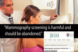
Mammography Is Harmful and Should Be Abandoned, Scientific Review Concludes


“I believe that if screening had been a drug, it would have been withdrawn from the market long ago.” ~ Peter C Gøtzsche (physician, medical researcher and author of Mammography Screening: Truth, Lies and Controversy.)

With Breast Cancer Awareness Month upon us again, a new study promises to undermine the multi-billion dollar cause-marketing campaign that shepherds millions of women in to have their breasts scanned for cancer with x-rays that themselves are known to contribute to breast cancer.

mammography_should_be_abandoned

If you have followed my work for any length of time, you know that I have often reported on the adverse effects of mammography, of which there are many. From the radiobiological and psychological risks of the procedure itself, to the tremendous harms of overdiagnosis and overtreatment, it is becoming clearer every day that those who subject themselves to screening as a “preventive measure” are actually putting themselves directly into harms way, unnecessarily.

Now, a new study conducted by Peter C Gøtzsche, of the Nordic Cochrane Centre, published in the Journal of the Royal Society of Medicine and titled “Mammography screening is harmful and should be abandoned,” strikes to the heart of the matter by showing the actual effect of decades of screening has not been to reduce breast cancer specific mortality, despite the generation of millions of new so-called “early stage” or “stage zero” breast cancer diagnoses.

Previous investigation on the subject by Gotzsche resulted in the discovery that over-diagnosis occurs in a staggering 52% of patients offered organized mammography screening, which equates to “one in three breast cancers being over-diagnosed.” The problem with over-diagnosis is that it almost always goes unrecognized. This then results in over-treatment with aggressive interventions such as lumpectomy, mastectomy, chemotherapy and radiation; over-treatment is a euphemistic term that describes being severely harmed and/or having one’s life shortened by unnecessary medical treatment. Some of these treatments, such as chemotherapy and radiation, can actually enrich cancer stem cells within tumors, essentially altering cells from benign to malignant, or transforming already cancerous cells into far deadlier phenotypes.

Other recent research has determined that the past 30 years of breast cancer screening has lead to the over-diagnosis and over-treatment of about 1.3 million U.S. women, i.e. tumors were detected on screening that would never have led to clinical symptoms, and should never have been termed “cancers” in the first place. Truth be told, the physical and psycho-physical suffering wrought by the harms of breast cancer screening can not even begin to be quantified.

Gøtzsche is very clear about the implications of his review on the decision to undergo mammography. He opines that the effect of screening on mortality, which is the only true measure of whether a medical intervention is worth undertaking, is to increase total mortality.

Mammography Is Harmful and Should Be Abandoned, Review Concludes

Gøtzsche summarizes his findings powerfully:

“Mammography screening has been promoted to the public with three simple promises that all appear to be wrong: It saves lives and breasts by catching the cancers early. Screening does not seem to make the women live longer; it increases mastectomies; and cancers are not caught early, they are caught very late. They are also caught in too great numbers. There is so much overdiagnosis that the best thing a women can do to lower her risk of becoming a breast cancer patient is to avoid going to screening, which will lower her risk by one-third. We have written an information leaflet that exists in 16 languages on cochrane.dk, which we hope will make it easier for a woman to make an informed decision about whether or not to go to screening.

“I believe that if screening had been a drug, it would have been withdrawn from the market long ago. Many drugs are withdrawn although they benefit many patients, when serious harms are reported in rather few patients. The situation with mammography screening is the opposite: Very few, if any, will benefit, whereas many will be harmed. I therefore believe it is appropriate that a nationally appointed body in Switzerland has now recommended that mammography screening should be stopped because it is harmful.”

In the midst of Breast Cancer Awareness Month, a cause marketing orgy bedecked with pink ribbons, and infused with a pinkwashed mentality that has entirely removed the word “carcinogen” (i.e. the cause of cancer) from the discussion. All the better to raise billions more to find the “cure” everyone is told does not yet exist.

Women need to break free from the medical industrial complex’s ironclad hold on their bodies and minds, and take back control of their health through self-education and self-empowerment.

Mammography Is Harmful and Should Be Abandoned, Scientific Review Concludes


“I believe that if screening had been a drug, it would have been withdrawn from the market long ago.” ~ Peter C Gøtzsche (physician, medical researcher and author of Mammography Screening: Truth, Lies and Controversy.)

mammography_should_be_abandoned

With Breast Cancer Awareness Month upon us again, a new study promises to undermine the multi-billion dollar cause-marketing campaign that shepherds millions of women in to have their breasts scanned for cancer with x-rays that themselves are known to contribute to breast cancer.

If you have followed my work for any length of time, you know that I have often reported on the adverse effects of mammography, of which there are many. From the radiobiological and psychological risks of the procedure itself, to the tremendous harms of overdiagnosis and overtreatment, it is becoming clearer every day that those who subject themselves to screening as a “preventive measure” are actually putting themselves directly into harms way, unnecessarily.

 Now, a new study conducted by Peter C Gøtzsche, of the Nordic Cochrane Centre, published in the Journal of the Royal Society of Medicine and titled “Mammography screening is harmful and should be abandoned,” strikes to the heart of the matter by showing the actual effect of decades of screening has not been to reduce breast cancer specific mortality, despite the generation of millions of new so-called “early stage” or “stage zero” breast cancer diagnoses.

Previous investigation on the subject by Gotzsche resulted in the discovery that over-diagnosis occurs in a staggering 52% of patients offered organized mammography screening, which equates to “one in three breast cancers being over-diagnosed.” The problem with over-diagnosis is that it almost always goes unrecognized. This then results in over-treatment with aggressive interventions such as lumpectomy, mastectomy, chemotherapy and radiation; over-treatment is a euphemistic term that describes being severely harmed and/or having one’s life shortened by unnecessary medical treatment. Some of these treatments, such as chemotherapy and radiation, can actually enrich cancer stem cells within tumors, essentially altering cells from benign to malignant, or transforming already cancerous cells into far deadlier phenotypes.

Other recent research has determined that the past 30 years of breast cancer screening has lead to the over-diagnosis and over-treatment of about 1.3 million U.S. women, i.e. tumors were detected on screening that would never have led to clinical symptoms, and should never have been termed “cancers” in the first place. Truth be told, the physical and psycho-physical suffering wrought by the harms of breast cancer screening can not even begin to be quantified.

Gøtzsche is very clear about the implications of his review on the decision to undergo mammography. He opines that the effect of screening on mortality, which is the only true measure of whether a medical intervention is worth undertaking, is to increase total mortality.

Mammography Is Harmful and Should Be Abandoned, Review Concludes

Gøtzsche summarizes his findings powerfully:

“Mammography screening has been promoted to the public with three simple promises that all appear to be wrong: It saves lives and breasts by catching the cancers early. Screening does not seem to make the women live longer; it increases mastectomies; and cancers are not caught early, they are caught very late. They are also caught in too great numbers. There is so much overdiagnosis that the best thing a women can do to lower her risk of becoming a breast cancer patient is to avoid going to screening, which will lower her risk by one-third. We have written an information leaflet that exists in 16 languages on cochrane.dk, which we hope will make it easier for a woman to make an informed decision about whether or not to go to screening.

“I believe that if screening had been a drug, it would have been withdrawn from the market long ago. Many drugs are withdrawn although they benefit many patients, when serious harms are reported in rather few patients. The situation with mammography screening is the opposite: Very few, if any, will benefit, whereas many will be harmed. I therefore believe it is appropriate that a nationally appointed body in Switzerland has now recommended that mammography screening should be stopped because it is harmful.”

In the midst of Breast Cancer Awareness Month, a cause marketing orgy bedecked with pink ribbons, and infused with a pinkwashed mentality that has entirely removed the word “carcinogen” (i.e. the cause of cancer) from the discussion. All the better to raise billions more to find the “cure” everyone is told does not yet exist.

Women need to break free from the medical industrial complex’s ironclad hold on their bodies and minds, and take back control of their health through self-education and self-empowerment.

Mammography Is Harmful and Should Be Abandoned, Scientific Review Concludes


“I believe that if screening had been a drug, it would have been withdrawn from the market long ago.” ~ Peter C Gøtzsche (physician, medical researcher and author of Mammography Screening: Truth, Lies and Controversy.)

With Breast Cancer Awareness Month upon us again, a new study promises to undermine the multi-billion dollar cause-marketing campaign that shepherds millions of women in to have their breasts scanned for cancer with x-rays that themselves are known to contribute to breast cancer.

If you have followed my work for any length of time, you know that I have often reported on the adverse effects of mammography, of which there are many. From the radiobiological and psychological risks of the procedure itself, to the tremendous harms of overdiagnosis and overtreatment, it is becoming clearer every day that those who subject themselves to screening as a “preventive measure” are actually putting themselves directly into harms way, unnecessarily.

Now, a new study conducted by Peter C Gøtzsche, of the Nordic Cochrane Centre, published in the Journal of the Royal Society of Medicine and titled “Mammography screening is harmful and should be abandoned,” strikes to the heart of the matter by showing the actual effect of decades of screening has not been to reduce breast cancer specific mortality, despite the generation of millions of new so-called “early stage” or “stage zero” breast cancer diagnoses.

Previous investigation on the subject by Gotzsche resulted in the discovery that over-diagnosis occurs in a staggering 52% of patients offered organized mammography screening, which equates to “one in three breast cancers being over-diagnosed.” The problem with over-diagnosis is that it almost always goes unrecognized. This then results in over-treatment with aggressive interventions such as lumpectomy, mastectomy, chemotherapy and radiation; over-treatment is a euphemistic term that describes being severely harmed and/or having one’s life shortened by unnecessary medical treatment. Some of these treatments, such as chemotherapy and radiation,can actually enrich cancer stem cells within tumors, essentially altering cells from benign to malignant, or transforming already cancerous cells into far deadlier phenotypes.

Other recent research has determined that the past 30 years of breast cancer screening has lead to the over-diagnosis and over-treatment of about 1.3 million U.S. women, i.e. tumors were detected on screening that would never have led to clinical symptoms, and should never have been termed “cancers” in the first place. Truth be told, the physical and psycho-physical suffering wrought by the harms of breast cancer screening can not even begin to be quantified.

Gøtzsche is very clear about the implications of his review on the decision to undergo mammography. He opines that the effect of screening on mortality, which is the only true measure of whether a medical intervention is worth undertaking, is to increase total mortality.

Mammography Is Harmful and Should Be Abandoned, Review Concludes

Gøtzsche summarizes his findings powerfully:

“Mammography screening has been promoted to the public with three simple promises that all appear to be wrong: It saves lives and breasts by catching the cancers early. Screening does not seem to make the women live longer; it increases mastectomies; and cancers are not caught early, they are caught very late. They are also caught in too great numbers. There is so much overdiagnosis that the best thing a women can do to lower her risk of becoming a breast cancer patient is to avoid going to screening, which will lower her risk by one-third. We have written an information leaflet that exists in 16 languages on cochrane.dk, which we hope will make it easier for a woman to make an informed decision about whether or not to go to screening.

“I believe that if screening had been a drug, it would have been withdrawn from the market long ago. Many drugs are withdrawn although they benefit many patients, when serious harms are reported in rather few patients. The situation with mammography screening is the opposite: Very few, if any, will benefit, whereas many will be harmed. I therefore believe it is appropriate that a nationally appointed body in Switzerland has now recommended that mammography screening should be stopped because it is harmful.”

In the midst of Breast Cancer Awareness Month, a cause marketing orgy bedecked with pink ribbons, and infused with apinkwashed mentality that has entirely removed the word “carcinogen” (i.e. the cause of cancer) from the discussion. All the better to raise billions more to find the “cure” everyone is told does not yet exist.

Women need to break free from the medical industrial complex’s ironclad hold on their bodies and minds, and take back control of their health through self-education and self-empowerment.

When it comes to detection, can ultrasound be mammography’s equal?


A study of more than 2,500 women has found ultrasound just as good as mammography at detecting breast cancer.

On the downside, ultrasound generated more false positives than mammography. However, the radiation-free modality also spotlighted more invasive and node-negative cancers—and the number of women recalled for additional testing post-ultrasound became comparable on incidence-screening rounds.

 - BreastMalig

The study’s authors, led by Wendie Berg, MD, of the University of Pittsburgh, conclude that their results suggest ultrasound could satisfactorily substitute for mammography in developing countries.

In the U.S., they state in their study discussion, ultrasound might supplement standard mammography for women with dense breasts who don’t meet high-risk criteria for screening MRI, as well as for high-risk women with dense breasts who balk at or refuse MRI.

The research team looked at 2,662 participants who were enrolled at 20 sites in the U.S., Canada and Argentina in the American College of Radiology Imaging Network  (ACRIN) 6666  trial and who completed three annual screens (7,473 exams) with ultrasound and film-screen (n = 4,351) or digital (n = 3,122) mammography and had biopsy or 12-month follow-up.

Noting cancer detection, recall and positive predictive values, they found:

  • 110 women had 111 breast cancer events; 89 (80.2 percent) were invasive cancers, median size 12mm.
  • The number of ultrasound screens to detect one cancer was 129, and for mammography the number of needed screens was 127.
  • Cancer detection was comparable for each of ultrasound and mammography at 58 of 111 (52.3 percent) vs. 59 of 111 (53.2 percent), with ultrasound-detected cancers more likely invasive (53 of 58, 91.4 percent, median size 12mm) vs. mammography at 41 of 59 (69.5 percent, median size 13mm).
  • Invasive cancers detected by ultrasound were more frequently node-negative, 34 of 53 (64.2 percent) vs. 18 of 41 (43.9 percent) on mammography.

Meanwhile, in 4,814 incidence screens in years two and three, ultrasound had higher recall and biopsy rates and a lower positive predictive value for biopsy than mammography.

“Training would be necessary for any facility planning to offer screening ultrasound, also true for developing countries,” Berg et al. write in their study discussion. “With appropriate training, ultrasound is no more operator dependent than interpreting mammography.”

The authors add that, while they previously showed in ACRIN 6666 that invasive lobular cancer and low-grade invasive ductal carcinoma are overrepresented among cancers seen only on ultrasound, they did not glean detailed molecular subtype results for the cancers in the present study.

Breast Ultrasound, Mammography May Be Equally Effective


breast exam

Ultrasound and mammography appear equally likely to detect breast cancer, a new study says.

The finding is good news, particularly for women who live in developing countries that typically have more access to ultrasound than to mammography, the researchers said.

While the detection rate with ultrasound was comparable to that of mammography, “it looks like ultrasound does better than mammography for node-negative invasive cancer,” said study leader Dr. Wendie Berg, professor of radiology at Magee-Womens Hospital of UPMC in Pittsburgh. Node-negative invasive cancer is cancer that hasn’t invaded the lymph nodes, but has grown past the initial tumor, according to the U.S. National Cancer Institute.

“The downside [to ultrasound] is, there were more false positives,” Berg said.

At least one expert doesn’t expect this study to change current screening practice in the United States.

“For U.S. patients, what [this study] really confirms is, ultrasound should be used as a supplemental screening exam in dense breast patients,” said Dr. Lusi Tumyan, a radiologist and assistant clinical professor at the City of Hope Cancer Center, in Duarte, Calif. She reviewed the findings but was not involved in the study.

“At this time we do not have enough data to support or refute ultrasound as a screening tool for average-risk patients,” Tumyan said. The take-home message for women in the United States, she added, is to discuss their specific risks with their physician and decide together which screening test is best for them.

The study was published Dec. 28 in the Journal of the National Cancer Institute.

Ultrasound is generally used as a follow-up test once a potential breast tumor has been discovered through a mammogram or a physical exam, according to the American Cancer Society (ACS). The ACS says that ultrasound is a valuable tool that’s widely available and noninvasive.

The new study involved 2,600 women living in the United States, Canada and Argentina who had ultrasound and mammogram annually for three years. They had no symptoms of breast cancer at the study’s start, but they did have dense breast tissue — considered a risk factor for breast cancer — plus at least one other risk factor for breast cancer.

Separate radiologists interpreted each of the two scans the women received.

At the end of the study, 110 women were diagnosed with breast cancer. Detection rates were similar between the two tests. Rates of false-positive results (where a scan erroneously suggests a tumor) were higher for ultrasound compared to mammography, the researchers reported.

Overall, the researchers found that 32 percent of more than 2,500 women without cancer were asked to come back for additional testing at least once after an ultrasound. That compared to 23 percent of women who’d had mammography, the study said.

The findings suggest that for women who don’t have a high risk of breast cancer but have dense breasts, “we find many more cancers if we do ultrasound in addition to mammography,” Berg said.

Berg said the cost of mammography and ultrasound are comparable in the United States. “The issue is: what are the cancers we most need to find,” she said. “The cancers you need to find are the invasive, node-negative ones. More of the cancers found with ultrasound were invasive and node-negative than those found with mammography.”

Guidelines about breast cancer screening vary among organizations. Current ACS guidelines advise women to consider beginning screening at age 40, depending on individual risk factors. They then recommend that women undergo annual screening with mammography from ages 45 to 54. At age 55, the ACS suggests continuing annual screening or switching to screening every two years, depending on risk factors. Some women, due to family history or other risk factors, should also be screened with MRIs, according to the ACS.

Insurance coverage for breast ultrasounds also varies, according to Tumyan.

“Ultrasound coverage varies with different insurance companies and different state laws. California has passed a law that requires radiologists to inform patients if they have dense breasts. But California law does not require insurance companies to pay for supplemental screening. However, in other states, the dense breast law requires insurance companies to pay for supplemental screening,” she explained.

What Now for Mammography?


Now that the ACS has weighed in, how will that change practice?

The American Cancer Society came out with new recommendations for breast cancer screening, calling for annual mammograms beginning at age 45 for average-risk women — even though the U.S. Preventive Services Task Force has said it isn’t justified until age 50, while radiologists’ groups have stood firm on age 40 as the time to start. The recommendations also differ in other respects.

We asked women’s health experts and a variety of healthcare professionals:

What will you tell women in their 40s who ask whether they should get annual mammograms?

How have patients been reacting to the previous controversies around mammography?

How much of a problem are these divergent recommendations, and what should be done about it?

The participants this week are:

Rachel Brem, MD, Vice Chair of Radiology and Director of the Breast Imaging and Intervention Center at the George Washington University School of Medicine and Health Sciences in Washington D.C.

Annina Wilkes, clinical assistant professor at the Sidney Kimmel Medical College at Thomas Jefferson University and Interim Director of Breast Imaging at the Jefferson Honickman Breast Imaging Center in Philadelphia

Kathryn A. Boling, MD, a primary care physician at Lutherville Personal Physicians, part of Mercy Medical Center, in Lutherville, Md.

Karen Oh, MD, associate professor within Diagnostic Radiology and Director of Breast Imaging and sub specialization in Obstetric and Gynecologic ultrasound at Oregon Health & Science University in Portland

Angela DeRosa, DO, MBA, clinical assistant professor at Midwestern University, Arizona College of Osteopathic Medicine, in Glendale and founder, DeRosa Medicine

No Change in Recommendations

Rachel Brem, MD: I will unequivocally recommend that women begin annual screening mammography at age 40 and continue annually thereafter. The new recommendations of the American Cancer Society (ACS) are a compromise between the benefits of screening mammography, lives saved, and the harms, which is the anxiety from mammography callbacks and the possibility of a biopsy for a benign finding. As a physician and a breast cancer survivor myself, the benefits clearly outweigh any “harms,” which are fleeting. My goal is to assure women the right to obtain screening mammograms to optimally diagnose early curable breast cancer. It is also important that women understand that the new ACS recommendations are a compromise. The ACS recommends that women continue to have the opportunity to decide to have screening mammograms beginning at 40 and annually thereafter.

Annika Wilkes, MD: In our practice we will continue to recommend annual mammography beginning at age 40. We will tell our patients that we will continue our recommendation because while it is a good idea to discuss personal risk factors for breast cancer and family history with their doctors, it should not change the interval of screening. Most women who develop breast cancer have no risk factors at all and annual mammography is the best way to catch a cancer when it is at its earliest, most treatable stage. We certainly agree that women should be familiar with the limitations of mammography including the potential need to be called back for additional views, possible short interval follow-up, and biopsy. These so called ” harms” do not occur for most patients. I would doubt that these factors would deter a patient from the possibility of early detection.

Dealing with Different Guidelines

Kathryn Boling, MD: I discuss with patients the fact that there are differing recommendations regarding when to start mammogram screening and how often to have screening. I do discuss with patients the risk of false positive results and attendant testing. I do ask women to have their mammograms done at the same facility as this allows easier comparison with earlier mammograms and may reduce false positive readings. I find in my practice that most women are not aware of the differing recommendations. Virtually all of my patients choose to begin mammograms at age 40. I believe that is a reasonable choice and that women should, at the very least, have that option. I admit, I do have a bias. In my almost 40 years in healthcare, I have seen many women diagnosed with breast cancer in their 40s.

Karen Oh, MD: If they ask me personally, I usually tell them that there is a mortality benefit — meaning mammography does save lives — and the decision to screen or not screen in their 40s is kind of personal, so based on your family history or sometimes your breast density or your desire to start screening versus the risk of false positives. I generally encourage people, personally, to at least consider starting to screen in your 40s just because even though it’s not as frequent in that decade to have breast cancer as in the other decades, the difference it can make for you personally if you catch it early is a big difference. I usually counsel people to think about those issues and then direct them back to their provider to make that decision with them.

Angela DeRosa, DO: I will still recommend women start getting mammo’s at age 40 every other year and then yearly at age 50.The problem is any time in medicine when you go looking for things you often find things. Many women with early breast cancer screening (and increased frequency of mammo/other imaging) DCIS are getting treated very aggressively and active surveillance may be better. But there is a huge divide amongst specialist on this as well. The data and my gut tells me that starting later (45) and doing them less often makes sense, but if any women get breast cancer and I didn’t follow the more aggressive guidelines, I am in the line of fire for lawsuits and board sanctions. Also patient compliance is often a problem…..so I am asking every year, I may be lucky to get them to go every 2 years. That is the practicality.

Potential Patient Confusion

Oh: We actually polled some of our patients about that because you don’t need an order from a provider to get screening, so they’re making their own decisions and a lot of people do look on the Internet. But a lot of people still ask their providers, and if they still have questions, it tends to be when they come in and we see them for something then they ask us as well in Radiology in the Breast Center. So I think there are a lot of questions about when to start and I generally guide them back to “Do you want to screen and have the possibility that you will have a false positive” (couched in different terms, but that’s basically the issue) versus “You will know a little bit earlier, and there is a benefit to knowing earlier,” especially in the younger age group.

Brem: Women are confused. They are unclear what the recommendations actually are and simply cannot understand why three organizations recommend different screening schedules. We need to educate women and we need to partner with the media to reassure women. Mammography saves lives, more lives if screening begins at 40 and continues annually thereafter. However, there is a price which is callbacks and the possibility of benign biopsy. However, it should be the woman’s choice to decide whether the benefits outweigh the “harms” for her.

DeRosa: They are confused and not sure what to think. They look to me for guidance. Many women are terrified (because we have made them that way) about getting DCIS or cancer and want to be aggressive in screening. However, I am seeing a surge of women who believe that the radiation is more problematic.

Recommendations Always Changing

Boling: I believe changing guidelines are more of a problem for physicians, other providers of women’s healthcare, and the medical system in general. When you care for patients, and you are constantly changing your recommendations, I think it erodes trust in the physician-patient relationship. Also, if you follow the “begin at 45” guideline and a late stage breast cancer is diagnosed at a future date, it becomes harder to defend your decision when there are conflicting recommendations. Then there is the question of how insurers will respond to differing recommendations. And of course, it is confusing to patients when they read all these different recommendations.

DeRosa: There are divergent recommendations/clinical guidelines in many areas and this is no different. It has become a tyranny. Many insurance organizations are using these tonot pay for services. Also clinical guidelines are often different depending on the specialty focus. Also they tend to be 5-10 years behind emerging research. So those on the forefront may be punished or considered heretics for practicing outside of these guidelines….even if they are leaders and forging new exciting paths. Anyone can follow guidelines……medicine is an art form and the individual patient needs to be considered.

Wilkes: I strongly disagree with the recommendation against clinical breast examination in average risk women. There has been an increase in breast cancer in young women, younger than age 40. For those women, breast examination, whether it is self breast exam or an exam by a clinician is the only way for cancer to be detected. This is also true for women above age 40 with dense breasts. Recommending breast examination, with familiarity of it’s benefits and limitations should continue.

Mammography Is Harmful and Should Be Abandoned, Scientific Review Concludes.


“I believe that if screening had been a drug, it would have been withdrawn from the market long ago.” ~ Peter C Gøtzsche (physician, medical researcher and author of Mammography Screening: Truth, Lies and Controversy.)

With Breast Cancer Awareness Month upon us again, a new study promises to undermine the multi-billion dollar cause-marketing campaign that shepherds millions of women in to have their breasts scanned for cancer with x-rays that themselves are known to contribute to breast cancer.

If you have followed my work for any length of time, you know that I have often reported on theadverse effects of mammography, of which there are many. From the radiobiological and psychological risks of the procedure itself, to the tremendous harms of overdiagnosis and overtreatment, it is becoming clearer every day that those who subject themselves to screening as a “preventive measure” are actually putting themselves directly into harms way, unnecessarily.

Now, a new study conducted by Peter C Gøtzsche, of the Nordic Cochrane Centre, published in the Journal of the Royal Society of Medicine and titled “Mammography screening is harmful and should be abandoned,” strikes to the heart of the matter by showing the actual effect of decades of screening has not been to reduce breast cancer specific mortality, despite the generation of millions of new so-called “early stage” or “stage zero” breast cancer diagnoses.

Previous investigation on the subject by Gotzsche resulted in the discovery that over-diagnosis occurs in a staggering 52% of patients offered organized mammography screening, which equates to “one in three breast cancers being over-diagnosed.” The problem with over-diagnosis is that it almost always goes unrecognized. This then results in over-treatment with aggressive interventions such as lumpectomy, mastectomy, chemotherapy and radiation; over-treatment is a euphemistic term that describes being severely harmed and/or having one’s life shortened by unnecessary medical treatment. Some of these treatments, such as chemotherapy and radiation, can actually enrich cancer stem cells within tumors, essentially altering cells from benign to malignant, or transforming already cancerous cells into far deadlier phenotypes.

Other recent research has determined that the past 30 years of breast cancer screening has lead to the over-diagnosis and over-treatment of about 1.3 million U.S. women, i.e. tumors were detected on screening that would never have led to clinical symptoms, and should never have been termed “cancers” in the first place. Truth be told, the physical and psycho-physical suffering wrought by the harms of breast cancer screening can not even begin to be quantified.

Gøtzsche is very clear about the implications of his review on the decision to undergo mammography. He opines that the effect of screening on mortality, which is the only true measure of whether a medical intervention is worth undertaking, is to increase total mortality.

Mammography Is Harmful and Should Be Abandoned, Review Concludes

Gøtzsche summarizes his findings powerfully:

“Mammography screening has been promoted to the public with three simple promises that all appear to be wrong: It saves lives and breasts by catching the cancers early. Screening does not seem to make the women live longer; it increases mastectomies; and cancers are not caught early, they are caught very late. They are also caught in too great numbers. There is so much overdiagnosis that the best thing a women can do to lower her risk of becoming a breast cancer patient is to avoid going to screening, which will lower her risk by one-third. We have written an information leaflet that exists in 16 languages on cochrane.dk, which we hope will make it easier for a woman to make an informed decision about whether or not to go to screening.

“I believe that if screening had been a drug, it would have been withdrawn from the market long ago. Many drugs are withdrawn although they benefit many patients, when serious harms are reported in rather few patients. The situation with mammography screening is the opposite: Very few, if any, will benefit, whereas many will be harmed. I therefore believe it is appropriate that a nationally appointed body in Switzerland has now recommended that mammography screening should be stopped because it is harmful.”

In the midst of Breast Cancer Awareness Month, a cause marketing orgy bedecked with pink ribbons, and infused with a pinkwashed mentality that has entirely removed the word “carcinogen” (i.e. the cause of cancer) from the discussion. All the better to raise billions more to find the “cure” everyone is told does not yet exist.

Women need to break free from the medical industrial complex’s ironclad hold on their bodies and minds, and take back control of their health through self-education and self-empowerment.