Lung-Sparing Surgery Is Effective for Some with Early-Stage Lung Cancer


When lung cancer is found early, before it has spread beyond the lungs, people usually have surgery to remove the tumor. For more than 25 years, the standard of surgical care for such patients—even those with very small tumors—has been to remove the entire large section, or lobe, of the lung that contains the tumor. This is done to reduce the chances of the cancer coming back.

A surgical team of 2 men and 2 women standing over a patient on a table in a surgery suite.
For certain people with lung cancer, a less extensive surgery to remove the tumor in their lung is an effective treatment option, according to a new study.

But some lung surgeons have suggested that certain patients may do just as well or even better with an operation to remove only part of the affected lobe.

Now, results of a large international clinical trial show that, for certain people with early-stage non-small cell lung cancer (NSCLC), surgery to remove a piece of the affected lobe is as effective as surgery to remove the whole lobe.

In the trial, people with early-stage NSCLC who had the more limited surgery lived as long without their cancer coming back as those who had an entire lobe of the lung removed. In addition, the portion of people still living after 5 years was about 80% in both groups.

Results of the new study were reported February 9 in the New England Journal of Medicine.

The new findings signal “a tremendous change in how we approach this type of lung cancer,” said Nasser Altorki, M.D., chief of thoracic surgery at Weill Cornell Medicine and New York-Presbyterian Hospital, who led the trial.

“This groundbreaking study confirms the results of a similar large study from Japan” reported last year, said David Tom Cooke, M.D., chief of general thoracic surgery at UC Davis Health, who enrolled patients in the international study and performed surgeries on some participants.

Together, the two trials show that carefully selected patients “can benefit from having less of their lung tissue removed to give them the same chance of a cure,” Dr. Cooke said. And removing less lung tissue “might mean better lung function long term.”

Standards for lung cancer surgery have changed over time

Each lung is made up of large sections called lobes, with three lobes in the right lung and two in the left. Each lobe is further divided into several anatomical units called segments.

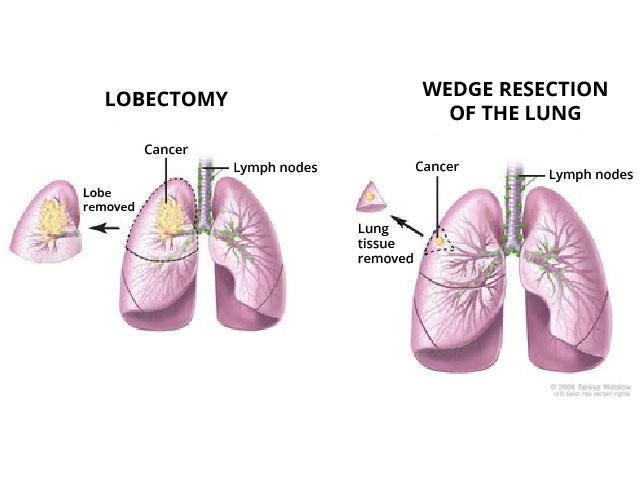
Surgery for lung cancer originally involved removing an entire lung. Removing a whole lobe is called a lobectomy. In sublobar surgery, sometimes called lung-sparing surgery, surgeons may remove an entire segment of a lobe, which is akin to removing a segment of an orange. Or they may remove only a wedge-shaped piece of the lung, which is like taking a bite out of the orange, Dr. Cooke explained.

Side by side illustration of a lobectomy and a wedge resection.
With a lobectomy (left), the entire lobe of the lung where the tumor developed is removed. In a wedge resection (right), only a smaller piece with the tumor is removed. 

In 1995, results from a key clinical trial showed that, for people with early-stage NSCLC, removing a lobe was better than removing part of a lobe. Trial participants who had only part of the affected lobe removed were three times more likely than those who had a lobectomy have their cancer come back (recur).

“Thereafter, lobectomy was the standard of surgical care for this group of patients,” explained thoracic surgeon Valerie Rusch, M.D., of Memorial Sloan Kettering Cancer Center, in an editorial accompanying the new study results. Lung-sparing surgery, Dr. Rusch noted, was used only for patients with limited lung function.

However, in recent decades, advances in imaging technology and new staging methods have enabled the detection and diagnosis of smaller, earlier-stage lung cancers, Dr. Altorki said. So, his team and others wanted to revisit the question of whether lung-sparing surgery may be a good option for some patients. 

Surgeons had a choice of lung-sparing surgery type

For the trial—which was conducted by the NCI-supported Alliance for Clinical Trials in Oncology—Dr. Altorki and his colleagues enrolled 697 people with early-stage NSCLC from June 2007 to March 2017.

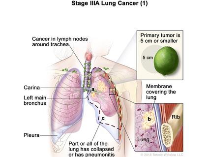
To participate in the trial, people had to meet stringent criteria. They had to have a single lung tumor that was 2 cm or less in size (about the diameter of a US penny), was in the outer third of the lung, and had not spread to nearby lymph nodes or elsewhere in the body. Participants also could not have been previously treated with chemotherapy or radiation therapy for the cancer.

Half of the participants were randomly assigned to have the entire lobe containing the tumor removed, and half were assigned to have lung-sparing surgery. For patients receiving lung-sparing surgery, each surgeon could decide whether to remove a segment of the lobe or only a wedge-shaped piece.

All participants were followed for at least 5 years, and the median follow-up time was 7 years. After 5 years, 63.6% of patients who underwent lung-sparing surgery and 64.1% who underwent lobectomy had not had their cancer come back. The 5-year overall survival in the two groups was 80.3% and 78.9%, respectively.

Similar but not identical studies

In the clinical trial from Japan, 5-year overall survival in both the lung-sparing surgery and lobectomy groups was more than 90%. The higher overall survival seen in that trial may be due to subtle differences between the two studies, such as the specific types of NSCLC that patients had, Dr. Rusch wrote.

Also noteworthy, in the Japanese trial all lung-sparing surgeries were done the same way, by removing a whole segment of the lung. Some surgeons think that surgery to take out a segment of the lobe, known as segmentectomy, “is a better operation” than removing a wedge-shaped piece, Dr. Cooke said, because segmentectomy also removes the associated lymph nodes in that portion of lung. 

However, at least for patients with limited lung function, wedge surgery “is the most frequently practiced method of sublobar surgery in North America and Europe,” Dr. Altorki and his colleagues wrote. In their trial, which included sites in the United States, Canada, and Australia, about 60% of patients assigned to lung-sparing surgery underwent wedge surgery, making the trial “more representative of a ‘real-world’ setting,” they wrote.

One possible concern about the trial, Dr. Cooke said, is that it did not compare the two types of lung-sparing surgery directly. However, Dr. Altorki said, his team will soon have data to address this question.

Benefits of having less lung tissue removed

“Now that lung cancer screening for people at high risk is generally accepted, and as more people get screened, we expect that there will be more people who have these types of very early–stage tumors and who would benefit from sublobar surgery,” Dr. Altorki said.

“Although all these operations are now safe, lung-sparing surgery is associated with an even lower risk [of serious complications, including death], than lobectomy,” Dr. Rusch wrote.

Perhaps most important, removing less lung tissue is believed to preserve lung function. However, neither trial found clinically meaningful differences in lung function between people who had a lobectomy and those who had lung-sparing surgery.

FDA Immunotherapy Approval Marks a First for Lung Cancer Atezolizumab is approved to treat some people with non-small cell lung cancer after surgery.

That could be because the studies assessed lung function using instruments that measure the amount of air entering and leaving a person’s lungs, Dr. Cooke said. Neither trial included other, more sensitive measures of lung function or used standard tests to assess people’s aerobic fitness. 

In addition, he noted, investigators did not ask patients to report how they felt after undergoing the two types of lung surgery.

Removing only a segment or a wedge offers another advantage for patients, Dr. Altorki said. People who are cured of their first non-small cell lung cancer are often at high risk of developing a second primary non-small cell lung cancer. And if a second lung cancer is found, people who initially had less lung tissue removed have more treatment options than those who had an entire lobe removed.

The era of precision lung cancer surgery

Despite these benefits, Dr. Altorki stressed, lung-sparing surgery won’t be the best option for everyone. “We need to do whatever is necessary to get a cure, or to get rid of the cancer and reduce the chance of recurrence as much as possible,” he said.

“When appropriate, lobectomy should still be performed,” Dr. Rusch wrote. “The era of ‘precision’ surgery for non-small cell lung cancer has arrived.”

Dr. Cooke compared the changing views on surgery for lung cancer to the evolution of surgical approaches for treating breast cancer. Over the years, breast surgeons have moved from doing radical mastectomies to lumpectomy or other breast-conserving surgery for some people with early-stage breast cancer.

When it comes to treating lung cancer, Dr. Cooke said, “We can’t stop here. The question now is, where does sublobar surgery fit in when we talk about precision medicine” for lung cancer? That could include treating people with immunotherapy or drugs that target specific genetic changes in a tumor before or after surgery, he said.

“This is an exciting time in the surgical care of patients with lung cancer, and it will be exciting to see where we go next,” Dr. Cooke added.

Lung Cancer Growth Suppressed by Anticancer and Antifibrotic Drug Combo


Using a new 3D cell culture system, researchers at Academia Sinica, National Yang Ming Chiao Tung University, National Taiwan University Hospital, and National Taiwan University, have shown how blocking the activity of fibroblasts can help to treat lung cancer. The team’s studies found that efficacy of the anticancer drug cisplatin was increased when treatment was combined with the antifibrotic drug nintedanib. Addition of nintedanib improved cisplatin’s effects on suppressing the growth of cancer-cell spheroids and the invasion of cancer cells. Analyses indicated that nintedanib therapy was linked to altered expression of fibroblast genes associated with cell adhesion, invasion, and ECM degradation.

“Our results suggest that the combination of nintedanib and cisplatin could be an effective treatment strategy for lung cancer by targeting both cancer cells and cancer-associated fibroblast activation surrounding the tumor,” said research lead Chau-Hwang Lee, PhD. at Academia Sinica, and National Yang Ming Chaio Tung University. The authors reported on their findings in APL Bioengineering, in a paper titledA 3D culture system for evaluating the combined effects of cisplatin and anti-fibrotic drugs on the growth and invasion of lung cancer cells co-cultured with fibroblasts.”

Fibroblasts build and maintain the extracellular matrix (ECM), or physical scaffolding for cells, in the connective tissues within the body. It is believed that cancerous tumors can recruit nearby fibroblasts and use them to promote their own growth and invasion. This process, called cancer-associated fibroblast (CAF) activation, can also protect tumors from chemotherapy and make treatment difficult. “The extracellular matrix (ECM) surrounding a solid tumor plays important roles in tumor progression and therapeutic efficacy,” the authors wrote. “In the tumor microenvironment (TME), cancer-associated fibroblasts (CAFs) facilitate the production of various cytokines in favor of cancer cell proliferation and metastasis. CAFs also alter the ECM’s properties and affect tumor progression and therapy resistance.”

Lee added, “Nearly 90% of late-stage lung cancer patient deaths are caused by the spread of tumors to other organs, rather than the primary tumor. Therefore, it is crucial to find ways to inhibit lung cancer metastasis to prolong the lives of lung cancer patients.”

Assessing the effects of changes to the ECM modulations on anticancer drug effects in vivo is challenging, however, the team noted. “Therefore, an in vitro model with a satisfactory predictive capacity is highly desirable for evaluating the efficacy of antitumor therapies.”

To simulate the tumor microenvironment and mimic real tissues, the team co-cultured lung cancer cells and fibroblasts in a 3D matrix comprised of Matrigel and fibrin, which simulated the tissue microenvironment around a solid tumor. In this 3D environment the cancer cells formed spheroids, around which the fibroblasts were randomly distributed.

The researchers tested the effects on the cancer cells of treatment using cisplatin (CDDP) with and without two anti-fibrotic drugs, nintedanib (NTD) and pirfenidone (PFD). Measuring indicators of tumor growth and invasiveness, they found that nintedanib improved the anti-cancer efficacy of cisplatin. Pirfenidone did not show a similar effect.

“Our results suggest that the combination of nintedanib and cisplatin could be an effective treatment strategy for lung cancer by targeting both cancer cells and cancer-associated fibroblast activation surrounding the tumor,” said Lee.

The studies found that the antifibrotic drug changed the expression of certain fibroblast genes. “We identified the expression of four genes relevant to cell adhesion, invasion, or ECM degradation that were reduced by NTD but increased or unaffected by PFD,” the investigators wrote. And while further studies will be needed to look more closely at the signalling pathways of proteins encoded by the genes under NTD or PFD treatment, the scientists noted, “RNA sequencing data suggest that the effects of NTD on CAFs could be more suppressive than PFD in terms of cell adhesion, invasion, or ECM degradation.”

Clinical and preclinical in vivo studies have previously indicated that NTD can improve the effects of certain anticancer drugs against lung cancers, the authors further noted. “The results of these clinical trials and the in vivo test are in agreement with that of our 3D co-culture experiments, supporting that NTD could improve the efficacy of anticancer drugs.”

The researchers believe that their 3D cell culture system will represent a valuable tool for assessing the efficacy of other drug combinations on tumor growth and invasion, and could feasibly eliminate the need for animal testing, while still reliably evaluating drug efficacy and safety. “This study could pave the way for the development of more effective treatment strategies for lung cancer and other solid tumors,” said Lee. “It is our hope that this study will introduce a new, promising tool for preclinical drug testing.”

The authors plan to study other types of cancers, such as liver and oral cancer, using the same system. “This fibrin/Matrigel co-culture system could be useful for evaluating the effects of drug combinations on various types of tumors related to tissue fibrosis, such as lung cancer, liver cancer, and oral cancer,” they concluded. “Further studies on drug-combination evaluations with patient-derived tissues cultured in fibrin/Matrigel are required.” The team also hope to improve the culture to better mimic the tumor microenvironment.

Lung Cancer Risks: Myths and Facts


Myth: It’s Too Late if You've Smoked for Years

Myth: It’s Too Late if You’ve Smoked for Years

1/10

Fact:  Quitting has almost-immediate benefits. Your circulation will improve and your lungs will work better. Your lung cancer risk will start to drop over time. Ten years after you kick the habit, your risk of dying from the disease drops by 50% compared to people who continue to smoke.

Myth: Low-Tar or 'Light' Cigarettes Are Safer Than Regular

Myth: Low-Tar or ‘Light’ Cigarettes Are Safer Than Regular

2/10

Fact: They’re just as risky. And beware of menthol: Some research suggests that menthol cigarettes may be more dangerous and harder to quit. Their cooling sensation prompts some people to inhale more deeply.

Myth: It’s OK to Smoke Pot

Myth: It’s OK to Smoke Pot

3/10

Fact: Marijuana smoking may raise your lung cancer risk. Many people who use pot also smoke cigarettes. Some research shows that people who do both could be even more likely to get lung cancer.  

Myth: Antioxidant Supplements Protect You

Myth: Antioxidant Supplements Protect You

4/10

Fact: When researchers tested these products, they unexpectedly found a higher risk of lung cancer among smokers who took beta-carotene. Talk to your doctor first. It’s OK to get antioxidants from fruits and vegetables.

Myth: Pipes and Cigars Aren’t a Problem

Myth: Pipes and Cigars Aren’t a Problem

5/10

Fact: Just like cigarettes, they’ll put you at risk for cancers of the mouth, throat, esophagus, and lungs. Cigar smoking, in particular, makes you much more likely to get heart disease and lung disease.

Myth: Smoking Is the Only Risk

Myth: Smoking Is the Only Risk

6/10

Fact: It’s the biggest one, but there are others. The No. 2 cause of lung cancer is an odorless radioactive gas called radon. Given off by rock and soil, it can seep up into homes and other buildings. You can test your house or office for it. Call your state or county health department for information.

Myth: Talcum Powder Is a Cause

Myth: Talcum Powder Is a Cause

7/10

Fact: Research shows no clear link between lung cancer and accidentally breathing in talcum powder. People who work with other chemicals, including asbestos and vinyl chloride, are more likely to get the disease.

Myth: If You Have Lung Cancer, Quitting Is Pointless

Myth: If You Have Lung Cancer, Quitting Is Pointless

8/10

Fact: If you stop, your treatment may work better and your side effects could be milder. And if you need surgery, ex-smokers tend to heal better than smokers. If you need radiation for cancer of the larynx, you’re less likely to become hoarse if you don’t light up. And in some cases, quitting makes a second cancer less likely to start.

Myth: Exercise Doesn't Affect Your Risk

Myth: Exercise Doesn’t Affect Your Risk

9/10

Fact: People who get regular physical activity may be less likely to get lung cancer, studies show. Working out also helps your lungs work better and helps prevent heart disease, strokes, and many other serious conditions.

Myth: Air Pollution Isn’t a Cause

Myth: Air Pollution Isn’t a Cause

10/10

Fact: Tobacco is by far the biggest threat, but air pollution is a risk factor, too. People who live in areas with a lot of it are more likely to get lung cancer than those who live where the air is cleaner. Many U.S. cities have cut down on air pollution in recent years, but there are still dangerous levels in other parts of the world.

Lung cancer: New drug combination reduces tumors in animal study


Image credt: Bjarte Rettedal/Getty Images.

  • Lung cancer is the second most commonTrusted Source form of cancer worldwide.
  • Each year, around 2 million people receive a lung cancer diagnosis, and 1.8 million people die of the disease.
  • There are two types of lung cancer — small cell lung cancer (SCLC) and non-small cell lung cancer (NSCLC) — with NSCLC causing the majority of cases.
  • NSCLC can be treated with surgery, radiation, and chemotherapy, but is rarely cured.
  • Now, research has found that, in mice with NSCLC, a combination of two drugs reduces tumor size and number, a finding that may lead to clinical trials in people.

According to the American Cancer SocietyTrusted Source, lung cancer is the second most common cancer diagnosed in the United States.

It is also the leading cause of cancer deaths, accounting for one in five deaths from cancer‚ more than colon, breast, and prostate cancers combined.

Most lung cancer cases are diagnosed in older people, with the average age of diagnosis being 70 years. The good news is that cases are falling as fewer people are smoking tobacco, which causes more than 80%Trusted Source of lung cancers.

However, some 2 million peopleTrusted Source around the world still receive a lung cancer diagnosis each year.

About 85%Trusted Source of cases are non-small cell lung cancers (NSCLCs). As chemotherapy and immunotherapy are not particularly effective against this type of lung cancer, new treatments are urgently required.

Now, a team from the Salk Institute and Northwestern University has found that a combination of two drugs, one already licensed by the Food and Drug Administration (FDA), and the other currently in clinical trials, reduced tumor size and number in mice with NSCLC.

The research is published in Science Advances.

Need for new treatments

“This treatment would be effective for patients with KRAS/LKB1 mutant lung adenocarcinomaTrusted Source,” said Dr. Lillian Eichner, a lead author on the study, and assistant professor of biochemistry and molecular genetics at Northwestern University.

“Each year in the U.S., there are [about] 20,000 new cases of this molecular subtype of the disease,” she noted. “Current survival time is on average 10 months from initial diagnosis for patients with this devastating disease, and improved therapeutic approaches are sorely needed.”

The researchers identified histone deacetylase (HDAC) inhibitors as a potential treatment for this type of lung cancer. HDAC inhibitorsTrusted Source are epigenetic regulatorsTrusted Source that have been shown to slow tumor growth in animals.

The study authors established that HDAC3 was critical for the growth of hard-to-treat LKB1-mutant tumors, so they then looked at whether pharmacologically blocking HDAC3 might affect tumor growth.

In this study, they used two different drugs — the HDAC1/HDAC3 inhibitor entinostatTrusted Source, currently in trials, and a MEK inhibitorTrusted Source, trametinib, already FDA-approved — to treat KRAS/LKB1 mutant NSCLC in mice.

Drug combo significantly reduced tumors

The researchers gave the mice entinostat, trametinib, or a combination of the two drugs by oral gavageTrusted Source — a tube directly into the stomach or small intestine — for 42 days.

At the end of the 42 days, they assessed tumor growth.

They found that neither drug alone reduced the growth of tumors. However, the combined treatment significantly reduced tumor burden compared to all other treatment groups.

As Dr. Eichner told Medical News Today: “We found that two clinically well-tolerated targeted therapies, a MEK inhibitor and an HDAC inhibitor — neither of which are chemotherapy — together elicit therapeutic benefit, i.e., significantly slow tumor growth. Both drugs are clinically well-tolerated, but neither [is] effective at slowing lung cancer growth when administered individually.”

Mice given the combined treatment had 79% less tumor area and 63% fewer tumors in their lungs than untreated mice.

Human trials needed

These drugs are already being used to treat cancers. Trametinib first received FDA approvalTrusted Source in 2013 for the treatment of metastatic malignant melanomaTrusted Source. It was approved for the treatment of NSCLC in 2017Trusted Source.

Entinostat has not yet been approved by the FDA for clinical use, but has undergone both phase 1 and 2 clinical trialsTrusted Source. There are also ongoing phase 3 trials in breast cancer patients. In the trials, people have generally tolerated the drug well.

The drugs have not yet been used together in people. Dr. Eichner outlined the next steps in assessing the combined therapy.

“A phase 1 clinical trial would come first, to assess the safety of this combination therapy approach,” she explained.

“Our preclinical studies were very encouraging with regard to [the] safety of this drug combination, and based on the established safety profiles of both drugs, we are hopeful that this would also be the case in people,” said Dr. Eichner.

“A phase 2 study would subsequently assess whether this combination slows tumor growth and extends [the] lifespan of this group of patients,” she added.

New hope for cancer patients

Drug development is a lengthy and costly process, with a recent studyTrusted Source reporting an average investment of $1.3 billion to bring a new drug to market.

And new cancer drugs take, on average, 6 to 12 yearsTrusted Source from discovery to approval. So finding new ways to use existing drugs is both cost-effective and faster.

“Our study finds that existing cancer treatments that didn’t originally work may be effective if repurposed. Our work provides an example of how understanding basic tumor biology can lead to new approaches for cancer treatment, sometimes even without the necessity to develop new drugs, which can be a slow process.”

– Dr. Lillian Eichner

Although it is early days, their findings could lead to new treatments for this hard-to-treat lung cancer. More research is needed to confirm the findings, but Dr. Eichner is optimistic.

“Our findings suggest that treating patients simultaneously with both of these existing and available drugs could help slow the growth of lung tumors for this group of patients,” she told MNT.

Extrachromosomal DNA promotes aggressiveness of small cell lung cancer


Researchers from CCR have discovered that extrachromosomal DNA — small particles of DNA that exist outside of chromosomes — can intensify tumor cell diversity and aggressiveness. The finding, published January 30, 2023, in Cancer Discovery

, helps explain how tumor cells can take on different characteristics, even within the same patient, that make the cancer more difficult to treat.

Typically, two chromosome copies are divided equally into each new cell during cell division, but ecDNA can segregate unequally, explains Anish Thomas, M.D., MBBS, Lasker Clinical Research Scholar in the Developmental Therapeutics Branch. Thus, one daughter cell can get more copies of the ecDNA than the other.

It was this trait that made ecDNAs intriguing to Thomas, whose group studies small cell lung cancer (SCLC), an aggressive form of lung cancer marked by resistance to chemotherapy and poor patient prognosis. Postdoctoral fellow Lorinc S. Pongor, Ph.D., had observed that certain SCLC cells contained abundant copies of a cancer-associated gene called MYC, which acts like a volume control for a broad range of genes that regulate tumor growth.

In collaboration with research biologist Darawalee Wangsa, Ph.D., and Thomas Ried, M.D., Senior Investigator in the Genetics Branch, the team confirmed that the extra copies of MYC in SCLC were housed outside the chromosome in ecDNA. They also found that the most common gene rearrangement in SCLC, called an RLF-MYCL fusion, is housed on ecDNA.

To investigate further, the researchers developed a non-invasive method to identify the presence of ecDNA in the blood plasma of patients with SCLC. They then looked at the number of MYC copies in multiple tumors from a patient with detectable ecDNA shortly after the patient passed away.

The patient’s tumor cells from different metastatic sites varied in their ecDNA content. Importantly, the tumor cells that had evolved into a more aggressive cell type contained ecDNA that allowed for many extra copies of MYC.

The researchers next step is to decipher how these genetic factors work together with the tumor’s environment to influence tumor cell aggressiveness. They also plan to investigate how ecDNAs might actively drive tumor metastasis in SCLC, with a continued focus on the amount of MYC in cells.

Thomashopes that their non-invasive detection of tumor ecDNA can one day be used to identify patients who are more likely to develop aggressive disease. While this is the first study to describe ecDNAs in SCLC, “fundamentally, it provides more insight into how tumors become diverse and aggressive, and how a single treatment might not be effective at hitting all the different types of tumor cells,” Thomas says.

EGFR and Lung Cancer


When tumor tissue is looked at under a microscope, physicians can see what type of cancer it is. But physicians can also look for changes in the DNA of the tumor that might be causing the tumor to grow. Sometimes these changes are called biomarkers or molecular markers.

Watch the brief video. URL https://youtu.be/CgcjlB1P7zI

One way to think about it is that our DNA is like an instruction manual. If there is a typo in the instruction manual, the cell receives wrong instructions and can grow into cancer. Biomarker testing looks for those typos, so physicians know if you are a candidate to receive a targeted therapy that directly addresses those typos. 

A mutation in the EGFR gene is one biomarker that physicians look for in non-small cell lung cancer. If you have non-small cell lung cancer, it is important to talk to your doctor about comprehensive biomarker testing to see if you have an EGFR mutation or another biomarker. The results of this testing influence your treatment options. To learn more about biomarker testing, visit Lung.org/biomarker-testing

What is EGFR-positive cancer?

EGFR (epidermal growth factor receptor) is a protein on cells that helps them grow. A mutation in the gene for EGFR can make it grow too much, which can cause cancer.  

There are different types of EGFR mutations. If you think of a mutation as a typo in the DNA, you can have missing or added words in the DNA, sometimes called deletions or insertions. You can also have places where the DNA is misspelled, which is called a point mutation. 

On your test results you might see that you have an EGFR 19 deletion or an EGFR L858R point mutation (the most common type of EGFR mutations that we know of). That information helps tell physicians exactly where the typo is in the DNA. EGFR 19 deletions and EGFR L858R point mutations are typically treated the same way. 

Amongst the EGFR mutations that are tested for in lung cancer, there are a few rare types that are treated differently than the more common EGFR mutations. The major example of this in lung cancer is EGFR exon 20 insertions. This is a type of EGFR mutation that doesn’t respond to the typical treatment for EGFR-positive lung cancer, which are called tyrosine kinase inhibitors, or TKIs. 

How do you know if you have EGFR-positive lung cancer?

In general, there are two ways to detect EGFR mutations. The best way is through comprehensive next-generation sequencing (NGS). This type of testing places tissue from a patient’s tumor (gathered from a biopsy) in a machine that looks for a large number of possible biomarkers at one time. There may be some situations where a patient can’t undergo the biopsy needed to perform NGS, and so liquid biopsy is recommended. A liquid biopsy can look for certain biomarkers in a patient’s blood. Talk to your doctor to make sure one of these tests was performed.  

Learn more about the different types of biomarker tests here.

Who is likely to have EGFR-positive lung cancer?

EGFR-positive lung cancer represents about 10-15% of lung cancer in the United States and generally appears in  adenocarcinoma subtype of non-small cell lung cancer. Patients with lung cancers with EGFR mutations tend to have minimal to no smoking history. But EGFR mutations can appear in lung cancer patients with different subtypes and smoking histories.  

What are the treatment options for someone with EGFR-positive cancer?

Knowing if you have EGFR-positive lung cancer has the most treatment implications for stage four patients. Most patients with stage four EGFR-positive lung cancer will likely be prescribed a pill called an EGFR targeted tyrosine kinase inhibitor (TKI) or EGFR-inhibitor to begin with. In addition to patients with stage 4 lung cancer, patients with stage IB-III lung cancer who have had their lung cancer removed through surgery are also eligible to receive an EGFR inhibitor after surgery.

The choice of a particular EGFR inhibitor depends on your oncologist’s preference, your specific type of lung cancer, and your treatment goals. Talk through the pros and cons of each option with your doctor. In general, based on studies showing that osimertinib is better tolerated and more effective than other EGFR inhibitors (erlotinib, gefitinib), most patients will receive osimertinib as their first EGFR inhibitor.

EGFR inhibitors can often control the cancer for several months or even multiple years depending on the patient and the drug, but it will not cure the cancer. Ultimately the cancer learns away around these treatments, which is called “acquired resistance”. When this happens, it is recommended that your doctor repeat biomarker testing, either through a tissue or liquid biopsy to see if you have a new biomarker or mutation that will help your doctor know which treatment to suggest next. 

Below are the treatments available for EGFR-positive lung cancer patients. 

Tyrosine Kinase Inhibitors (TKIs)

  • Afatinib (approved for stage 4 lung cancer)
  • Dacomitinib (approved for stage 4 lung cancer)
  • Erlotinib (approved for stage 4 lung cancer)
  • Gefitinib (approved for stage 4 lung cancer)
  • Osimertinib (approved for stage 1-4 lung cancer)

Others treatment options for EGFR-positive lung cancer include:

  • Ramucirumab with Erlotinib. 

Ramucirumab is a medication given through an IV that works with the EGFR-inhibitor to control the cancer. Ramucirumab blocks proteins needed by cancer cells to form blood vessels to help support their growth and spread.

Exon 20 Insertions

  • Patients with EGFR exon 20 insertions may receive chemotherapy, immunotherapy or the targeted therapy drug Amivantamab-vmjw.

EGFR Inhibitors after Surgery

  • Some EGFR-positive patients whose lung cancer is caught early may be eligible for surgery to remove the tumor. 
  • To keep the cancer from coming back, some patients may be given chemotherapy followed-by osimertinib (an EGFR inhibitor pill). 

Work with your doctor to discuss your goals and options each time you have to make a treatment decision. The three big questions to ask are:

  1. What is the goal of this treatment?
  2. What are the potential side effects?
  3. What other options do I have?

Research is happening at a rapid pace and your doctor should be up to date on the recommendations for your specific type of lung cancer. If you don’t feel comfortable with the answers you are receiving, do not hesitate to seek out a second opinion.  

FDG PET/CT Lowers Risk of Death in Patients with Resectable Non-Small Cell Lung Cancer


A study published in Radiology has found that the use of preoperative PET/CT is associated with lower risk of death in patients with stage IIIA–IIIB non–small cell lung cancer compared with those without preoperative PET/CT.

The retrospective study used data from the Health and Welfare Data Center established by Taiwan’s Ministry of Health and Welfare. Patients with resectable stage I–IIIB NSCLC who underwent thoracic surgery from January 1, 2009, to December 31, 2018, from the Taiwan Cancer Registry were included.

In the matched cohort, 6754 patients (3349 men and 3405 women) underwent PET/CT and 6754 (3362 men and 3405 women) did not. In adjusted analysis, patients with stage IIIA or IIIB NSCLC and preoperative PET/CT had a lower risk of death versus those without PET/CT (for stage IIIA: hazard ratio [HR] = 0.90 [95% CI: 0.79, 0.94], P = .02; for stage IIIB: HR = 0.80 [95% CI: 0.71, 0.90], P , .01). There was no improvement in a lower risk of death for patients with stage I–II NSCLC (after multivariable adjustment, the HR was 1.19 [95% CI: 0.89, 1.30], P = .65).

In the paper, the authors noted that prior randomized clinical trials with smaller sample sizes have suggested that the use of PET/CT reduced the number of futile thoracotomies, although PET/CT did not improve survival. Their results were in agreement with prior studies, likely due to the relatively low sensitivity and specificity of PET, which can result in false-negative results and miss occult cancer, especially in patients with early-stage NSCLC.

The authors conclude, “The longer overall survival of the patients with stage IIIA–IIIB NSCLC in the preoperative PET/CT group can be attributed to a higher staging accuracy for advanced stages compared with those in the non–preoperative FDG PET/CT group. The recurrence-free survival in the PET/CT group is also superior to that in the non–PET/CT group.” They found no benefit for the use of 18F-FDG PET/CT in clinical stage I and II disease.

Researchers find why immunotherapy doesn’t work well for lung cancer treatment


https://timesofindia.indiatimes.com/world/rest-of-world/researchers-find-why-immunotherapy-doesnt-work-well-for-lung-cancer-treatment/articleshow/97585744.cms?from=mdr

Impact of interstitial lung abnormalities on postoperative pulmonary complications and survival of lung cancer


Abstract

Background Interstitial lung abnormalities (ILAs) are associated with the risk of lung cancer and its mortality. However, the impact of ILA on treatment-related complications and survival in patients who underwent curative surgery is still unknown.

Research question This study aimed to evaluate the significance of the presence of computed tomography-diagnosed ILA and histopathologically matched interstitial abnormalities on postoperative pulmonary complications (PPCs) and the long-term survival of patients who underwent surgical treatment for lung cancer.

Study design and methods A matched case–control study was designed to compare PPCs and mortality among 50 patients with ILA, 50 patients with idiopathic pulmonary fibrosis (IPF) and 200 controls. Cases and controls were matched by sex, age, smoking history, tumour location, the extent of surgery, tumour histology and pathological TNM stage.

Results Compared with the control group, the OR of the prevalence of PPCs increased to 9.56 (95% CI 2.85 to 32.1, p<0.001) in the ILA group and 56.50 (95% CI 17.92 to 178.1, p<0.001) in the IPF group. The 5-year overall survival (OS) rates of the control, ILA and IPF groups were 76% (95% CI 71% to 83%), 52% (95% CI 37% to 74%) and 32% (95% CI 19% to 53%), respectively (log-rank p<0.001). Patients with ILA had better 5-year OS than those with IPF (log-rank p=0.046) but had worse 5-year OS than those in the control group (log-rank p=0.002).

Conclusions The presence of radiological and pathological features of ILA in patients with lung cancer undergoing curative surgery was associated with frequent complications and decreased survival.

FDA Approves Expanded Indication for CYTALUX Molecular Imaging Agent in Lung Cancer Detection


On Target Laboratories, Inc. announced that the US FDA has approved an expanded indication for the use of CYTALUX in lung cancer. According to the company, CYTALUX is the first and only targeted molecular imaging agent that illuminates lung and ovarian cancer intraoperatively, enabling the detection of more cancer for removal. The new indication will provide surgeons the ability to integrate CYTALUX into their treatment plan for adult patients with known or suspected lung cancer, where it previously was only approved for adults with ovarian cancer.

The label expansion is based on safety and efficacy evidence demonstrated in the ELUCIDATE Trial, a Phase 3, multi-center, single dose, open-label trial that investigated the use of CYTALUX in patients scheduled to undergo thoracic surgery for confirmed or suspected lung cancer.  

While surgery is a gold-standard treatment, up to 55% of people with lung cancer who undergo surgery with curative intent have a recurrence. During surgery for lung cancer, some lesions can be difficult for surgeons to visualize, particularly if they are small, beneath the surface of the lung, or a type of lesion called a ground glass opacity, which is becoming increasingly common as the rates of lung cancer screenings rise.

“Today’s approval of an expanded indication for CYTALUX marks an important step forward in the treatment landscape for lung cancer and for the more than 220,000 people in the U.S. who receive a lung cancer diagnosis each year,” said Chris Barys, President and Chief Executive Officer of On Target Laboratories, Inc. “We are proud to continue pioneering development of targeted intraoperative molecular imaging agents that illuminate cancers intraoperatively to enhance surgeons’ ability to see cancer in real time as they operate.”

“During the ELUCIDATE Trial, CYTALUX proved to be a valuable surgical tool with its ability to localize lung lesions that may have otherwise been missed,” said Dr. Linda Martin, Chief of Thoracic Surgery, University of Virginia School of Medicine. “CYTALUX has potential to become standard of care in thoracic surgery because of the safety and efficacy demonstrated in the trial.  It is an important tool for surgeons to consider for patients.”

CYTALUX was approved in November 2021 for use in adult patients with ovarian cancer as an adjunct for intraoperative identification of malignant lesions. CYTALUX is the first targeted molecular imaging agent that illuminates ovarian and lung cancer intraoperatively, enabling the detection of more cancer for removal. CYTALUX, administered by standard IV in as little as one hour before surgery, binds to folate receptors that are overexpressed in certain cancers4,5 and illuminates intraoperatively under near-infrared light.