Efficacy and quality of life for FOLFOX/bevacizumab +/− irinotecan in first-line metastatic colorectal cancer—final results of the AIO CHARTA trial


Abstract

Background

FOLFOXIRI plus bevacizumab has demonstrated benefits for metastatic colorectal cancer (mCRC) patients. However, challenges arise in its clinical implementation due to expected side effects and a lack of stratification criteria.

Methods

The AIO “CHARTA” trial randomised mCRC patients into clinical Group 1 (potentially resectable), 2 (unresectable/risk of rapid progression), or 3 (asymptomatic). They received FOLFOX/bevacizumab +/− irinotecan. The primary endpoint was the 9-month progression-free survival rate (PFSR@9). Secondary endpoints included efficacy in stratified groups, QoL, PFS, OS, ORR, secondary resection rate, and toxicity.

Results

The addition of irinotecan to FOLFOX/bevacizumab increased PFSR@9 from 56 to 67%, meeting the primary endpoint. The objective response rate was 61% vs. 69% (P = 0.21) and median PFS was 10.3 vs. 12 months (HR 0.83; P = 0.17). The PFS was (11.4 vs. 12.9 months; HR 0.83; P = 0.46) in potentially resectable patients, with a secondary resection rate of 37% vs. 51%. Moreover, Group 3 (asymptomatic) patients had a PFS of 11.1 vs. 16.1 months (HR 0.6; P = 0.14). The addition of irinotecan did not diminish QoL.

Conclusion

The CHARTA trial, along with other studies, confirms the efficacy and tolerability of FOLFOXIRI/bevacizumab as a first-line treatment for mCRC. Importantly, clinical stratification may lead to its implementation.

Slider with three content items shown per slide. Use the Previous and Next buttons to navigate the slides or the slide controller buttons at the end to navigate through each slide.

Abstract

Background

FOLFOXIRI plus bevacizumab has demonstrated benefits for metastatic colorectal cancer (mCRC) patients. However, challenges arise in its clinical implementation due to expected side effects and a lack of stratification criteria.

Methods

The AIO “CHARTA” trial randomised mCRC patients into clinical Group 1 (potentially resectable), 2 (unresectable/risk of rapid progression), or 3 (asymptomatic). They received FOLFOX/bevacizumab +/− irinotecan. The primary endpoint was the 9-month progression-free survival rate (PFSR@9). Secondary endpoints included efficacy in stratified groups, QoL, PFS, OS, ORR, secondary resection rate, and toxicity.

Results

The addition of irinotecan to FOLFOX/bevacizumab increased PFSR@9 from 56 to 67%, meeting the primary endpoint. The objective response rate was 61% vs. 69% (P = 0.21) and median PFS was 10.3 vs. 12 months (HR 0.83; P = 0.17). The PFS was (11.4 vs. 12.9 months; HR 0.83; P = 0.46) in potentially resectable patients, with a secondary resection rate of 37% vs. 51%. Moreover, Group 3 (asymptomatic) patients had a PFS of 11.1 vs. 16.1 months (HR 0.6; P = 0.14). The addition of irinotecan did not diminish QoL.

Conclusion

The CHARTA trial, along with other studies, confirms the efficacy and tolerability of FOLFOXIRI/bevacizumab as a first-line treatment for mCRC. Importantly, clinical stratification may lead to its implementation.

Background

Despite the relevant improvements in the last decades, median survival in mCRC is limited to about 25–30 months in clinical trials. After negative trials combining a chemo-doublet with alternative angiogenetic inhibitors or the combination of bevacizumab and EGFR antibodies, the focus switched to triplet chemotherapy combinations with a targeted drug [1,2,3,4,5,6,7,8,9,10,11,12,13,14,15]. The CHARTA trial was initiated in parallel to other trials (TRIBE 1 + 2, STEAM, OLIVIA) to evaluate the addition of irinotecan to a FOLFOX and bevacizumab regimen in first-line mCRC [16,17,18,19,20]. Treatment guidelines for mCRC recommend the upfront stratification of patients based on the general constitution (e.g., co-morbidity or biological age) and disease-specific factors (e.g., number and extent of organ involvement or growth dynamics) to define the overall treatment aim [21]. The herein-used clinical grouping system categorises patients accordingly as Group 1: unresectable liver and/or lung metastases, potentially resectable after downsizing, comorbidities allowing surgery; Group 2: multiple metastases, rapid progression, risk of rapid deterioration, unlikely to become or never resectable; and Group 3: no symptoms or risk of rapid deterioration. However, its clinical application and the prognostic or predictive value have not been demonstrated yet. Consequently, the CHARTA trial was prospectively stratified to assess the clinical impact of such grouping in this disease setting. Furthermore, defining the right choice of treatment in this palliative setting requires to consider the quality of life (QoL) of the treated patients particularly with an intensified treatment regimen.

Methods

Study design and participants

The CHARTA trial (AIO KRK 0209) was an open-labelled, randomised, multicenter Phase II trial comparing FOLFOX/bevacizumab with or without irinotecan in patients with metastatic colorectal cancer. Patients were recruited from 51 centres in Germany.

Eligible patients were at least 18 years with a histologically confirmed unresectable metastatic colorectal cancer with or without primary tumour in situ. Patients were required to have measurable disease according to RECIST v1.1 (Response Evaluation Criteria in Solid Tumours) [22]. Other inclusion criteria were ECOG performance status (PS) of 0 to 2 (ECOG PS of 2, only if tumour-related), adequate baseline haematology and clinical chemistry. The study was conducted in accordance with the principles of good clinical practice and the Declaration of Helsinki. The study protocol was approved by the local ethics committees and was subject to authorisation by the competent authority. All participants provided written informed consent.

Randomisation and masking

After obtaining informed consent, eligible patients were randomly assigned in a 1:1 ratio to FOLFOX and bevacizumab with or without irinotecan. Allocation was done centrally with a randomisation procedure and a stratification for the locally assessed clinical groups (1, 2 or 3) [21].

Procedures

Screening assessments were completed within 28 days before the first dose of study treatment. The treatment was separated in two phases. The induction phase with a modified FOLFOX regimen (oxaliplatin at a dose of 85 mg/m2 iv over 2 h (day 1), LV at a dose of 200 mg/m2 iv over 2 h (day 1) and 5-FU at a dose of 3200 mg/m2 iv over 48 h (day 1–3)) and bevacizumab at a dose of 5 mg/kg iv over 30 to 90 min (day 1) with (FOLFOXIRI/bevacizumab) or without irinotecan (FOLFOX/bevacizumab) at a dose of 165 mg/m2 iv over 1 h (day 1) in a biweekly schedule for a maximum of 12 cycles (6 months) was followed by a maintenance phase with either 5-FU/LV and bevacizumab (same dosage and schedule as above) or capecitabine at a dose of 1600 mg/m2 in two doses orally day 1–14 and bevacizumab at a dose of 7.5 mg/kg iv over 30–90 min (day 1) every 3 weeks for up to 12 months. At the discretion of the investigator the first cycle of FOLFOXIRI/bevacizumab could be reduced to 75% of 5-FU/LV and irinotecan. Treatment was administered until progression, intolerable toxicity, secondary resection or for a maximum duration of 18 months. Dose modifications and reductions were based on toxicities causally related to the respective drug. Adverse events were coded according to the Common Terminology Criteria for Adverse Events (CTCAE) version 4.0. All patients who received at least one dose of the study drug were included in the safety analyses. Disease assessment was performed every 4 cycles (8 weeks) until 6 months (induction phase), followed by assessments every 3 month. Quality of life was assessed in parallel to disease assessment using the EORTC QLQ C30 and CR 29 questionnaire.

Outcomes

The primary endpoint was the progression-free survival rate at 9 months (PFSR@9). Secondary endpoints included the efficacy in clinical groups, quality of life, progression-free survival (PFS), overall survival (OS), overall response rate (ORR) (according to RECIST v1.1), secondary resection rate and toxicity (according to NCI-CTCAE v4.0).

Statistical analysis

The sample size calculation was based on a two-sided (continuity corrected) chi-square test for two independent groups, with PFSR@9 as the primary endpoint. First-line therapy with FOLFOX and bevacizumab displayed a median PFS of 9.4 months [23], leading to a PFSR@9 of ~55%. The four-drug combination treatment was expected to result in a PFSR@9 of at least 71.3%. The risk of estimating the four-drug combination treatment as active although the PFSR@9 was less than 71.3% should be 10%. The risk of rejecting the therapy although the PFSR@9 was more than 71.3% should be 20%, which leads to a power of 80%. With a dropout rate of 4% the number to be included was 125 patients per arm. Response rates and secondary resection rates were summarised using frequency tables and compared by logistic regression to adjust for the strata. For the time-to-event variables PFS and OS, the Kaplan–Meier method was used to estimate the event-free survival, and the log-rank test was conducted to compare the two treatment groups. Cox’s proportional hazard model was used to adjust for the influences of the three strata. Toxicity was documented in a descriptive manner. Quality of life was analysed according to the respective scoring manuals. For the mean comparison between treatment arms of respective clinical groups at the timepoint 0 or 24 weeks, the Mann–Whitney U Test was used. For the overall comparison of both treatment arms for clinically relevant changes (at least 10-point difference) of quality of life and dose reduction of chemotherapeutics, the two-sided chi-squared test was conducted. For comparisons of mean dose concentrations, the unpaired t test was used. The threshold for significance was set to P = 0.1.

Results

Patient characteristics

Between July 2011 and December 2014, a total of 250 patients with mCRC were enrolled and randomised to FOLFOX/bevacizumab or FOLFOXIRI/bevacizumab. Overall, 6 patients received no treatment and thus, 244 patients were treated and formed the safety population. The modified (eligible) intent to treat (mITT) population comprised 242 patients after the exclusion of 8 patients due to withdrawal of consent before treatment (n = 4) and a major violation of selection criteria (neuroendocrine carcinoma n = 2 and prior systemic chemotherapy for metastatic disease n = 2). Patient disposition is displayed in the CONSORT diagram in Fig. 1. Baseline characteristics of the mITT population were well balanced between both treatment arms in the respective clinical groups (Table 1). The median age was 62 and 60 years, female gender was 35% and 36% of patients, and 95% and 97% of patients had ECOG 0/1, for the control compared to the experimental group, respectively. Notably, 90% and 83% of patients had synchronous metastases, and Köhne risk score was intermediate in 70% (in both arms) or high in 17% and 15%, indicating a prognostically rather poor patient population. Patients had overall RAS/BRAF wild-type tumours in 35% and left-sided primary tumours in 70%. The clinical group distribution was 29% in Group 1, 55% in Group 2 and 16% in Group 3. Clinical Group 3 patients had the highest median age of 64 or 68 years and the highest proportion of ECOG PS 2 patients with 11% and 5%, respectively.

figure 1
Fig. 1: Consort diagram showing patients’ disposition.

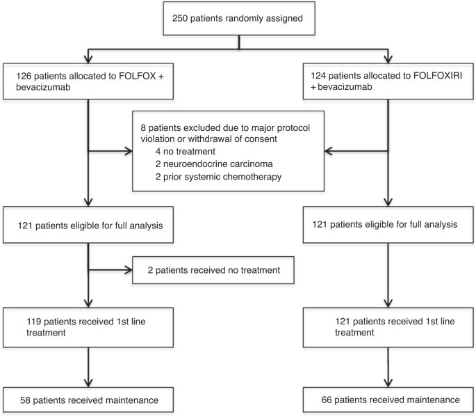
Table 1 Patient characteristics (mITT) according to clinical groups.

Full size table

Treatment intensity and duration

The median duration of the induction phase treatment was 5.6 months (range 0.5–13) in both arms, ranging between median 5.3 months in clinical Group 1 (potentially resectable) and 5.8 months in clinical Group 3 (asymptomatic) patients (Fig. 2a). The median number of cycles administered was similar in both arms with 12 cycles (range 1–12) in the induction phase (first 6 months of treatment) and 8 cycles (range 1–26) in the maintenance phase. Dose reductions were significantly more often required for FOLFOXIRI/bevacizumab (8.9% vs. 17.1% of cycles, P < 0.00001) considering the total mITT population.

figure 2
Fig. 2: Dose intensity and duration of treatment according to clinical groups.

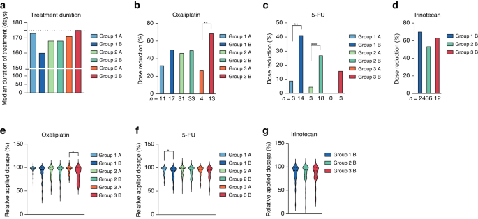
Particularly, clinical Group 1 and 2 patients receiving FOLFOXIRI/bevacizumab required more 5-FU dose reductions during the induction phase (P = 0.0021 and P = 0.0004; Fig. 2b) and clinical Group 3 patients required more oxaliplatin dose reductions (P = 0.0093; Fig. 2c). Irinotecan was reduced in 70% (Group 1), 53% (Group 2) or 63% (Group 3) of patients (Fig. 2d). The per protocol defined initial dose reduction was applied in 21 patients (Group 1: 10 patients (14%), Group 2: 10 patients (7.4%) and Group 3: 1 patient (2.6%)) and maintained in 13 patients in the second and third cycle. In contrast to Groups 1 and 2, the mean applied dosage of oxaliplatin (95%) was higher in Group 3 patients treated with FOLFOX/bevacizumab compared to FOLFOXIRI/bevacizumab (86%, P = 0.0198; Fig. 2e). In Group 1 patients, the mean applied dosage of 5-FU was higher with FOLFOX/bevacizumab (94%) compared to FOLFOXIRI/bevacizumab (89%, P = 0.0318; Fig. 2f) and the mean irinotecan dosages where comparable in all the groups treated with irinotecan (Fig. 2g).

Efficacy

After a median follow-up of 87.6 months (inverse Kaplan–Meier method), the efficacy was determined in the mITT population of eligible patients (n = 242). The progression-free survival rate at 9 months was significantly improved from 56.2% with FOLFOX/bevacizumab to 66.9% with FOLFOXIRI/bevacizumab on the predetermined significance level of 10% (P = 0.086—stratified logistic regression; Fig. 3a and Table 2). Thus, the primary endpoint was met. The objective response rate (ORR) was 61% and 69% (P = 0.21), secondary R0/1-resection rate was 15% vs. 20% (P = 0.39) and with the inclusion of patients with complete remission (CR) the rate was 18% vs. 24% (P = 0.34). Median PFS was 10.3 vs. 12.0 months (HR 0.83, 95% CI 0.64–1.08, P = 0.19), and the median OS was 24.0 vs. 28.0 months (HR 0.82, 95% CI 0.62–1.09, P = 0.24), for control and experimental group, respectively (Table 2 and Fig. 3b). Subsequent second-line treatments were applied in 67.8% vs. 73.6% of patients, and third line treatments in 40.5% vs. 43.8% of patients (Supplementary Table A1). The type of salvage regimen was comparable in both groups.

figure 3
Fig. 3: PFS and OS in total study cohort or divided by clinical Groups.

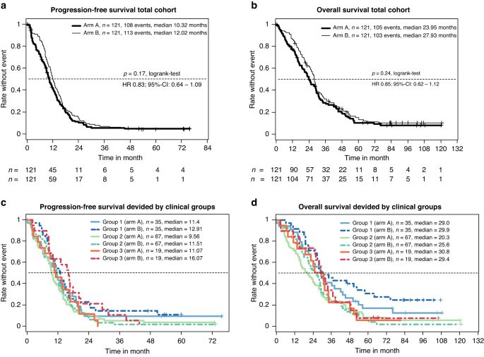
Table 2 Efficacy according to RECIST 1.1.

Full size table

Subgroup analyses

The clinical grouping, which was determined by the investigators and used as the stratification factor for randomisation, worked well to discriminate the three groups with different prognoses (Table 2 and Fig. 3c, d) and to differentiate the comparative effect of both treatment arms. Although not significantly different, the PFS benefit of the addition of irinotecan was mainly driven by clinical Group 3 (HR 0.6; P = 0.14) and to a bit lesser extent by clinical Group 1 (HR 0.83; P = 0.46), but very little by clinical Group 2 (HR 0.94; P = 0.73) (Table 2). In line with the improved PFSR@9, FOLFOXIRI/bevacizumab led to a trend of increased secondary R0/1-resection rates in clinical Group 1 patients from 37.1% to 51.4% (P = 0.33). Expectedly, patients achieving a “No Evidence of Disease” (NED) status by secondary resection or chemotherapy alone had a nearly doubled median OS (44.2 vs. 23.8 months, P < 0.0001) and showed a trend to improved PFS (P = 0.013) compared to patients not achieving NED status (Supplementary Fig. A1). Considering the mITT age below 61 (HR 0.57) and synchronous metastasis (HR 0.75) favoured irinotecan. Furthermore, a trend towards improved PFS in the FOLFOXIRI/bevacizumab arm was observed for RAS wild-type (HR 0.74) and left colon (HR 0.76), but not ECOG or BRAF status (Supplementary Fig. A2).

Toxicity

Treatment was generally given outpatient and was well tolerated. Adverse events (AE) are summarised in Supplementary Table A2. The grade 3/4 adverse events, which were numerically higher in the experimental arm were: neutropenia (14% vs. 20%), with only 1% febrile neutropenia in both arms, diarrhoea (12% vs. 16%), fatigue/asthenia (3% vs. 9%), treatment-related fatal (n = 3 vs. 1) and overall fatal (n = 5 vs. 2). Overall, treatment with FOLFOXIRI/bevacizumab was feasible, and the most occurring adverse events were mild to moderate. The adverse event-related discontinuation rate was 16.5% vs. 19.8%, the total serious adverse event rate was 85% vs. 89% and the total fatal outcome was 3 vs. 4, comparing FOLFOX/bevacizumab and FOLFOXIRI/bevacizumab.

Quality of life

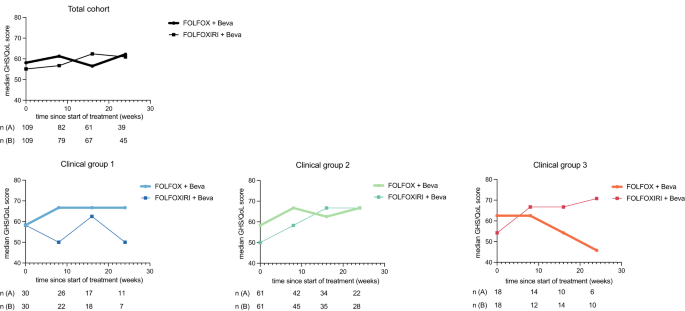
Health-related quality of life questionnaires were available from 95.4% at baseline, 72.6% at week 8, 59.5% at week 16 and 43.5% at week 24. No significant difference in the median Global health score (GHS) at week 0, 8, 16 and 24 were seen between both total population treatment groups (Fig. 4). Moreover, the GHS mean values pooled over all induction timepoints were similar between treatment groups (59.8 and 58.8, P = 0.726) (Supplementary Table A3). Clinically relevant changes in GHS score (at least 10 points) were similar between both groups, showing an improvement in 44.3% and 39.7% (P = 0.629) and deterioration in 21.5% and 26.9% (P = 0.461) for the control compared to the experimental group, respectively (Supplementary Table S4). No significant difference was observed in either clinical group comparing both treatment arms, however, the GHS mean values showed a trend towards improvement at a clinically relevant difference during therapy for patients treated with FOLFOXIRI/bevacizumab among clinical Group 3 patients (> 10 points, P = 0.185; Fig. 4).

figure 4
Fig. 4: Median GHS/QoL in total population or clinical Groups 1, 2 and 3 during treatment with FOLFOX/bevacizumab +/− irinotecan.

Discussion

The randomised CHARTA trial tested the addition of irinotecan to FOLFOX/bevacizumab in first-line metastatic colorectal cancer treatment and met its primary endpoint of an improved PFS rate at 9 months. In addition, numerically albeit not statistically significant improvements in ORR, PFS and OS were noted in favour of FOLFOXIRI/bevacizumab, in line with the results of other trials in this setting [24,25,26,27]. The control arm with the chemotherapy doublet and bevacizumab seemed to perform slightly better in “CHARTA” with a median PFS of 10.3 months compared to 9.7 months for FOLFIRI/ bevacizumab in “TRIBE” and 9.3 for FOLFOX/ bevacizumab in “STEAM” based on which the PFSR@9 month was selected as the primary endpoint. Thus, the hazard ratio for PFS with 0.84 is less pronounced in the “CHARTA” trial compared to “TRIBE 1 and 2” (HR 0.75). In addition, disease assessments were scheduled differently in “CHARTA”, every 2 months for 6 months, followed by 3 monthly assessments, compared to “TRIBE2”, where the assessments were conducted every 8 weeks even during the maintenance regimen. Furthermore, patient characteristics differed between the “TRIBE” trials and “CHARTA” particular regarding ECOG PS distribution (ECOG 0 90% vs. 50%). Overall, these small differences might have contributed to the slight difference in the hazard ratios and may explain why only the primary endpoint of PFSR@9 was different but not ORR, PFS and OS.

Even though the effectiveness of FOLFOXIRI/bevacizumab in treating mCRC is well-established, only a small percentage of all mCRC patients (less than 5%) and mCRC patients over the age of 50 (less than 2%) are currently receiving this combination therapy [28]. The CHARTA trial’s patient stratification based on predicted clinical outcomes presents an opportunity to customise treatments and ensure the implementation of this regimen in clinical practice. We prospectively categorised patients into clinical groups based on treatment aims (Group 1: potentially resectable after downsizing; Group 2: unlikely to become or never resectable; and Group 3: no symptoms or risk of rapid deterioration). Although we did not observe a significant difference in PFS or OS in either group, we observed a trend for the greatest PFS benefit in clinical Group 3 patients (HR 0.6). However, guidelines recommend a more conservative, sequential approach for this group due to factors such as absence of symptoms, advanced age, frailty, or lack of eligibility for post-chemotherapy metastasis resection. This benefit might be linked to a low rate of 5-FU dose reductions in this group (Fig. 2a, c, f). Considering OS, clinical Group 1 patients within the FOLFOXIRI/bevacizumab arm contained an extraordinary overall survival rate after 5 years. This is closely related to the achievement of a NED status (Fig. 3d and Table 1). Notably, clinical Group 2 patients derived no apparent benefit of the addition of irinotecan to FOLFOX/bevacizumab in terms of efficacy, however, also did not experience any decline in their QoL. In CHARTA, the clinical Group 2 (unresectable, symptomatic or risk of rapid progression) was larger than expected (55.3%) likely due to the selection of advanced patients for clinical trials testing the intensified regimen.

Besides clinical groups, other tumour-specific or patient’s specific factors correlated with improved progression-free survival such as “RAS wt status”, primary tumour sidedness, synchronous metastasis or age which is in line with other studies testing FOLFOXIRI/bevacizumab [26].

The main limitation of these results are the overall low numbers of patients who are particularly considered in the subgroup analysis and specifically as clinical Group 3. Hence, it is important to acknowledge the possibility that chance may have influenced the favourable outcomes observed in clinical Group 3. Consequently, additional studies are warranted to investigate the potential suitability of intensified treatment for patients with older age and asymptomatic disease.

The FOLFOXIRI/bevacizumab regimen demonstrated good tolerability with no unexpected safety findings. Dose modifications were primarily necessitated by haematological (36.4%) or gastrointestinal (26.7%) toxicity. The primary grade 3/4 adverse events experienced by patients on this combination included diarrhoea, fatigue, and nausea. However, these differences did not translate into noticeable impacts on patient-reported global health scores, suggesting only a minimal effect on overall QoL.

In summary, we provide further data showing that FOLFOXIRI/bevacizumab is superior to FOLFOX/Bevacizumab considering PFSR@9 in first-line mCRC treatment. Moreover, patient stratification according to clinical grouping is feasible and potentially defines patients who are more likely to benefit from the treatment. Such a classification may help to implement this effective but rather aggressive treatment regimen into clinical practice.

Conclusion

This multicentre randomised trial demonstrates a superior PFS rate at 9 months for the addition of irinotecan to FOLFOX/bevacizumab, with a good tolerability and QoL profile, and thus supports the value of FOLFOXIRI/bevacizumab in first-line treatment of mCRC. Furthermore, findings from the CHARTA trial suggest a clinical stratification that could identify patients who might particularly benefit from this regimen.

Japanese Study: Green Tea Extract Shows Promise for Chemo Patients


Green tea may relieve GI side effects of common cancer drug

Green tea has many health benefits. Now research shows it may be helpful for cancer patients.(studiolopa/iStock)

Green tea has many health benefits. Now research shows it may be helpful for cancer patients.(studiolopa/iStock)

Drinking green tea has many benefits for the human body. Not only is it a refreshing drink, it is also known for its health benefits. Among other beneficial effects, it is an antioxidant and inhibits hypertension.

A Japanese study has shown that green tea extract shows promise for cancer patients. On March 27, a study presented at the pharmaceutical society of Japan’s annual meeting in Nagoya, Japan, showed that green tea extract can alleviate severe diarrhea caused by the cancer drug irinotecan (CPT-11). The study was presented by Dr. Tomoo Hosoe of Tokyo’s Hoshi University.

Green Tea and Chemo Side Effects

Irinotecan is an anti-tumor drug used for many types of cancer, including colon cancer. However, the chemotherapy drug has serious side effects, including diarrhea.

Irinotecan causes diarrhea due to its SN-38G content. In the intestine, the bacteria β-glucuronidase converts SN-38G into SN-38, which inhibits tumor growth. However, the process also damages the intestinal mucosa.

In a study on mice, Dr. Hosoe’s team found that the high levels of catechin—a natural antioxidant—in green tea extract inhibit irinotecan-induced diarrhea.

The researchers divided the mice into two groups. One group was given irinotecan alone, and the other group was given irinotecan plus green tea extract. The results showed that the mice given only irinotecan had a 3.5-fold increase in fecal water content (diarrhea). In the other group, mice given a combination of green tea extract and irinotecan showed a 1.5-fold increase in diarrhea, which was significantly less than that of the irinotecan-only group.

In terms of the activity of β-glucuronidase-producing bacteria, the number of bacteria was significantly reduced in the group mixed with green tea extract compared to the group given irinotecan alone. Moreover, the increased dose of green tea extract was more effective in reducing the activity of β-glucuronidase.

Green Tea: More Good News for Cancer Patients

For years, Japanese researchers have studied the connection between green tea and cancer. While most studies focus on the role of green tea in preventing cancer, a 2004 study showed that green tea extract had an inhibitory effect on the growth of tumor angiogenesis. Researchers at Japan’s Osaka University studied the effect of green tea extract on blood vessel growth using human umbilical vein endothelial cells (HUVEC). Twenty-four hours after the addition of green tea extract (0-25 mg/ml) to endothelial cells cultured in the medium, cell viability, cell proliferation capacity, cell migration capacity, and cell formation capacity were analyzed.

The results showed that the cell viability of endothelial cells did not change, but cell proliferation, tube formation, and cell migration abilities were significantly reduced, depending on the concentration of green tea extract added. In addition, the activity of vascular endothelial growth factor (VEGF) in endothelial cells, was also reduced as the dose of green tea extract was increased.

The research team concluded that green tea extract inhibited tumor angiogenesis by reducing the expression of VEGF receptors. The results of the study were published in the Japanese Society for Home Economics Research Publications in May 2004.

The Long History of Tea in Japan

The history of tea began in China and spread throughout the world. During the Tang Dynasty (618-907 A.D.), tea was introduced from China to Japan. At that time, during Japan’s Nara period (710-1192 A.D.), the country was attempting to learn and adopt the more advanced Chinese systems and culture. Tea was extremely precious and only a very small number of nobles and monks could enjoy it.

During the Kamakura period (1192-1392 A.D.) in Japan, a Japanese Buddhist priest named Zen master Eisai visited China and brought back green tea leaves to Japan. Eisai wrote “How to Stay Healthy By Drinking Tea,” which recorded the effects of tea leaves and how to make tea. The first complete book on tea in Japan, Eisai’s book had a major impact on the spread of tea culture.

Nowadays, most of the tea roasted in Japan is green tea. In fact, Japanese tea is generally referred to as Japanese green tea. There are various types of green tea, including sencha, mint tea, green tea, and bancha. Green tea is grown extensively in Japan.

Effectiveness of Etoposide and Cisplatin vs Irinotecan and Cisplatin Therapy for Patients With Advanced Neuroendocrine Carcinoma of the Digestive System


Key Points

Question  For patients with advanced neuroendocrine carcinoma of the digestive system, which of the 2 community standard regimens is more effective: etoposide plus cisplatin (EP) or irinotecan plus cisplatin (IP)?

Findings  In this randomized clinical trial of 170 patients who were chemotherapy naive and had recurrent or unresectable neuroendocrine carcinoma of the digestive system, median overall survival was 12.5 months in the EP arm and 10.9 months in the IP arm.

Meaning  Both EP and IP therapy remain standard first-line chemotherapy options.

Abstract

Importance  Etoposide plus cisplatin (EP) and irinotecan plus cisplatin (IP) are commonly used as community standard regimens for advanced neuroendocrine carcinoma (NEC).

Objective  To identify whether EP or IP is a more effective regimen in terms of overall survival (OS) in patients with advanced NEC of the digestive system.

Design, Setting, and Participants  This open-label phase 3 randomized clinical trial enrolled chemotherapy-naive patients aged 20 to 75 years who had recurrent or unresectable NEC (according to the 2010 World Health Organization classification system) arising from the gastrointestinal tract, hepatobiliary system, or pancreas. Participants were enrolled across 50 institutions in Japan between August 8, 2014, and March 6, 2020.

Interventions  In the EP arm, etoposide (100 mg/m2/d on days 1, 2, and 3) and cisplatin (80 mg/m2/d on day 1) were administered every 3 weeks. In the IP arm, irinotecan (60 mg/m2/d on days 1, 8, and 15) and cisplatin (60 mg/m2/d on day 1) were administered every 4 weeks.

Main Outcomes and Measures  The primary end point was OS. In total, data from 170 patients were analyzed to detect a hazard ratio (HR) of 0.67 (median OS of 8 and 12 months in inferior and superior arms, respectively) with a 2-sided α of 10% and power of 80%. The pathologic findings were centrally reviewed following treatment initiation.

Results  Among the 170 patients included (median [range] age, 64 [29-75] years; 117 [68.8%] male), median OS was 12.5 months in the EP arm and 10.9 months in the IP arm (HR, 1.04; 90% CI, 0.79-1.37; P = .80). The median progression-free survival was 5.6 (95% CI, 4.1-6.9) months in the EP arm and 5.1 (95% CI, 3.3-5.7) months in the IP arm (HR, 1.06; 95% CI, 0.78-1.45). A subgroup analysis of OS demonstrated that EP produced more favorable OS in patients with poorly differentiated NEC of pancreatic origin (HR, 4.10; 95% CI, 1.26-13.31). The common grade 3 and 4 adverse events in the EP vs IP arms were neutropenia (75 of 82 [91.5%] patients vs 44 of 82 [53.7%] patients), leukocytopenia (50 of 82 [61.0%] patients vs 25 of 82 [30.5%] patients), and febrile neutropenia (FN) (22 of 82 [26.8%] patients vs 10 of 82 [12.2%] patients). While incidence of FN was initially high in the EP arm, primary prophylactic use of granulocyte colony-stimulating factor effectively reduced the incidence of FN.

Conclusions and Relevance  Results of this randomized clinical trial demonstrate that both EP and IP remain the standard first-line chemotherapy options. Although AEs were generally manageable, grade 3 and 4 AEs were more common in the EP arm.

Discussion

Evidence for treatment strategies of advanced NEC is lacking owing to the rarity of the disease. Recently, the results of a randomized phase 2 trial for NEC were reported16; however, enrollment was terminated early in 66 patients owing to the premature analysis, and no definitive conclusion was obtained. The present study represents the final results of a well-designed and, to our knowledge, the first phase 3 trial with high-quality pathological diagnoses and detailed information provided by a CPR.

The primary analysis of OS revealed no statistically significant difference between arms. Therefore, it is reasonable to continue using both regimens as the standard treatment for NEC. We were interested in the effect of pathological findings and primary organs on treatment efficacy. Although the results showed no statistically significant differences in any subgroups, the overall impression had a greater interaction by primary organ than by pathological findings in terms of OS and PFS (Figure 3 and eFigure 5 in Supplement 1). Because platinum regimens have been reported to be less effective in grade 3 NETs,5,8 and the WHO 2017 classification system9 and 2019 classification system10 formally declared grade 3 NETs as a separate entity from NECs,9,10 subgroup analyses of PDNECs only are of relevant clinical interest. Subgroup analysis of OS in PDNECs only indicated weak tendency of preferred results in the EP arm over the IP arm. A subgroup analysis of only PDNECs in each primary tumor indicated that the OS of the EP arm was better than that of the IP arm in the subset of pancreatic PDNEC. Although post hoc subgroup analyses should be interpreted with caution, these results have important implications. The CPR indicated that pancreatic NECs are characterized by a higher proportion of small cell carcinoma and a lower proportion of tumors with non-NEC components (eTable 2 in Supplement 1). Although these characteristics may have influenced the unique results in this subgroup, they cannot fully explain this phenomenon.

In terms of AEs, grade 3 and 4 AEs such as myelosuppression and FN were more common (>2 times) in the EP arm. Although these AEs were generally manageable, the high incidence of FN remains a point of caution. We took note of the high incidence of FN during this trial in the monitoring report; therefore, we revised the protocol to recommend primary prophylaxis with G-CSF32,33 after October 31, 2017. In the exploratory analysis of this study, prophylaxis with G-CSF seemed to effectively decrease the incidence of FN. However, considering higher planned/actual DI and lower relative DI in the EP arm, and higher rate of treatment termination owing to AE or patient’s refusal (third dose reduction owing to neutropenia), lower initial dose of EP therapy may be appropriate.

In addition to the outcome of the clinical trial, this study is very valuable in its provision of information on high-quality pathological diagnoses made by consistent CPR panel members throughout the entire period. Notably, the pathological diagnosis was not consistent with that of the participating institutions in nearly 10% of cases, indicating the difficulty of an accurate pathological diagnosis of NEC. Additionally, detailed information about the pathological findings was obtained for the cross-organ manner, which itself is an important asset in the field of NEC. In the forest plots of OS and PFS, the difference between EP and IP among the CPR subgroups was smaller than that of the primary organs (Figure 3 and eFigure 5 in Supplement 1).

Limitations

This study has some limitations. Although the primary analyses revealed no statistically significant difference in OS between the 2 arms, the study was not designed to assess the equivalence of the 2 regimens. Therefore, the 2 regimens should not be recognized as equivalent, but rather as they do not differ beyond a certain level. In addition, in patients diagnosed via results of biopsy specimens, accurate information of the entire tumor could not be obtained with respect to proliferative activity or the proportion of non-NEC components. In this study, patients diagnosed by biopsy harboring any proportions of non-NEC components, as long as a harboring NEC component was allowed to be enrolled, means that theoretically a certain percentage of patients with mixed adenoneuroendocrine carcinomas were enrolled. These limitations were unavoidable considering the diagnostic procedure, and this is also a universal issue in daily practice. Additionally, because further effects of immune checkpoint inhibitors on platinum regimens has been proven in small cell lung carcinoma,34,35 it is the next important clinical question in NECs.

Conclusions

Results of this phase 3 randomized clinical trial demonstrate that both EP and IP remain the standard first-line chemotherapy regimens for advanced digestive NECs. Post hoc subgroup analyses pointed to the superiority of EP in pancreatic PDNECs, and EP was safely given along with the use of primary prophylactic G-CSF.

Source: JAMA

Pancreatic cancer — a changing outlook if diagnosed early


Tumor DNA sequencing to identify appropriate targeted therapies is revolutionizing treatment of a variety of cancers, and pancreatic cancer — a notoriously deadly disease — is no exception.

Based on this capability and improved outcomes observed with such an approach, National Comprehensive Cancer Network guidelines issued last year recommend clinicians consider germline testing for all patients with pancreatic cancer and molecular tumor analysis for patients with metastatic disease.

Improvements to adjuvant chemotherapies have led to NCCN guidelines that also recommend adjuvant therapy with modified FOLFIRINOX (leucovorin, 5-FU, irinotecan and oxaliplatin) for patients who can tolerate it.

Despite these growing treatment options, pancreatic cancer is now the third-leading cause of cancer death, having recently surpassed breast cancer. By 2030, the disease is projected to surpass colorectal cancer to become the second most common cause of cancer mortality.

Profiling patients’ tumors to evaluate for mutations or to assess for microsatellite instability is reasonable because the newer therapeutics are more effective than standard pancreatic cancer treatments, according to Syma Iqbal, MD, FACP.
Profiling patients’ tumors to evaluate for mutations or to assess for microsatellite instability is reasonable because the newer therapeutics are more effective than standard pancreatic cancer treatments, according to Syma Iqbal, MD, FACP.

“The reasons for this are uncertain, but what has been thought to be contributing to the [increased death rate] is the aging population,” Syma Iqbal, MD, FACP, assistant professor of clinical medicine in the division of medical oncology at Keck School of Medicine of USC, told HemOnc Today. “People are living longer, and perhaps this changing of demographics, which includes various minority groups at risk for developing pancreatic cancer, plays a role. Although we have made incremental progress in this disease throughout the years, there is still a lot of progress to be made.”

HemOnc Today spoke with medical oncologists and surgeons about the current prognosis for patients with pancreatic cancer, how treatments and surgical techniques have evolved during the last few years, and what lies ahead for this difficult-to-treat patient population.

Prognosis, detection

The American Cancer Society estimated that, last year, 56,770 people in the U.S. would be diagnosed with pancreatic cancer and 45,750 would die of the disease.

These figures reflect an increase in pancreatic cancer incidence, the reason for which is uncertain. However, experts with whom HemOnc Today spoke suggest factors at play include the aging population, obesity and smoking.

“Pancreatic cancer, compared with breast cancer, tends to occur a bit later in life, so it may be that patients are simply living longer and not dying of other causes. Instead, they are living to be increasingly diagnosed with pancreatic cancer,” Steven D. Leach, MD, director of Norris Cotton Cancer Center and professor of surgery at Dartmouth Geisel School of Medicine, told HemOnc Today. “The obesity epidemic occurring in western societies may also be contributing to the increased risk. We are hopeful that some of the newer treatments will lead to significant improvements in OS and prognosis for patients with pancreatic cancer.”

The trend of increasing pancreatic cancer incidence is especially worrisome because outcomes are historically dismal for this disease.

Approximately 90% of patients diagnosed with pancreatic cancer do not survive 1 year, and 5-year survival rates are in the single digits. The likelihood for cure is less than 50%, even for those with localized disease.

David A. Iannitti Sr., MD
David A. Iannitti Sr.

“Cure is determined by whether or not the tumor can be removed by surgery as well as the molecular nature of the disease — whether it harbors specific mutations that render the tumor more or less sensitive to chemotherapy,” David A. Iannitti Sr., MD, chief of the division of hepatopancreaticobiliary surgery at Carolinas Medical Center and surgical oncologist at Levine Cancer Institute at Atrium Health, said during an interview with HemOnc Today. “These methods define the only patients who truly have a chance for cure — we can only operate on a small number of these patients, and about one-third will achieve long-term survival.”

For patients with early-stage disease who are able to undergo surgery, median OS is 36 months.

However, surgery is not possible for those with locally advanced pancreatic cancer, who face average survival of 1 year. Of the more than 80% of pancreatic cancer cases that are detected at an advanced stage — too late for surgery — 5-year OS rates range from 2% to 5%.

Early detection could dramatically improve outcomes but is not an option for the vast majority of the general population, according to David H. Ilson, MD, PhD, medical oncologist at Memorial Sloan Kettering Cancer Center and a HemOnc Today Editorial Board Member.

David H. Ilson, MD, PhD
David H. Ilson

“We are now seeing more than 50,000 American patients each year who develop newly diagnosed pancreatic cancer,” Ilson said. “Despite no early detection methods for the general population, there are screening measures for a small subgroup of individuals who have a genetic predisposition. Between 8% and 10% of pancreatic cancers appear to have familial predisposition; an even lesser percentage may have specific genes that can be tested for.”

Most notably, BRCA mutation carriers have a finite increased risk for pancreatic cancer, in addition to an increased risk for ovarian and breast cancers. Genetic syndromes, including hereditary pancreatitis, familial melanoma and Lynch syndrome, also are associated with an increased risk for pancreatic cancer.

Although many academic institutions screen these high-risk individuals, no guidelines exist on which screening techniques to use for these patients or how frequently they should undergo screening.

Moreover, pancreatic cancer among individuals without any of these risk factors may go undetected until it begins to present symptoms at its advanced stages.

Last year, the U.S. Preventive Services Task Force maintained its stance against screening for adults without family history, signs or symptoms of pancreatic cancer. The task force concluded there was no evidence that screening for pancreatic cancer or treatment of screen-detected disease improves mortality and morbidity.

The evidence report and systemic review that supported the USPSTF’s statement showed limited evidence to evaluate the benefits and harms of surgery for pancreatic cancer detected through screening.

Given that approximately 1.6% of people living in the U.S. will develop pancreatic cancer, even an ideal screening test with 99% sensitivity and 99% specificity would result in 1,000 false positives for every 100,000 individuals tested, according to an editorial by Aimee L. Lucas, MD, MS, associate professor of medicine in the Henry D. Janowitz division of gastroenterology at Icahn School of Medicine at Mount Sinai, and Fay Kastrinos, MD, MPH, assistant professor of medicine at Columbia University Irving Medical Center, that accompanied the USPSTF recommendations.

“These false-positive results would require subsequent diagnostic evaluation and accrue additional complications, costs and patient distress that would cause the risks of screening to outweigh any potential benefit,” they wrote.

Despite years of challenges with screening, technologies continue to evolve and may soon reduce these concerns about false-positive results. Liquid tumor biopsies to screen blood for mutations or abnormalities appear promising as potential detection methods that could dramatically change the diagnosis of pancreatic cancer, experts said.

Treatment evolution

Pancreatic cancer remains a difficult disease to treat.

For nearly a decade, treatment in the adjuvant setting has consisted of either gemcitabine or 5-FU alone. More recently, randomized clinical trials of combination chemotherapy have emerged.

For instance, Conroy and colleagues of the PRODIGE 24 trial compared the efficacy and safety of a modified FOLFIRINOX regimen with gemcitabine as adjuvant therapy for patients with resected pancreatic cancer.

The analysis included 493 adults with pancreatic ductal adenocarcinoma who underwent surgery and had no signs of metastatic disease, malignant ascites or pleural effusion.

Researchers randomly assigned patients 3 to 12 weeks after surgery to a modified FOLFIRINOX regimen (85 mg/m2 oxaliplatin, 400 mg/m2 leucovorin, 180 mg/m2 irinotecan [later reduced to 150 mg/m2] and 2,400 mg/m2 fluorouracil) every 2 weeks or 1,000 mg/m2 gemcitabine on days 1, 8 and 15 every 4 weeks for 24 weeks.

Results — published in 2018 in The New England Journal of Medicine — showed the modified FOLFIRINOX regimen led to significant improvements in DFS (21.6 months vs. 12.8 months; HR = 0.58; 95% CI, 0.46-0.73) and OS (54.4 months vs. 35 months; HR = 0.64; 95% CI, 0.48-0.86) compared with gemcitabine.

“This is probably the most important trial that has come out in recent years,” Ilson said. “FOLFIRINOX resulted in significantly better 5-year OS and PFS, with up to a 40% 5-year OS rate compared with gemcitabine alone. FOLFIRINOX is now considered a standard of care after pancreatic cancer resection.”

Beyond chemotherapy, treatment options also are emerging for patients with various biomarkers, including BRCA mutations or mismatch repair deficiency.

For instance, poly(ADP-ribose) polymerase, or PARP, inhibitors have demonstrated efficacy in several cancer types associated with BRCA mutations. PARP inhibitors work by blocking the enzymatic activity of PARP and trapping PARP at the sites of DNA damage. Thus, inhibition of PARP in cancers with a BRCA1 or BRCA2 mutation results in the accumulation of DNA damage that cannot be repaired, leading to tumor cell death.

Michael J. Hall, MD, MS, chairman and associate professor in the department of clinical genetics at Fox Chase Cancer Center, and colleagues presented data at last year’s ASCO Annual Meeting from the double-blind, placebo-controlled POLO study showing that patients with germline BRCA-mutated metastatic pancreatic cancer assigned olaparib (Lynparza; AstraZeneca, Merck) — a PARP inhibitor approved in the U.S. for treatment of certain patients with ovarian cancer and HER2-negative metastatic breast cancer — demonstrated significantly reduced risk for progression or death than those assigned placebo.

Olaparib conferred significantly longer PFS compared with placebo (7.4 months vs. 3.8 months; HR = 0.53; 95% CI, 0.35-0.82). At 6 months, the percentage of progression-free patients in the olaparib arm was double that of the placebo arm (53% vs. 23%). This PFS trend continued at 1 year (33.7% vs. 14.5%), 18 months (27.6% vs. 9.6%) and 2 years (22.1% vs. 9.6%).

“When I started in oncology 10 years ago, there was one drug for pancreatic cancer, and the disease was usually fatal within 6 to 8 months,” Hall previously told HemOnc Today. “The results were actually even better than I expected.”

In another study presented at ASCO, Pishvaian and colleagues assessed the PARP inhibitor veliparib (ABT-888, AbbVie) plus FOLFOX chemotherapy among patients with metastatic pancreatic cancer.

According to study results, the combination regimen appeared safe, well-tolerated and effective — especially among patients who were platinum-naive, had a family history of breast or ovarian cancer, and/or harbored somatic DNA damage response mutations, including BRCAPALB2 or ATM.

Identifying molecular drivers

Such findings underscore the importance of efforts that have intensified in recent years to learn more about the molecular drivers of pancreatic cancer. Experts have uncovered mutations and now have a better understanding of the disease biology.

“Profiling patients’ tumors to evaluate for mutations or to assess for microsatellite instability is reasonable because the newer therapeutics that are more effective than our standard treatments have opened up treatment options for this population of patients,” Iqbal said.

For instance, in a study published in 2018 in JAMA, Hu and colleagues identified mutations in six genes that appeared associated with pancreatic cancer.

Comparing a cohort of 3,030 adults with pancreatic cancer enrolled at Mayo Clinic with data on 123,136 individuals who had exome sequence data from the Genome Aggregation Database and 53,105 individuals from the Exome Aggregation Consortium Database, researchers found significant associations between pancreatic cancer and CDKN2A (0.3% of cases vs. 0.02% of controls; OR = 12.33; 95% CI, 5.43-25.61), TP53 (0.2% vs. 0.02%; OR = 6.7; 95% CI, 2.52-14.95), MLH1 (0.13% vs. 0.02%; OR = 6.66; 95% CI, 1.94-17.53), BRCA2 (1.9% vs. 0.3%; OR = 6.2; 95% CI, 4.62-8.17); ATM (2.3% vs. 0.37%; OR = 5.71; 95% CI, 4.38-7.33) and BRCA1 (0.6% vs. 0.2%; OR = 2.58; 95% CI, 1.54-4.05).

Researchers observed a mutation in one of the six predisposition genes among 27 of 343 patients (7.9%; 95% CI, 5.3-11.2) with a family history of pancreatic cancer and 140 of 2,687 patients (5.2%; 95% CI, 4.4-6.1) with no family history, indicating family history of pancreatic cancer did not indicate the presence of 83.8% of mutations.

Based on these data and others, genomic profiling of pancreatic cancer is now routine practice.

“For the 8% to 10% of patients who may have a familial risk for developing pancreatic cancer, recent publications have indicated that we may unveil a genetic predisposition even in a family that does not have a clear family history of pancreatic cancer,” Ilson said. “Occasionally, we will identify a BRCA mutation or a Lynch syndrome DNA mismatch repair protein mutation that will lead to screening other family members. This has led to NCCN guidelines now recommending germline testing in all patients newly diagnosed with pancreatic cancer. We are also increasingly advocating for genomic sequencing of these patients.”

DNA repair deficiency can implicate a cluster of abnormalities or mutations that occur in 10% to 25% of patients with pancreatic cancer, potentially identifying a subgroup that may be more sensitive to platinum-based chemotherapy.

Further, a rare biomarker, microsatellite instability (MSI)-high status, which is present in only about 1% of the pancreatic cancer population, is associated with significant response to immune checkpoint inhibitors.

“Within the past several years, regulatory authorities in the United States approved checkpoint inhibitors to be used in any patient who has an MSI-high status cancer that is progressing on conventional chemotherapy, due to high rates of remission,” Ilson said. “We occasionally have patients with pancreatic cancer who have MSI-high status and are well-served with immune checkpoint inhibitors when first- or second-line chemotherapies were not effective. Although rare, we are obligated to test patients for these genomic abnormalities.”

Updates in surgical procedures

Progress also has been made in surgical procedures for patients with resectable disease.

“Surgery has come a long way within the past 2 decades in terms of refinement of surgical techniques,” Iannitti said. “Additionally, within the past 10 to 15 years, there has been a significant increase in minimally invasive approaches to these tumors — the easiest of which is pancreatectomy. Probably 90% of the pancreatectomies that we perform for cancer are done minimally invasively. From this point of view, perioperative morbidity and complication rates have decreased dramatically.”

The Whipple procedure — or pancreaticoduodenectomy, which Iannitti described as a “profoundly more complicated operation” — may be performed for certain patients based upon the size of the tumor.

“We perform about 121 of these procedures annually and about one-third are done minimally invasively,” Iannitti added. “It is overall more common to perform this procedure robotically rather than laparoscopically, because of the intricacies of surgery and reconstruction.”

Robotic procedures are increasingly performed in certain patient populations, Iannitti added.

“However, these procedures have not decreased the length of hospital stay for our patients, although there is a decrease in narcotic use and decrease in pain,” he said. “Overall, I would say robotic or minimally invasive Whipple procedures are an advantage for the patient with pancreatic cancer.”

Neoadjuvant therapies remain essential for patients with locally advanced disease.

“I personally have taken on the challenge of working with localized pancreatic cancer, because these patients have a difficult cancer to manage,” Iannitti said. “In these patients, we will advance their staging with laparoscopy before we start neoadjuvant therapy, but we strongly advocate for neoadjuvant chemotherapy for just a short period — six doses or so to make sure there is no progression of disease and so we can get a feel for the biology of the tumor. One of the problems with pancreatic cancer is that if we leave gross tumor behind, the survival is the same whether the patient undergoes surgery or not.”

Despite improvements in surgical techniques, about 80% of pancreatic cancers are detected at an advanced stage and not eligible for surgery.

For these patients, researchers have attempted a variety of approaches to make the tumors more amenable to surgery — from cryoablation and radiofrequency ablation to microwave ablation — few of which have demonstrated success.

Nearly a decade ago, researchers developed irreversible electroporation (IRE) — a nonthermal technique that allows for the destruction of cancer cells with less damage to the surrounding area.

“Most pancreatic tumors that we see are not surgically removable, and so I have spent my entire career working to develop technologies to destroy tumors that we cannot surgically remove,” Iannitti said. “Pancreatic cancer is a horrible disease that causes tremendous pain and suffering for patients and their loved ones. There are a lot of people on all sides — experimental and medical — who are not giving up and still pushing forward. We are making advances slowly, but steadily.”

Iannitti and colleagues are currently in the preclinical stages of developing another novel technique that builds on IRE. HFIRE — high-frequency irreversible electroporation — is intended to help patients with advanced-stage disease.

“Results from this study are anticipated within the next few years,” Iannitti said. “If I were asked at any point in my professional career, I would have said there is no chance on the planet we could ever cure hepatitis C in my lifetime, and yet we have. I am hopeful about the advancement of surgical procedures for pancreatic cancer. We are going to continue to work on this and I know we will make surgery better and less invasive for these patients.”

Ongoing research

Despite a lack of success so far, researchers with whom HemOnc Today spoke said it’s not yet time to give up on immunotherapy for pancreatic cancer.

“Immunotherapy has been assessed in this disease, but has not shown the activity that we had hoped for,” Iqbal said. “We are still holding out hope for immunotherapeutics and efforts to combine them with other agents to make pancreatic tumors more sensitive to immunotherapy — we have not yet abandoned immunotherapy. We are still hopeful that if we are able to modulate the tumor, then perhaps we can make the tumor more sensitive to immunotherapy.”

Various targets are under investigation in several ongoing clinical trials in the advanced-disease setting, Iqbal added.

“Although these checkpoint inhibitors are not practice changing for the majority of pancreatic cancer cases, they have made an impact in the small population that has MSI-high status,” she said.

Leach echoed this sentiment.

“The one area that patients with pancreatic cancer have not yet benefited from is immunotherapy,” Leach said. “Immune checkpoint inhibitors have revolutionized treatment for many solid tumor cancer types, but so far only between 1% and 2% of patients with pancreatic cancer — those with microsatellite instability — have responded to these agents. We realize this is because pancreatic cancer has a highly specialized tumor microenvironment.”

Research is just beginning to reveal the potential of various pancreatic cancer genes and signaling pathways to create an immunosuppressive tumor microenvironment, Leach added.

“There are now a variety of clinical trials underway looking at various small molecule-targeted drugs that include CCR2 inhibitors and other strategies,” Leach said.

Other targets under investigation include antimitochondrial agents and anti-CD40 therapy.

Experts unanimously agreed that for the first time, the research pipeline for therapeutic options for patients with advanced pancreatic cancer is robust.

The recently opened Stand Up to Cancer Pancreatic Cancer Collective-funded GENERATE study is one avenue by which first-degree relatives of mutation-positive index cases can get tested from the convenience of their homes. Mutation carriers then can be channeled into screening programs such as Cancer of the Pancreas Screening consortium for longitudinal follow-up.

“We are in an exciting time in pancreatic cancer research,” Leach said. “We still have a long way to go, but we have a vibrant collaborative community of physicians and researchers studying the problem. There is a real sense of excitement about the progress being made in the field of pancreatic cancer.” – by Jennifer Southall

Response to Cetuximab With or Without Irinotecan in Patients With Refractory Metastatic Colorectal Cancer Harboring the KRAS G13D Mutation: Australasian Gastro-Intestinal Trials Group ICECREAM Study


PURPOSE: RAS mutations predict lack of response to epidermal growth factor receptor monoclonal antibody therapy in patients with metastatic colorectal cancer (mCRC), but preclinical studies and retrospective clinical data suggest that patients with tumors harboring the exon 2 KRAS G13D mutation may benefit from cetuximab. We aimed to assess cetuximab monotherapy and cetuximab plus irinotecan in patients with molecularly selected (G13D mutation) chemotherapy-refractory mCRC in a randomized phase II trial of this rare molecular subtype.

PATIENTS AND METHODS: Patients with chemotherapy-refractory KRAS G13D mutation-positive mCRC who had progressed within 6 months of irinotecan therapy were randomly assigned to cetuximab 400 mg/m(2) loading dose and then 250 mg/m(2) once per week with or without irinotecan 180 mg/m(2) once every 2 weeks. The primary end point was 6-month progression-free survival; secondary end points were response rate, overall survival, quality of life, and toxicity.

RESULTS: Fifty-one of 53 patients recruited over 2 years were eligible. The 6-month progression-free survival rate was 10% (95% CI, 2% to 26%) for cetuximab versus 23% (95% CI, 9% to 40%) for cetuximab plus irinotecan with a hazard ratio of 0.74 (95% CI, 0.42 to 1.32). Response and stable disease rates were 0% and 58% for monotherapy versus 9% and 70% for combination treatment, respectively. Overall survival and quality of life were similar; toxicities were higher with combination therapy.

CONCLUSION: In patients with G13D-mutated chemotherapy-refractory mCRC, there was no statistically significant improvement in disease control at 6 months with either cetuximab monotherapy or cetuximab plus irinotecan. No responses were seen with single-agent cetuximab. The responses observed with the combination of cetuximab and irinotecan may reflect true drug synergy or persistent irinotecan sensitivity. The ICECREAM (Irinotecan Cetuximab Evaluation and Cetuximab Response Evaluation Among Patients with a G13D Mutation) study demonstrates the need to prospectively evaluate hypotheses that were previously supported by retrospective analyses and exemplifies the value of international collaboration in trials of rare molecular subtypes.