Though prosthetics have come a long way towards enabling people who have lost their limbs to fully function, they still lack in certain areas like fine motor skills and the sense of touch. Now, researchers at the University of Glasgow have developed new technology that could remedy the latter.
A team from the Glasgow School of Engineering has developed artificial skin that would allow amputees to regain their sense of touch when using a prosthetic. The prototype involves using a polymeric protective layer over the prosthetic’s surface, that sends back pressure and temperature signals to the wearer. For example, someone wearing this prosthetic could pick up a cup of coffee, and would be able to feel the grain of the ceramic as well as how hot the mug is.
The problem these researchers, headed by Dr Ravinder Dahiya, faced is that such a device needed its own power source, making it necessary to also attach a battery pack to the robotic arm. Of course, this made the prosthetic bulky and unwieldy. The solution then, was to develop a version of the touch-sensitive layer that could power itself.
As such, the team began experimenting with graphene, a material that remains transparent and flexible while also being incredibly durable. Using this, the researchers were able to make a new protective layer that was also capable of converting solar energy, 98 percent of the light hitting it to be exact. They did this by layering the graphene over a set of solar cells underneath the “skin”, which would generate energy while in the sun and power the wearer’s sense of touch.
RAVINDER DAHIYA WITH HIS PROTOTYPE PROSTHETIC
However, the researchers believe they still have a lot of improvements to make to the prototype. The device is still too bulky, so the next step is to miniaturise the technology further, in order to bring the prosthetic’s weight closer to that of a real human hand. The team is also researching a way to store the solar energy converted in a light-weight battery pack within the device. Doing that would give the wearer the full experience of a mobile, sensitive hand, without worrying about when it will run out of power.
Watch the video. URL:
While the technology has been designed with amputees in mind, it could also eventually find its way to applications in robotics. A touch-sensitive skin could give a huge boost to the development of caregiver robots. With a sense of touch feeding back data, these bots could exercise restraint when dealing with infants, the old, or infirm, while still allowing them utilise more strength when say, picking up and moving household objects.
Nishikant Deshmukh’s breakthrough technology, which gives “eyes to surgeons to find a cancerous tumor,” will be especially useful in the developing world.
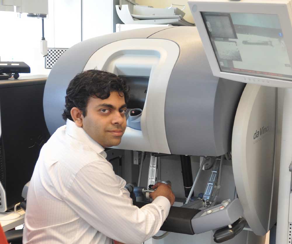
Dr. Nishikant Deshmukh with the da Vinci surgical console.
An Indian researcher at the Johns Hopkins University has developed the world’s first five-dimensional ultrasound system that will help surgeons detect and treat cancerous tumors.
Nishikant Deshmukh, 33, who just earned a doctoral degree from the prestigious university in Computer Science, developed the breakthrough system as part of his PhD thesis.
The ultrasound technology currently used by most surgeons is predominantly two-dimensional. Some hospitals also use a more advanced 3D computer graphics. However, the 3D model is not real-time, and it takes longer to generate images, making it difficult for surgeons to use information from it while conducting complex surgeries that require real-time decision-making.
“My technology can give vision to the surgeon for locating tumors while operating upon patients,” the Amravati, Maharashtra, -born researcher told The American Bazaar.
In a nutshell, Dr. Deshmukh’s technology combines 3D ultrasound B-mode and the 3D ultrasound elastography volumetric data and make them available in real-time.
Elastography is a medical imaging method that measures elastic properties of soft tissue and maps them as an image for diagnosing stiff regions such as cancer tumor. B-mode images are the ones we usually come across during a doctor’s sonography scan. Sonography, or diagnostic ultrasound, is a medical imaging technology where sound waves are used to produce images.
A view of the 5D Ultrasound. A denotes detected cancer like region and B denotes the background tissue like region. The images are of tissue and cancer mimicking material used for developing prototype systems.
The technology Dr. Deshmukh developed is termed as 5D ultrasound due to its ability to visualize and get the current combined data in real-time. The advanced imaging model that he developed can generate elastography using Graphic Processing Units at 60-70 frames per second, which enables combining elastography with real-time machine-generated B-mode images
Dr. Deshmukh presented the findings of his research for the first time at the 2015 Information Processing in Computer Assisted Interventions (IPCAI), a premier forum in the field. He has also published the research, along with his advisors and colleagues at the Laboratory of Computational Science and Robotics at the Johns Hopkins University and the National Institutes of Health, in two journals, the International Journal of Computer Assisted Radiology and Surgery and PLOS ONE.
Dr. Deshmukh, who has an undergraduate degree in Computer Engineering from the University of Pune, said his technology could be used for early stage cancer detection in areas such as prostate and breast. “It will help a radiologist to determine whether the abnormally grown tissue is a potentially fatal tumor, or a more benign cyst.”
The researcher said the technology would be especially useful in rural areas in the developing world where the more expensive Magnetic Resonance Imaging (MRI) is not available.
Dr. Deshmukh has also integrated the elastography system with the minimally invasive da Vinci robotic system, which has been used clinically since the year 2000.
“What we did was to accelerate it on GPUs to make it fast enough to be able to use it during surgery,” he said. “We also integrated it with the da Vinci system where the robot generates steady palpation motion for us.”
Dr. Deshmukh came to the Johns Hopkins University in 2008 to pursue his higher studies. Earlier, he worked at the National Stock Exchange of India in Mumbai for three years. His knowledge in parallel and distributed computing at NSE helped him to do advanced research in cancer imaging at The Johns Hopkins University, he said. The field is identified as Computer Integrated Surgery, which is a cross-disciplinary field of Computer Science, Medical Imaging, Biomedical Engineering, Robotics and Mechanical Engineering.
The researcher said he was motivated to pursue cancer diagnostic using computation power after seeing a family member die of cancer in rural India. “The disease could not be diagnosed at early stage,” he said. Dr. Deshmukh was also selected as the top 200 young scientists from 80 countries as part of the Heidelberg Laureate Forum and an INK Fellow, both in the year 2015.
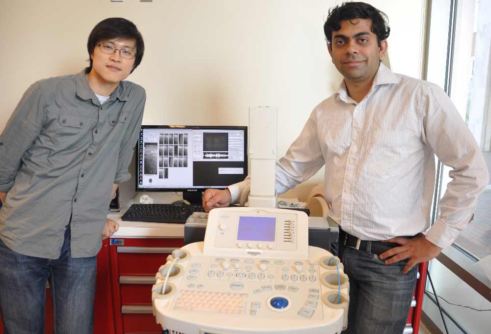
Dr. Nishikant Deshmukh (right) with his colleague and labmate Dr. Hyun Jae Kang with the altered ultrasound machine.
Dr. Deshmukh is also a supporter of organic farming movement in India, which is working to lower farmer suicide rate, afforestation, children education and reduce pesticide-affected food. As part of a nonprofit, he and his colleagues at Johns Hopkins have raised nearly $300,000 for various causes in India.