Head Injuries Could Be a Risk Factor for Developing Brain Cancer


Summary: Traumatic brain injuries may increase the risk of developing glioma brain cancer later in life, researchers report. The study found brain injury caused specific genetic mutations to synergize with inflammation, making brain cells more likely to become cancerous.

Source: UCL

Researchers from the UCL Cancer Institute have provided important molecular understanding of how injury may contribute to the development of a relatively rare but often aggressive form of brain tumor called a glioma.

Previous studies have suggested a possible link between head injury and increased rates of brain tumors, but the evidence is inconclusive. The UCL team have now identified a possible mechanism to explain this link, implicating genetic mutations acting in concert with brain tissue inflammation to change the behavior of cells, making them more likely to become cancerous.

Although this study was largely carried out in mice, it suggests that it would be important to explore the relevance of these findings to human gliomas.

The study was led by Professor Simona Parrinello (UCL Cancer Institute), Head of the Samantha Dickson Brain Cancer Unit and co-lead of the Cancer Research UK Brain Tumor Center of Excellence. She said, “Our research suggests that a brain trauma may contribute to an increased risk of developing brain cancer in later life.”

Gliomas are brain tumors that often arise in neural stem cells. More mature types of brain cells, such as astrocytes, have been considered less likely to give rise to tumors. However, recent findings have demonstrated that after injury astrocytes can exhibit stem cell behavior again.

Professor Parrinello and her team therefore set out to investigate whether this property may make astrocytes able to form a tumor following brain trauma using a pre-clinical mouse model.

Young adult mice with brain injury were injected with a substance which permanently labeled astrocytes in red and knocked out the function of a gene called p53—known to have a vital role in suppressing many different cancers. A control group was treated the same way, but the p53 gene was left intact. A second group of mice was subjected to p53 inactivation in the absence of injury.

Professor Parrinello said, “Normally astrocytes are highly branched—they take their name from stars—but what we found was that without p53 and only after an injury the astrocytes had retracted their branches and become more rounded. They weren’t quite stem cell-like, but something had changed. So we let the mice age, then looked at the cells again and saw that they had completely reverted to a stem-like state with markers of early glioma cells that could divide.”

This suggested to Professor Parrinello and team that mutations in certain genes synergized with brain inflammation, which is induced by acute injury and then increases over time during the natural process of aging to make astrocytes more likely to initiate a cancer. Indeed, this process of change to stem-cell like behavior accelerated when they injected mice with a solution known to cause inflammation.

The team then looked for evidence to support their hypothesis in human populations. Working with Dr. Alvina Lai in UCL’s Institute of Health Informatics, they consulted electronic medical records of more than 20,000 people who had been diagnosed with head injuries, comparing the rate of brain cancer with a control group, matched for age, sex and socioeconomic status.

They found that patients who experienced a head injury were nearly four times more likely to develop a brain cancer later in life, than those who had no head injury. It is important to keep in mind that the risk of developing a brain cancer is overall low, estimated at less than 1% over a lifetime, so even after an injury the risk remains modest.
This shows a brain
The UCL team have now identified a possible mechanism to explain this link, implicating genetic mutations acting in concert with brain tissue inflammation to change the behavior of cells, making them more likely to become cancerous.

Professor Parrinello said, “We know that normal tissues carry many mutations which seem to just sit there and not have any major effects. Our findings suggest that if on top of those mutations, an injury occurs, it creates a synergistic effect.

“In a young brain, basal inflammation is low so the mutations seem to be kept in check even after a serious brain injury. However, upon aging, our mouse work suggests that inflammation increases throughout the brain but more intensely at the site of the earlier injury. This may reach a certain threshold after which the mutation now begins to manifest itself.”

Abstract

Injury primes mutation bearing astrocytes for dedifferentiation in later life

Highlights

  • The tumor suppressor p53 restricts injury-induced plasticity of cortical astrocytes
  • p53 loss destabilizes astrocyte identity in the context of injury in early life
  • Increased neuroinflammation at the injury site drives dedifferentiation upon aging
  • EGFR activation by injury signals mediates dedifferentiation downstream of p53 loss

Summary

Despite their latent neurogenic potential, most normal parenchymal astrocytes fail to dedifferentiate to neural stem cells in response to injury. In contrast, aberrant lineage plasticity is a hallmark of gliomas, and this suggests that tumor suppressors may constrain astrocyte differentiation.

Here, we show that p53, one of the most commonly inactivated tumor suppressors in glioma, is a gatekeeper of astrocyte fate. In the context of stab-wound injury, p53 loss destabilized the identity of astrocytes, priming them to dedifferentiate in later life.

This resulted from persistent and age-exacerbated neuroinflammation at the injury site and EGFR activation in periwound astrocytes. Mechanistically, dedifferentiation was driven by the synergistic upregulation of mTOR signaling downstream of p53 loss and EGFR, which reinstates stemness programs via increased translation of neurodevelopmental transcription factors.

Thus, our findings suggest that first-hit mutations remove the barriers to injury-induced dedifferentiation by sensitizing somatic cells to inflammatory signals, with implications for tumorigenesis.

What Woodpecker Brains Can Tell Us About Head Injuries in the NFL


Superbowl Sunday is expected to draw 110 million viewers tuning in to watch the big game — not including, just off the field, four independent concussion specialists strategically placed on the sidelines to quickly spot, evaluate, and treat potential head injuries that can lead to chronic traumatic encephalopathy, or CTE.

Woodpecker 20040529 151837 1c

First diagnosed in an NFL player in the early 2000s, CTE has been the subject of a growing body of research on the degenerative condition.

On Friday, scientific journal PLOS ONE adds intriguing information in the growing field of research on CTE, although this one focuses not on football players, but rather on woodpeckers.

These hardy birds repeatedly bang their heads into wood with forces of 1200-1400 g (about ten times as much force as a football tackle that results in a concussion) when they are pecking wood — with no apparent brain damage.

At least, that’s what scientists have believed since an influential 1976 study. But because the study was based on an outdated brain-staining methodology, a team of researchers at Boston University decided to redo the research with modern technology.

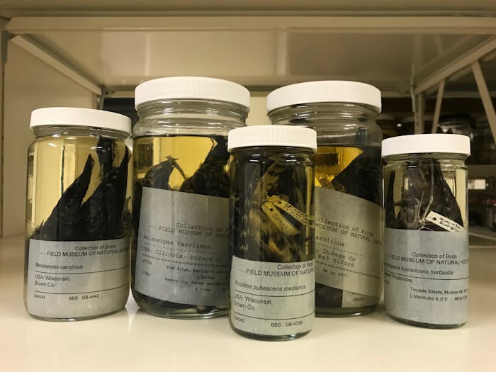
Preserved woodpecker stains
Preserved woodpeckers used for this study. 

The researchers, led by then BU graduate student George Farah, examined the preserved brains of ten woodpeckers of different species (six downy woodpeckers, one yellow-bellied sapsucker, one northern flicker, one pale-bellied woodpecker, and one lineated woodpecker) as well as a control group of the red-winged blackbird, which does not have a habit of banging its beak and head against anything.

This time around, the researchers found an abnormal buildup of the protein tau in the woodpeckers, but not in the non-pecking control group. In humans, comparable tau buildup would be an indication of brain damage. This throws into doubt the seminal results of the 1976 research that woodpeckers are not brain damaged.

According to researcher Peter Cummings, the team was surprised by the findings. “We didn’t think that we would find tau in the woodpeckers,” he tells Inverse. “From an evolutionary perspective, why would they have tau in the brains when they have been a species for thousands of years?”

Previously, researchers had believed that it was biophysical adaptations, including strong neck muscles and a long tongue that can brace the skull during the high impact pecking and hard bone towards the front of the skull (unlike birds’ usually lightweight, spongy skull, which is more aerodynamic).

But as Cummings wonders, “Why would the adaptations stop at the brain? You would think the brain itself would protect itself from impact.”

He also notes there are different types of tau, and that it is not necessarily pathologic. “Some are neuro-protective, and helps protect the neuron from becoming dysfunctional,” he says.

Of course, the scientists are quick to note that association is not causation, and that their findings require further research. Additionally, the scope of their project was limited, since the sample size was only fifteen birds, the ten woodpeckers and five blackbirds.

Woodpeckers are not the only animals that may be able to teach us more about CTE. Inverse has previously covered the lessons that zebrafish brains hold for brain injuries, while Farah says that they also considered studying the long-horned ram, which suffers similar head trauma during mating season.

Nevertheless, even the suggestion that the woodpeckers’ brains themselves may have evolved to accommodate regular head impact of such great force could provide further clues on how professional football players can continue to play America’s favorite sport — safely.

Abstract: Woodpeckers experience forces up to 1200–1400 g while pecking. It is assumed due to evolutionary adaptations, the woodpecker is immune to brain injury. This assumption has led to the use of the woodpecker as a model in the development of sports safety equipment such as football helmets. However, it is unknown at this time if the woodpecker brain develops neuro-trauma in relation to the high g-forces experienced during pecking. The brains of 10 ethanol preserved woodpeckers and 5 ethanol preserved red-winged black bird experimental controls were examined using Gallyas silver stain and anti-phospho-tau. The results demonstrated perivascular and white matter tract silver-positive deposits in eight out of the 10 woodpecker brains. The tau positive accumulations were seen in white matter tracts in 2 of the 3 woodpeckers examined. No staining was identified in control birds. The negative staining of controls birds contrasted with the diffuse positive staining woodpecker sections suggest the possibility that pecking may induce the accumulation of tau in the woodpecker brain. Further research is needed to better understand the relationship.

Mild Brain Injury Leaves Lasting Scar.


At Sunday’s World Cup Final, German soccer player Christoph Kramer knocked his head against an Argentine opponent’s shoulder with such force that Kramer spun to the ground and fell face down. The blow was one of many at this year’s competition, which further fueled a rising debate about concussion, the damages of fútbol versus football and the best response to head injuries.

Part of the challenge in understanding these injuries is how varied they can be. Although much attention has gone to severe forms of traumatic brain injury (TBI) such as concussion-induced coma, far more common are the milder impacts that come from falling off a bicycle, a low-speed car accident or taking a weak punch in a fistfight. These injuries may not entail losing consciousness but rather just a brief lack in responsiveness before recovering.

Now a group of researchers in the U.K. at Newcastle University, the University of Aberdeen and the University of Edinburgh have released results of a longer-term investigation of individuals who have suffered such first-time, minor head injuries. Their findings hint that the contusions leave a lasting trace in the brain.

The team, led by Newcastle imaging physicist Andrew Blamire, scanned the brains of 53 individuals with mild or moderate TBI within two weeks of the injury. They mapped the tracts of fibers connecting brain regions in the patients as well as in 33 healthy subjects. Blamire and colleagues discovered distinct differences between the two groups. “Even in patients with mild injury, you can detect a marker of that injury,” Blamire says. That marker may distinguish mild injuries from more forceful impacts. In cases of severe TBI, brain tissue known as white matter that envelops the tracts deteriorates, effectively mashed by the impact. But Blamire identified an opposite trend in the mild and moderate cases. For these patients, the white matter fibers became even more structured. He and his colleagues hypothesize that this organization may be caused by an inflammatory response in which the brain’s glial cells leap into action, perhaps repairing damage or blocking further injury.

Along with scanning, the team also tested thinking and memory in their subjects. Compared with healthy subjects injured patients had lower scores on multiple tests in the two-week interval after their injuries, including an average 25 percent drop on a verbal fluency test, in which an individual thinks of as many words as possible that start with a given letter. The brain changes identified in imaging correlated strongly to this test, suggesting some overlap between the affected area’s function and the test’s cognitive target.

To take their findings a step further Blamire and colleagues repeated their procedure with 23 of their head injury patients one year later. As they report in Neurology on July 16, the variability between patients was high but on average the test scores had returned to the levels of healthy individuals. The brain changes, meanwhile, remained. This suggests that once the brain has sustained damage, the scars persist.

Blamire observes that lingering signs of damage could help certain patients identify a source of their earlier mental troubles—such as memory problems—in the absence of other symptoms. For people with ongoing cognitive difficulties, it could even be useful in legal scenarios, providing evidence of head trauma even after months have past.

Neuroradiologist Michael Lipton at Albert Einstein College of Medicine, who did not participate in this research, explains that the study’s results echo findings from his and other research groups. He notes, however, that the one-year follow-up is unique and agrees that the imaging findings could reflect an inflammatory response. Lipton’s group has further hypothesized that the brain changes may even predict better recovery—in essence, that the brain has rewired itself to compensate for the damage. Only further research will reveal if this is the case, however.

Over the longer term, the findings could contribute to a growing understanding of how the brain responds to collisions and concussions. “Work like this is really essential to understanding how we can intervene,” Lipton says.

Blamire agrees, explaining that eventually brain imaging could help doctors distinguish between which patients will require counseling and treatment and which will make a full recovery on their own.