Winner of Prestigious Science Photo Competition Shows Near-Impossible Shot


By the end of high school, most of us had some idea of what an atom is. They’re the most basic forms of the elements on the periodic table. They’re tiny spheres that theoretically make up everything in the universe, from the cells in our bodies to the air that we breathe. They can be split to create an atomic bomb. Sometimes they’re drawn like solar systems of round protons and orbiting electrons, though of course that’s not actually how they look. As with so many concepts in science, the tricky thing about atoms is that we can’t really see them.

best science photography single atom

But the winning photo in a prestigious, U.K.-wide science photography competition organized by the nation’s Engineering and Physical Sciences Research Council (EPSRC) has changed that. The photo, taken by Oxford University quantum physicist David Nadlinger, Ph.D., is entitled ‘Single Atom in an Ion Trap.” The title is self explanatory: Nadlinger literally captured a photo of a single atom in a device called an ion trap. The closest scientists have come to doing this was when Griffith University researchers photographed the shadow of an atom in 2012.

But here’s Nadlinger’s atom, in all its minuscule glory. You might have to squint.

atom science photography winner u.k.
‘Single Atom in an Ion Trap’ by David Nadlinger.

Zooming into the space between the trap shows a slightly better view of the atom, which Nadlinger described with a reference to Carl Sagan’s timeless words about our planet.

“The idea of being able to see a single atom with the naked eye had struck me as a wonderfully direct and visceral bridge between the miniscule quantum world and our macroscopic reality,” Nadlinger said in an EPSRC statement. “A back-of-the-envelope calculation showed the numbers to be on my side, and when I set off to the lab with camera and tripods one quiet Sunday afternoon, I was rewarded with this particular picture of a small, pale blue dot.”

single atom photography
Nadlinger’s “pale blue dot.”

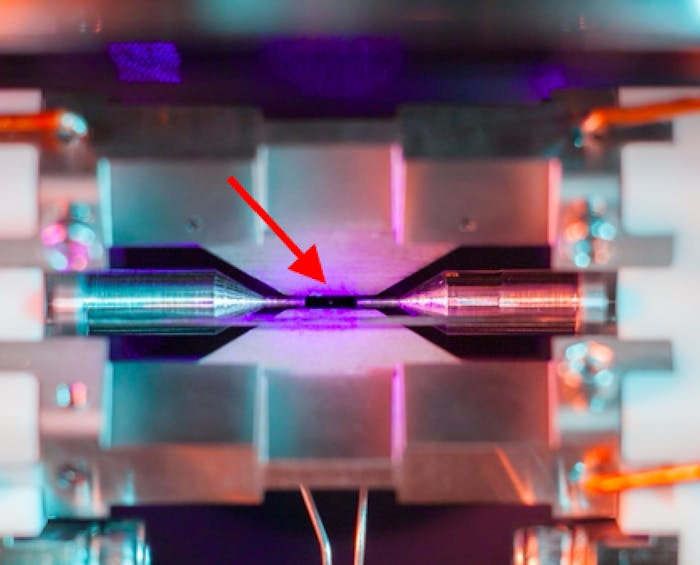
Ion traps are a family of devices that use magnetic and electric fields to capture individual charged particles (ions are just atoms that don’t have a stable number of electrons), which are useful to quantum physicists studying time and quantum computing. To prevent the atom from zooming off, the trap employs an ultra-high vacuum chamber. Nadlinger took this photo by pointing his camera through a window of this chamber, capturing the atom trapped in the 2-millimeter space between two needles.

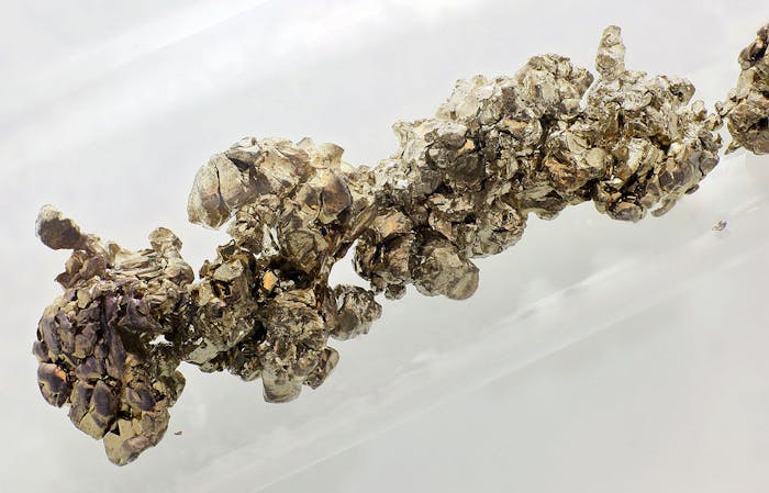
The atom in this photo is a positively charged ion of strontium; when enough of these ions network together, they form what we know as the silvery metal strontium. But since we can only see things that reflect light, Nadlinger illuminated the atom with a specific blue-violet laser, which caused the atom to absorb and re-emit enough light for a long exposure photograph to capture.

strontium
A whole lot of strontium atoms connected to each other in crystal form.

Nadlinger’s photograph may not make it any easier for students of science to understand the quantum structure of the atom, which requires breaking it down to even tinier, impossible-to-photograph parts. But at least it provides our struggling brains with something tangible to work with.

What can a graphene sandwich reveal about proteins?


Stronger than steel, but only one atom thick – latest research using the 2D miracle material graphene could be the key to unlocking the mysteries around the structure and behaviour of proteins in the very near future.

https://i0.wp.com/cdn.physorg.com/newman/gfx/news/2013/whatcanagrap.jpg

Scientists at The University of Manchester and the SuperSTEM facility, which is located at STFC’s Daresbury Laboratory and funded by the Engineering and Physical Sciences Research Council (EPSRC), have discovered that the most fragile, microscopic materials can be protected from the harmful effects of radiation when under the microscope if they are ‘sandwiched’ between two sheets of . The technique could soon be the key to enabling the direct study of every single individual atom in a , something yet to be achieved, and revolutionise our understanding of cell structure, how the immune system reacts to viruses and aid in the design of new antiviral drugs.

Observing the structure of some the tiniest of objects, such as proteins and other sensitive 2D materials, at the atomic scale requires a powerful electron microscope. This is exceptionally difficult because the radiation from the can destroy the highly fragile object being imaged before any useful data can be accurately recorded. However, by protecting fragile objects between two sheets of graphene it means they can be imaged for longer without damage under the electron beam, making it possible to quantitatively identify every single atom within the structure. This technique has proven very successful on the test case of a fragile in-organic 2D crystal and the results published in the journal ACS Nano.

During this research, the team of scientists, which included Sir Kostya Novoselov, who shared a Nobel Prize in Physics in 2010 for exploiting the remarkable properties of graphene, were able to observe the effects of encapsulating a microscopic crystal of another highly fragile 2D material, molybdenum di-sulfide, between two sheets of graphene. They found that they were able to apply a high electron beam to directly image, identify and obtain complete chemical analysis of each and every atom within the molybdenum di-sulfide sheet, without causing any defects to the material through radiation.

The University of Manchester’s Dr Recep Zan, who led the research team, said: “Graphene is a million times thinner than paper, yet stronger than steel, with fantastic potential in areas from electronics to energy. But this research shows its potential in biochemistry could also be just as significant, and could eventually open up all sorts of applications in the biotechnology arena.”

Professor Quentin Ramasse, Scientific Director at SuperSTEM added: “What this research demonstrates is not so much about graphene itself, but how it can impact the detail and accuracy at which we can directly study other inorganic 2D materials or highly fragile molecules. Until now this has mostly been possible through less direct and often complicated methods such as protein crystallography which do not provide a direct visualisation of the object in question. This new capability is particularly exciting because it could pave the way to being able to image every single atom in a protein chain for example, something which could significantly impact our development of treatments for conditions such as cancer, Alzheimer’s and HIV.”

Urine-powered mobile phone charger lets you spend a penny to make a call.


New microbial fuel cells contain bacteria that produce electricity from urine as part of their natural life cycle

A group of researchers from the University of the West of England have invented a method of charging mobile phones using urine.

Key to the breakthrough is the creation of a new microbial fuel cell (MFC) that turns organic matter – in the case, urine – into electricity.

v2-Ioannis

The MFCs are full of specially-grown bacteria that break down the chemicals in urine as part of their normal metabolic process. The bacteria produce electrons as they consume the matter and it this natural process that creates a small electrical charge to be stored in the MFC.

“No one has harnessed power from urine to do this so it’s an exciting discovery,” said Dr Ioannis Ieropoulos, an engineer at the Bristol Robotics Laboratory where the fuel cells were developed.

“The beauty of this fuel source is that we are not relying on the erratic nature of the wind or the sun; we are actually reusing waste to create energy. One product that we can be sure of an unending supply is our own urine.”

After the urine has been processed by the MFCs the electrical charge is stored in a capacitor. In the first test of the new invention, researchers simply plugged in a commercial Samsung phone charger and were able to charge up the handset.

Although the amount of electricity produced by the fuel cell is relatively small – only enough for a single call on the mobile – researcher believe it might be installed in bathrooms in the future, helping to power electric razors, toothbrushes and lights.

The device is about the size of a car battery, but engineers believe that future versions will be smaller and more portable. With each fuel cell only costing around £1 to produce such devices could provide a new, cheaper way of generating power.

The research was sponsored by public money from the Engineering and Physical Sciences Research Council and the Gates Foundation (the charity run by Microsoft-founder Bill Gates), with the scientists hopeful that the technology could be beneficial in developing countries.

“One [use] would be to put these into domestic situations or it could be used in remote regions of the developing world,” said Dr Ieropoulos.

“The fuel cells we have used to charge a mobile phone with hold around 50ml of urine but the smallest we have had working in the laboratory hold 1ml, so we can make them a lot smaller. Our aim is to have something that can be carried around easily.”

“The concept has been tested and it works – it’s now for us to develop and refine the process so that we can develop MFCs to fully charge a battery.”

Source: http://www.independent.co.uk