Rapid Weight Loss a Diabetic Retinopathy Risk?


While research is conflicting, GLP-1 therapies might cause transient worsening of retinal disease.Share on Facebook. Opens in a new tab or window

A photo of a retina affected by diabetic retinopathy

While rapid weight-loss with bariatric surgery or drugs might temporarily worsen diabetic retinopathy as blood sugar levels are rapidly corrected, the low overall risk likely doesn’t outweigh the benefits of weight loss, according to a review.

Altogether, the studies are conflicting and the evidence insufficient, Basil K. Williams Jr., MD, of the University of Miami, and colleagues concluded in Current Opinion in Ophthalmologyopens in a new tab or window.

For example, in a 1998 randomized studyopens in a new tab or window, diabetic retinopathy worsened at 6 months in 3.5% (25 of 711) of patients treated with intensive insulin therapy compared with 1.2% (nine of 728) of those on conventional insulin therapy (OR 2.98, P=0.006). At 4-year follow-up, though, retinopathy wasn’t worse than at baseline in either group.

But a 2020 multicenter case-control studyopens in a new tab or window of 3,145 patients with type 2 diabetes found no link between the use of GLP-1 agonists — a category that includes semaglutide (Ozempic, or Wegovy for weight loss) — and worsening diabetic retinopathy (P=0.47).

“The goals for diabetic retinopathy treatment are to get blood sugars, blood pressure, and weight under control. This is by far the most important thing to do for the long term, so whatever approach is right for the patient is going to be the ideal treatment,” Williams told MedPage Today. “However, it is really important to have a conversation with the patient upfront to let them know that this may worsen retinopathy temporarily. But in the long run, it’s going to be beneficial for them.”

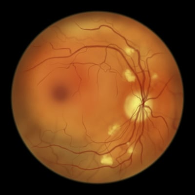
According to the review, an estimated 9.6 million people in the U.S. have diabetic retinopathy, including about one-quarter of patients with diabetes mellitus.

Clinicians have long suspected that rapidly improving blood sugar can make eye health worse. Back in 1998, the insulin therapy study noted that “there have been many reports of the curious, unanticipated, and seemingly paradoxical worsening of diabetic retinopathy after rapid improvement of blood glucose control.”

For the new review, researchers wanted to better understand the effect of rapid weight loss and improvement of HbA1c in light of the new generation of GLP-1 agonists, Williams said.

Some research did show that “when you get the diabetes controlled very, very rapidly, you can get some transient worsening of the diabetic retinopathy that improves over time,” he said.

The mechanism appeared to be related to changes in osmotic pressure in the vessels in the vascular system, he said. As blood sugar control improves, “the pressure gradient between inside the vessels and outside the vessels is different. There are more proteins now outside the vessels, and that pulls more fluid outside the vessels. That causes a little bit of additional leakage.”

This change stabilizes over time, he said. The review suggested that a sudden 2% or greater drop in HbA1c could impact retinopathy progression for 6 to 12 months. “Then things would be improving from there,” Williams noted.

Moving forward, Williams predicted that the new generation of weight-loss drugs “will be really valuable and decrease the long-term implications of diabetic retinopathy on our population. But we do have to consider that there’s a small percentage of people who will have some transient worsening. Navigating those small negatives with the overall greater benefit is something we’re going to have to deal with more and more.”

For now, the review authors recommended that patients undergo a baseline retinal examination before intensive glycemic control that leads to a rapid decrease in weight, followed by continued monitoring.

The review authors examined studies into tight insulin control, bariatric surgery, and GLP-1 agonists. They highlighted a 2022 systematic review and meta-analysisopens in a new tab or window that found that four major randomized controlled trials linked GLP-1 agonists to rapidly worsening diabetic retinopathy but also to cardiovascular benefits.

Also, a 2016 studyopens in a new tab or window of semaglutide linked the drug to a higher risk of retinopathy complications (HR 1.76, 95% CI 1.11-2.78, P=0.02), although the numbers of patients affected were small (3% [50 of 1,648] with semaglutide vs 1.8% [146 of 1,649] with placebo).

The review did not include a matched cohort study presented last year at the annual meeting of the American Society of Retina Specialists. Ehsan Rahimi, MD, of Stanford University in California, reportedopens in a new tab or window that treatment with GLP-1 agonists almost doubled the likelihood of progression from nonproliferative to proliferative diabetic retinopathy after 3 years (RR 1.585, 95% CI 1.337-1.881, P<0.0001). The drugs were also linked to significantly higher rates of progression to diabetic macular edema.

“We see these patients in our clinics all the time,” Rahimi said at the 2023 conference. “They go on these medicines, and their HbA1c crashes, goes down very quickly. That rapid reduction is thought to play some role. But if you look at the basic science literature, it’s suggested that there are direct effects of these medications on the retina. That being said, it’s also been suggested that there may be a protective effect on the retina. We’re getting a lot of mixed signals.”

Vitamin D and Diabetic Retinopathy


Low levels of vitamin D may be associated with an enhanced risk of diabetic retinopathy, according to a new study. It is just the latest in a history of work to identify a connection between the two conditions, though the potential mechanism linking the two remains mysterious.

The new study, published in Clinical Nutrition ESPEN, analyzed the data from 402 adults with type 2 diabetes, half of whom had diabetic retinopathy (DR). The two groups were very similar in most ways: There was no real difference in average age, weight, A1C, cholesterol, or use of diabetic medications. The researchers found three differences common to those who had developed diabetic retinopathy:

  • Higher systolic blood pressure (142 mmHg vs. 130 mmHg). Blood pressure is known to be one of the major causes of diabetic retinopathy, in concert with high blood sugar.
  • Longer duration of diabetes (8.5 years vs. 6.0 years). Duration of diabetes is another known risk factor for diabetic retinopathy and other complications; the longer the body endures diabetes, the more time high blood sugar levels have to cause damage and dysfunction.
  • Higher likelihood of vitamin D deficiency. 58.9 percent of those with DR had vitamin D deficiency, compared with only 33.3 percent of those without DR. Those with DR were also less than half as likely to have normal vitamin D status.

Dozens of similar studies have found similar connections. In a large 2022 review, researchers considered 36 studies to have investigated the link between DR and vitamin D, and found that 30 of them identified a correlation between low vitamin D levels and the complication — including studies of people with both type 1 and type 2 diabetes. Only a handful of the 36 studies reported no association at all.

Nevertheless, in a statement to the American Academy of Ophthalmology’s EyeNet Magazine, a spokesman from the team of researchers said that they were reluctant to draw conclusions as the research has been hampered by small sample sizes and a lack of standardized protocols. Larger and more robust experiments are still needed.

How might it work? The mechanism by which low vitamin D levels could hasten retinopathy is still a matter of conjecture. A 2023 review outlines some of the possibilities. Among other theories: Vitamin D has anti-inflammatory properties, which could help quell an overactive immune system in the eyes, and the nutrient may help prevent the formation of abnormal new blood vessels that scar the retina in late vision-threatening DR.

As yet, though, this is all conjecture. Vitamin D deficiency is not listed as a cause of DR in an American Academy of Ophthalmology article on the subject. And there is also a possibility that the causation moves in the opposite direction: Perhaps people with diabetic retinopathy are less likely to spend time outside in the sun, and that’s the cause of their lower vitamin D levels?

Vitamin D and Diabetes

The broader connections between vitamin D and diabetes have been a focus of intense research for many years. People with type 1 and type 2 diabetes generally have lower levels of vitamin D than the general public, and vitamin D deficiency has been investigated as a potential contributing factor for both conditions.

Vitamin D is important for a huge number of bodily functions and has a very complex relationship with the metabolic system. Many different studies have found that vitamin D supplementation can improve glucose management, but diabetes authorities have been extremely reluctant to validate vitamin D as an effective treatment. Despite an apparent wealth of evidence in favor of prioritizing vitamin D levels, most experts believe that science remains largely unconvincing.

In 2022, after a major study showed that vitamin D supplementation had no impact on bone health — one of the few vitamin D benefits that was widely accepted as legitimate — The New England Journal of Medicine published an editorial (PDF) arguing that doctors should stop screening vitamin D levels and that people should “stop taking vitamin D supplements to prevent major diseases or extend life.”

To be clear, severe vitamin D deficiency can have real consequences, such as rickets and osteomalacia, and supplementation may still be important for certain people, such as infants, breastfeeding mothers, or those with conditions that cause nutrient malabsorption. People with darker skin produce less vitamin D in sunlight and therefore are more likely to have lower amounts of vitamin D in circulation — Black Americans, for example, are at a sharply elevated risk of vitamin D deficiency.

Major authorities such as the Centers for Disease Control and Prevention and the National Institutes of Health continue to treat vitamin D as a nutrient of concern, making broad recommendations for vitamin D intake. On the other hand, there’s also a downside to excessive vitamin D supplementation: vitamin D toxicity.

The Bottom Line

Studies linking low vitamin D levels with diabetes and diabetic retinopathy keep piling up, but we still don’t know if the connection is real. Many experts recommend that people both with and without diabetes should prioritize vitamin D intake — whether through healthy eating, getting sunlight, or using supplements. The evidence of benefits, however, is shaky. If you’re concerned, consider asking your doctor if you should be monitoring your vitamin D levels.

How AI Is Detecting Diabetic Retinopathy


Diabetic retinopathy is often preventable if caught early. Advancements in artificial intelligence and eye-scanning technology have made it possible for more people with diabetes to get evaluated for diabetic retinopathy. 

One in three people with diabetes has diabetic retinopathy, making it one of the most common diabetes complications. 

“It steals their joy, it steals their autonomy, and it deserves a solution,” said Dr. Robert Levine, chairman of the Mary Tyler Moore Vision Initiative.

At the ADA’s Scientific Sessions in San Diego, we heard about the latest advancements in the field of diabetic retinopathy ranging from new developments in artificial intelligence to discussions on whether GLP-1 medications worsen the condition.

How artificial intelligence helps detect eye issues

The traditional approach to diabetic retinopathy screenings involves the manual interpretation of retinal images by an ophthalmologist. However, due to a shortage of eye doctors and the growing number of people with diabetes, there is an urgent need for a more accessible solution.

Eyenuk, a pioneering company in medical AI, recently gained FDA approval for an innovative technology that analyzes images of the back of your eye and immediately detects if you have some form of diabetic retinopathy. 

With the integration of artificial intelligence technology, healthcare providers can capture retinal images of their patients using what’s called a fundus camera. These images are then securely transmitted to Eyenuk’s cloud-based platform, which looks for signs of diabetic retinopathy. For those living in rural areas with limited access to ophthalmologists, this technology dramatically increases access to eye screenings.

Gaurav Agarwal, head of product and design at Eyenuk, said that Eyenuk stands out from its competitors as it’s the only device FDA-approved to detect moderate to severe vision-threatening stages of diabetic retinopathy. This is also an important preventative tool as detecting eye issues sooner allows for early intervention and better health outcomes.

How do GLP-1s affect diabetic retinopathy?

Apart from advancements in artificial intelligence, ADA panelists like Dr. Risa Wolf, a pediatric endocrinologist at Johns Hopkins University, discussed the role that GLP-1s may have on the progression of diabetic retinopathy. 

The SUSTAIN-6 clinical trial found an increased risk of retinopathy-related complications from the GLP-1 semaglutide compared to a placebo, while others like the LEADER trial and AngioSafe type 2 diabetes study found that taking GLP-1s was not associated with diabetic retinopathy. Although the data is still inconclusive, it appears that GLP-1s may lead to a temporary, short-term worsening of retinopathy that stabilizes over time. 

“My belief is that we will see long-term benefits rather than worsening,” said Dr. Blake Cooper, who works at Retina Associates of Greater Kansas City.

He talked about another ongoing clinical study, the FOCUS trial, which is investigating the long-term effects of semaglutide on diabetic retinopathy and is expected to conclude in 2027. He said current data from earlier studies found that using semaglutide for one year was not associated with an increased risk of retinopathy.

These Medications Might Make Diabetic Retinopathy Worse


Ksenia Chernaya/Pexels

By Ross Wollen

December 19th, 2022

Diabetic retinopathy is a common complication of diabetes that affects the blood vessels in the retina, the light-sensitive tissue at the back of the eye. If left untreated, diabetic retinopathy can cause severe vision loss or even complete blindness.

The good news is that diabetic retinopathy (DR) can be diagnosed long before it actually begins to impact your vision. The condition is very treatable, and potentially even reversible, especially when caught early.

Doctors have a good understanding of what causes DR: high blood sugar, high blood pressure, and, to a lesser extent, high cholesterol. Diabetic retinopathy is very sensitive to metabolic health, and many of the medications that people with diabetes commonly take can affect its progression and development — for better or for worse.

This article will review the drugs that we know (or suspect) might worsen diabetic retinopathy.

Warning

A quick word of warning: This article shouldn’t be taken as medical advice, and no patient is qualified to decide for themselves whether to avoid any drug listed here. Many of these medications are important for the health of millions of adults, and their benefits may easily outweigh whatever effect they might have on DR.

If you’re concerned that you may be taking a medication that could make your diabetic retinopathy worse, please talk to your doctor. Only a medical professional is qualified to assess the unique totality of your health conditions to recommend medication adjustments.

The Two Diabetes Drugs That (Might) Make DR Worse

Most diabetes drugs have beneficial effects for diabetic retinopathy, slowing its progression and potentially even helping to reverse the damage. Taking your glucose-lowering medications as prescribed by your doctor is absolutely one of the best ways to protect yourself from DR.

There are, however, one and possibly two exceptions to that rule, as described by a recent survey of the topic in the medical journal Eye.

Thiazolidinediones (TZDs)

TZDs have been called “the forgotten diabetes medication.” These pills directly improve insulin resistance, a root cause of type 2 diabetes, but have been deemphasized by authorities due to concerns over harmful side effects, including cardiovascular disease.

Although these drugs are increasingly out of fashion, they are still commonly prescribed. As of 2019, about 8 percent of people with type 2 diabetes used a TZD.

TZDs carry a known risk of diabetic macular edema (DME), an especially damaging form of diabetic retinopathy that affects our keenest vision in the center of our eyesight. TZDs can cause fluid retention, which appears to exacerbate the swelling of blood vessels that characterizes DME. It only happens in a small number of cases — fewer than 3 percent of those that use the drug.

There are now two types of TZDs on the market:

  • Rosiglitazone (Avandia)
  • Pioglitazone (Actos)

Luckily, cessation is associated with rapid eye improvement.

Ozempic

There is some evidence that semaglutide (Ozempic), a GLP-1RA, may increase the incidence of diabetic retinopathy. One of several pivotal studies of semaglutide found an increased risk of DR, and the FDA has reported that a significantly higher percentage of Ozempic users have DR in comparison with users of similar drugs like dulaglutide (Trulicity) and liraglutide (Victoza). The connection is disputed, however, as another large study of semaglutide found no such risk.

Even if the association is real, it’s very possible that your own doctor would conclude that semaglutide is worth the risk. Ozempic is a very effective drug for people with diabetes, typically conferring both rapid glycemic improvements and weight loss.

It seems possible that Ozempic’s effectiveness, in fact, explains its negative effect on the eyes. Contrary to all expectations, rapid improvement in glucose control can actually worsen diabetic retinopathy. This is called “early worsening,” because the eyes will get worse before the major long-term benefits of better blood sugar control become evident.

Experts don’t believe that this is necessarily a reason to avoid Ozempic. A recent discussion of the issue by experts from the American Academy of Ophthalmology suggested that “early worsening” from Ozempic is both “temporary and manageable,” although it does call for increased scrutiny from eye doctors.

Other Drugs With Negative Metabolic Effects

Drugs That Increase Blood Sugar

Some medications are known to cause blood sugar spikes:

  • Steroids, including hydrocortisone and prednisone, can have a dramatic effect on blood sugar. (Steroid creams and inhalers do not have the same effect).
  • Hormonal birth control, including the pill, the shot, the patch, and the IUD
  • Beta-blockers, which are used to treat hypertension, irregular heartbeat, and anxiety
  • Anti-psychotic drugs, which are used to treat schizophrenia and related mental illnesses

If you take any of the above drugs, it might be wise to make sure your main diabetes healthcare provider is aware of it. They may or may not suggest an adjustment.

  • Statins are also associated with rising blood sugar levels — but even taking that factor into account, experts still recommend statins for most adults with diabetes because they reduce the risk of cardiovascular disease.

Finally, if you have advanced diabetic retinopathy that requires treatment, you may be given steroid injections. These drugs can have a powerful anti-inflammatory effect within the eyeball that directly improves DR symptoms, even if they exert a negative effect on insulin sensitivity.

Drugs That Increase Blood Pressure

Hypertension (high blood pressure) is the other big factor that speeds the development and progression of diabetic retinopathy. Accordingly, medications that are known to raise your blood pressure can be considered risk factors for DR.

An incomplete list of some common drugs that can increase blood pressure includes:

  • Pain relievers, including acetaminophen (Tylenol) and nonsteroidal anti-inflammatory drugs (NSAIDs), such as ibuprofen (Motrin, Advil) and naproxen (Aleve)
  • Antidepressants, including fluoxetine (Prozac), monoamine oxidase inhibitors, and tricyclic antidepressants
  • Decongestants, including pseudoephedrine (Sudafed, Contac) and phenylephrine (Sudafed PE)
  • Hormonal birth control, including the pill, the shot, the patch, and the IUD
  • Stimulants, such as methylphenidate (Ritalin)

Some supplements, including caffeine and ginseng, can have a similar effect. The Mayo Clinic has a full article on the topic.

Drugs That Increase (Bad) Cholesterol

Experts are very confident that high blood sugar and high blood pressure both lead to diabetic retinopathy. The evidence linking high cholesterol with DR is not quite as strong, although we do know that some cholesterol-lowering drugs (especially fibrates) significantly reduce the incidence of DR.

These are some of the most common drugs that are believed to elevate “bad” cholesterol (LDL and/or triglycerides), which may or may not be risk factors for DR:

  • Steroids
  • Hormonal birth control, including the pill, the shot, the patch, and the IUD
  • Retinoids (used to treat acne)
  • Beta-blockers, which are used to treat hypertension, irregular heartbeat, and anxiety
  • Diuretics

Drugs That Increase Weight

It should be no surprise that weight gain, which is so highly related to the development and progression of type 2 diabetes, is associated with an increased risk of diabetic retinopathy. A 2021 study in Korea found that patients with recent diagnoses of type 2 diabetes that lost 10 percent of their body weight cut their risk of DR in half, whereas those that had 10 percent weight gain tripled their risk.

It’s therefore probably fair to consider any drugs that cause weight gain as potential risk factors for DR.

Some of the most common drugs associated with weight gain include:

  • Tricyclic antidepressants, including amitriptyline (Elavil), doxepin (Silenor), and nortriptyline (Pamelor)
  • Selective serotonin reuptake inhibitors (SSRIs), another type of antidepressant, including escitalopram (Lexapro), paroxetine (Paxil), sertraline (Zoloft)
  • Anti-psychotics, particularly olanzapine (Zyprexa)
  • Anti-seizure medications, including gabapentin (Gralise), pregabalin (Lyrica), and vigabatrin (Sabril).
  • Steroids
  • Beta-blockers
  • Antihistamines

The diabetes medications insulin and sulfonylureas are also associated with weight gain, which is why diabetes authorities have recently begun to prefer other options for glucose control.

Takeaways

Diabetic retinopathy is largely caused by poor metabolic health: high blood sugar, high blood pressure, and possibly high cholesterol. Many prescription medications have undesirable metabolic side effects and can therefore be considered potential contributors to DR.

Furthermore, two types of diabetes drugs in particular are associated with worsening diabetic retinopathy: the family of thiazolidinediones (TZDs), which includes rosiglitazone (Avandia) and pioglitazone (Actos), and semaglutide (Ozempic). Your ophthalmologist should be aware that you are taking one of these drugs, but will not necessarily advise a change, even if you are at high risk of vision loss from DR.

Many of the drugs discussed in this article are vital to the health of millions; therefore, it’s impossible to say whether or not readers should avoid them. As always, the guiding hand of a doctor that understands your unique health status is critical. It’s sometimes up to you to make sure that your various specialists are all on the same page. We encourage you to ensure that your main diabetes care provider and ophthalmologist are aware of every medicine you take.

This article has concentrated on the effects that various medications have on diabetic retinopathy. Medicine, of course, is only one factor of many — diet, exercise, glycemic control, and many other decisions play an immense role in the development and progression of diabetes complications.

Oral drug shows favorable safety profile in patients with diabetic retinopathy


Oral drug candidate APX3330 showed a “favorable safety and tolerability profile” in patients with diabetes and diabetic retinopathy, according to research presented in a poster at Academy 2022.

APX3330 is an oral, small-molecule drug that targets Ref-1, co-author Ronil Patel, MS, vice president of business development and market strategy at Ocuphire Pharma, told Healio.

Source: Adobe Stock.
An experimental oral drug for diabetic retinopathy showed a good safety profile.
Source: Adobe Stock

Originally developed for hepatic inflammation by Eisai, then for cancer by Apexian, Ocuphire licensed APX3330 from Apexian primarily for diabetic retinopathy, he said.

The researchers reported safety and tolerability in the 24-week, randomized, placebo-controlled, double-masked, multicenter ZETA-1 phase 2b clinical trial. Participants in the treatment group received 600 mg daily of APX3330.

Ninety-one out of 103 patients completed the trial, Patel said.

Adverse events were reported in 46 participants across both study arms, according to the poster. Twenty-two were thought to be related to the treatment, 18 of which were mild and included pruritis, rash, headache, dyspnea, vitreous floaters, various gastrointestinal symptoms, vertigo, urticaria and fatigue.

“We do not know yet if the adverse events were due to APX3330 or placebo,” Patel said.

Seven severe adverse events were reported in six participants, but they were deemed unrelated to either APX3330 or placebo, according to the study. Researchers also said that no major organ toxicities or vital sign abnormalities were seen.

“APX3330 has a good safety profile,” Patel said. “We just have to wait and see what the efficacy is.”

The efficacy endpoint is a two-step improvement in diabetic retinopathy severity score, he said.

“We’ll share the results soon,” Patel added. “We want to intervene before a patient needs injections.”

Watch Out for Diabetic Retinopathy


Diabetic retinopathy is one of several common eye diseases, but is the most common cause of vision impairment and blindness among working-age adults in the United States. From 2010 to 2050, the number of Americans with diabetic retinopathy is expected to nearly double, from 7.7 million to 14.6 million. Diabetic retinopathy occurs when diabetes affects the blood vessels in the retina (the light-sensitive tissue in the back of the eye), causing them to leak and distort vision. If not found and treated early, diabetic retinopathy can cause permanent vision loss.

Man holding hands over eyes

Stay Alert

Diabetic retinopathy may not have any symptoms in the early stages. So if you have diabetes, be sure to schedule a comprehensive dilated eye exam once a year. Diabetic retinopathy can be diagnosed and treated before you notice any vision problems.

Symptoms that could indicate that the disease has progressed to a more advanced stage include:

  • Blurry vision
  • Spots that “float” in your vision
  • Halos around lights
  • Loss of central vision
  • Loss of color vision

Anyone with type 1 or type 2 diabetes, or women who had diabetes during pregnancy (gestational diabetes), can develop diabetic retinopathy. The risk increases the longer a person has diabetes and when blood sugar, blood pressure, and cholesterol levels are hard to control.

Fruit, measuring tape, dumbbell, glucose monitoring meterHealthy eating can reduce your risk for diabetic retinopathy.

Stay on Top of It

There are simple steps you can take to keep your eyes healthy and make sure you’re seeing your best. Taking an active role in managing your diabetes is critical:

  • Make healthy eating and physical activity part of your daily routine. This can help control blood pressure, blood sugar, and cholesterol, which can reduce your risk for developing diabetic retinopathy.
  • Quit smoking or never start. Smoking increases your risk for developing many complications from diabetes, including diabetic retinopathy.
  • If you have diabetes, schedule an annual comprehensive dilated eye exam. This can help catch vision problems early.
  • Closely follow your doctor’s instructions on how often to check your blood sugar. Keep your blood sugar as close as possible to the target range your doctor recommends.
  • If you notice any changes in your vision in one or both eyes, contact an eye doctor (ophthalmologist or optometrist) right away.

Stay Positive

Although there is no cure for diabetic retinopathy, some treatments can prevent permanent vision loss. Your eye doctor may recommend laser treatment that can help shrink blood vessels, injections that can reduce swelling, or surgery. It’s important for you to go to all follow-up appointments that your doctor schedules.

If you have diabetic retinopathy, low-vision rehabilitation and aids such as magnifying glasses, large-print newspapers, and telescopic lenses can help you stay independent. Ask your eye doctor about seeing a low-vision specialist.

Source:www.cdc.gov

For all book lovers please visit my friend’s website.
URL: http://www.romancewithbooks.com

Diabetic Retinopathy Risk Drops With Diet Rich in Marine PUFAs


Eating at least two weekly servings of oily fish, rich in omega-3 polyunsaturated fatty acids (PUFAs), can help middle-aged and older people with type 2 diabetes reduce their risk for diabetic retinopathy, suggests a post hoc analysis of a major diet trial.

After adjusting for factors including age, sex, and intervention group, researchers from the PREDIMED trial found that participants who were 55 years or older and consumed at least 500 mg/day of omega-3 PUFAs showed a 48% reduced risk for incident diabetic retinopathy compared with those who consumed less than 500 mg/day (hazard ratio, 0.52; P = .001).

“Higher risk reductions were observed in participants with hypertension, those with diabetes of greater than 5 years’ duration, and those treated with insulin at baseline,” according to the report from Aleix Sala-Vila, DPharm, PhD, a researcher at CIBER-Fisiopatología de la Obesidad y Nutrición, Institut d’investigacions Biomèdiques August Pi i Sunyer, in Barcelona, Spain, and colleagues, published online August 18 in JAMA Ophthalmology.

Results align with findings from experimental models and with what researchers already know about how omega-3 cells affect diabetic retinopathy, according to the group.

“It is a fact that the amount of omega-3 in our body, and therefore in our retina, can be modulated by our diet,” Dr Sala-Vila told Medscape Medical News. “A sustained consumption of two weekly servings of fatty fish will increase the levels of omega-3 in cells. This would prevent or at least counteract inflammation in our body, a key player in the onset and progression of diabetic retinopathy. Our data reinforce a notion to date only explored in animals.”

Dr Sala-Vila said it is unclear whether supplements might have the same effect as eating fish and pointed to possible doubts raise by the ORIGIN trial (N Engl J Med. 2012;367:309-318). The trial showed no reduction of cardiovascular events over 6 years for participants initially at high cardiovascular risk who took 1000 mg/day of omega-3 PUFAs in a supplement.

Authors of the current study summarize its contribution to the literature: “Our findings support the view that regular consumption of oily fish might be beneficial to delay the onset or progression of vascular diseases in arterial beds other than the coronary and cerebrovascular ones.”

Study Based on PREDIMED Data

Data were analyzed from people with type 2 diabetes in PREDIMED, a nutrition intervention trial conducted in Spain that tested Mediterranean diets supplemented with extra virgin olive oil or nuts vs a low-fat control diet for primary cardiovascular prevention.
The current analysis is based on a PREDIMED subcohort of 3614 persons with type 2 diabetes at baseline; their aged ranged from 55 to 80 years. Full data were available for 3482 participants (48% men; average age, 67 years). Food intake was assessed at baseline and yearly for 6 years of follow-up using a 137-item food-frequency validated for the PREDIMED study. Then researchers interviewed participants on frequency of consumption of each food item in the past year and asked about usual portion sizes.

The main outcome was incident diabetic retinopathy requiring laser photocoagulation, vitrectomy, and/or antiangiogenic therapy. After a follow-up of an average 6 months, researchers found 69 new diabetic-retinopathy events.

Even More Benefit in United States?

In an accompanying editorial, Michael Larsen, MD, DMSc, department of ophthalmology, Rigshospitalet-Glostrup and University of Copenhagen in Glostrup, Denmark, notes that the study was conducted in Spain, primarily in large urban centers, where fish is a mealtime staple.

“Fish and nuts are already part of the food culture, available in every supermarket, cafeteria, and restaurant and in most households.” It is no surprise, he says, that 75% (2611 participants) met target omega-3 consumption levels at baseline.

He told Medscape Medical News that the potential for change in preventing diabetic retinopathy is greater in the United States, where consumption of fish and tree nuts (also high in omega-3 PUFAs) is much lower.

“If the results can be verified and if they extrapolate to the bottom stratum of the [omega-3] scale, where many of us roam, the implications for public health will be considerable,” he writes. “The potential value of a large-scale switch to a diet rich in [omega-3] fatty acids merits serious attention.”

Vitamin D deficiency may confer risk for retinopathy in diabetes


Patients with diabetic retinopathy are more likely to have lower 25-hydroxyvitamin D levels vs. healthy controls, according to a speaker here.

“Vitamin D may have a role in the pathophysiology of creating diabetic retinopathy,” Anawin Sanguankeo, MD, of the department of medicine at Bassett Medical Center in Cooperstown, New York, said during a media briefing at the AACE Scientific and Clinical Congress. “In the future, there should be studies that assess [whether] giving vitamin D to patients that have diabetes will prevent diabetic retinopathy or slow progression in patients who already have [diabetic retinopathy] compared to patients who do not have optimal vitamin D.”

Anawin Sanguankeo

Anawin Sanguankeo

Vitamin D deficiency, Sanguankeo said, has been associated with several cardiometabolic complications, including insulin secretion, metabolic syndrome and systemic diabetes progression. The role of vitamin D in the pathogenesis of diabetic retinopathy in humans remains an area of debate, though studies in rat models suggest an association, he said.

Sanguankeo, Sikarin Upala, MD, also of Bassett Medical Center, and colleagues completed a systematic review and meta-analysis of 11 observational studies conducted through July 2015, assessing the relationship between vitamin D deficiency (serum 25-(OH)D 20 ng/mL or less) and diabetic retinopathy (n = 9,350). Researchers calculated the pooled effect estimate of diabetic retinopathy, comparing patients with optimal vitamin D levels and vitamin D-deficient groups, and calculated the pooled mean difference of 25(OH)D levels between patients with diabetic retinopathy and healthy controls, using a random-effect, Mantel-Haenszel analysis.

Researchers found an association between diabetic retinopathy and vitamin D deficiency (OR = 1.27; 95% CI, 1.17-1.37). In patient subgroups, researchers also found that patients with diabetic retinopathy had lower serum 25-(OH)D levels vs. controls, with a mean difference of –2.22 ng/mL; 95% CI, –2.78 to –1.67).

“I think that evidence is strong enough to have future studies in this area,” Sanguankeo said. “Before there is a recommendation for clinicians [ to supplement with vitamin D], there should be evidence stronger than this, in randomized controlled trials or in prospective cohort studies. This is just the beginning of future studies that should be done.”

Randomized Clinical Trial Evaluating Intravitreal Ranibizumab or Saline for Vitreous Hemorrhage From Proliferative Diabetic Retinopathy.


Importance  Vascular endothelial growth factor plays a role in proliferative diabetic retinopathy (PDR). Intravitreal injection of saline has been shown potentially to lead to improved visual acuity compared with observation alone in eyes with vitreous hemorrhage. Therefore, it is important to determine if intravitreal anti–vascular endothelial growth factor can reduce vitrectomy rates (and risks associated with vitrectomy) compared with saline for vitreous hemorrhage from PDR that precludes placement or confirmation of complete panretinal photocoagulation.

Objective  To evaluate intravitreal ranibizumab compared with intravitreal saline injections on vitrectomy rates for vitreous hemorrhage from PDR.

Design  Phase 3, double-masked, randomized, multicenter clinical trial. Data reported were collected from June 2010 to March 2012 and include 16 weeks of follow-up.

Setting  Community-based and academic-based ophthalmology practices specializing in retinal diseases.

Participants  Two hundred sixty-one eyes of 261 study participants, who were at least 18 years of age with type 1 or type 2 diabetes mellitus. Study eyes had vitreous hemorrhage from PDR precluding panretinal photocoagulation completion.

Intervention  Eyes were randomly assigned to 0.5-mg intravitreal ranibizumab (n = 125) or intravitreal saline (n = 136) at baseline and 4 and 8 weeks.

Main Outcome Measure  Cumulative probability of vitrectomy within 16 weeks.

Results  Cumulative probability of vitrectomy by 16 weeks was 12% with ranibizumab vs 17% with saline (difference, 4%; 95% CI, −4% to 13%) and of complete panretinal photocoagulation without vitrectomy by 16 weeks was 44% and 31%, respectively (P = .05). The mean (SD) visual acuity improvement from baseline to 12 weeks was 22 (23) letters and 16 (31) letters, respectively (P = .04). Recurrent vitreous hemorrhage occurred within 16 weeks in 6% and 17%, respectively (P = .01). One eye developed endophthalmitis after saline injection.

Conclusions and Relevance  Overall, the 16-week vitrectomy rates were lower than expected in both groups. This study suggests little likelihood of a clinically important difference between ranibizumab and saline on the rate of vitrectomy by 16 weeks in eyes with vitreous hemorrhage from PDR. Short-term secondary outcomes including visual acuity improvement, increased panretinal photocoagulation completion rates, and reduced recurrent vitreous hemorrhage rates suggest biologic activity of ranibizumab. Long-term benefits remain unknown. Whether vitrectomy rates after saline or ranibizumab injection are different than observation alone cannot be determined from this study.


Source: JAMA