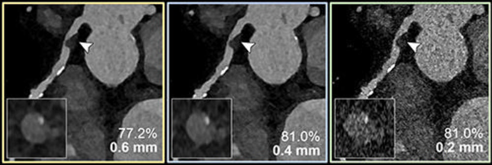
CAD Assessment Improves With Photon-Counting CT


Ultrahigh-spatial-resolution photon-counting detector CT improved assessment of coronary artery disease (CAD), allowing for reclassification to a lower disease category in 54% of patients. According to a new study published in Radiology, the technology has the potential to improve patient management and reduce unnecessary interventions.

Coronary CT angiography is a first-line test in the assessment of coronary artery disease. However, its diagnostic value is limited in patients with severe calcifications, or calcium buildup in the plaque of the coronary arteries.

Ultrahigh-spatial-resolution photon-counting detector CT (PCD-CT) improves image quality compared to conventional CT. Additionally, it provides better spatial resolution, or the ability to differentiate two adjacent structures as being distinct from one another.

“Our study provides a glimpse into the potential impact of performing coronary CT angiography using ultrahigh spatial resolution technology on risk reclassification and recommended downstream testing,” said study co-author Tilman Emrich, MD, attending radiologist at the University Medical Center Mainz in Germany, and assistant professor of radiology at the Medical University of South Carolina in Charleston.

For the study, researchers evaluated coronary stenoses, or narrowing in the coronary arteries, in a vessel phantom (in-vitro) containing two different stenosis grades (25%, 50%), and retrospectively in 114 patients (in-vivo) who underwent ultrahigh-spatial-resolution cardiac PCD-CT for the evaluation of coronary artery disease. In-vitro values were compared to the phantom’s manufacturer specifications, and patient results were assessed regarding effects on coronary artery disease reporting and data system reclassification (CAD-RADS).

“The study used a combination of artificial vessel models and real-world patient data,” Dr Emrich said. “It simulated three types of reconstructions from a single PCD-CT scan, resembling conventional CT, high-resolution, and ultrahigh-spatial-resolution scans. Observers evaluated the severity of stenosis and generated CAD-RADS classifications, guiding further patient management decisions.”

In-vitro results demonstrated a reduced overestimation of the stenosis by ultrahigh-spatial-resolution scans by reducing the adverse effects of the calcifications on the image.

Results from the patients with suspected or diagnosed coronary artery disease confirmed a lower median degree of stenosis for calcified plaques (29% vs. 42%) with ultrahigh-spatial-resolution PCD-CT compared to standard CT. Ultrahigh-spatial-resolution often led to patients being reclassified to a lower CAD-RADS category. Of the 114 patients, 54% were given a lower CAD-RADS classification than they were originally assigned. The researchers found in-vitro quantification of the 193 coronary CT angiography-based stenoses was also more accurate using ultrahigh-spatial-resolution than standard resolution.

“We found that ultrahigh-spatial-resolution reconstructions resulted in significant changes in recommendations for over 50% of patients,” Dr Emrich said. “The impact was particularly notable in cases with calcified plaques, where ultrahigh-spatial-resolution reduced the overestimation of stenosis.”

Dr Emrich explained that ultrahigh-spatial-resolution may address the current limitations of conventional cardiac CT angiography by reducing the overestimation of stenosis due to calcium blooming, an effect which can cause small, high-density structures—such as calcifications—to appear larger than their true size.

“This could significantly alter recommendations for downstream testing, potentially leading to a reduction of unnecessary procedures (and their potential complications) and reduced healthcare costs,” he said.

No substantial benefits of ultrahigh-spatial-resolution were observed for mixed and non-calcified plaques.

“It is important to note that these findings are from a simulation study, and further validation is needed in real-world comparisons,” Dr Emrich said.

Coronary CT Angiography Can Track Regression of Noncalcified Plaques by Statin Therapy


Moderate and intensive statin treatment can retard or even bring about regression of noncalcified coronary plaque as shown by coronary CT angiography (CCTA), according to new research from China[1].

“Clinicians should know the potential usage of CT scans for high-risk patients, and if noncalcified plaques are found, strengthened statin therapy should be considered,” lead author Dr Bin Lu (Fuwai Hospital, Beijing) told heartwire from Medscape by email. “If clinicians know patients have positive coronary noncalcified plaques based on CT images, they should recommend to their patients to take statins regularly and follow up the disease after 1 or 2 years.”

Extensive clinical trial evidence points to serial coronary intravascular ultrasound (IVUS) as an imaging method to show that statin therapy can stop progression of coronary plaque or induce regression, but IVUS is invasive and unsuitable for nonischemic patients. However, CCTA has emerged as an imaging tool that can accurately and noninvasively measure luminal narrowing and characterize coronary plaques, according to the researchers.

They conducted a prospective multicenter observational study involving 206 consecutive patients with mild noncalcified plaque and undergoing CCTA.

The results were published online July 13, 2016 in the American Heart Journal.

The researchers divided patients into three groups: intensive statin therapy (n=55), moderate statin therapy (n=85), or no statin therapy (n=66). Patients in the intensive group took 20 to 40 mg/d atorvastatin or 10 to 20 mg/d rosuvastatin. Patients in the moderate group took 10 to 20 mg/d atorvastatin, 5 to 10 mg/d rosuvastatin, 20 to 40 mg/d fluvastatin, or 10 to 20 mg/d simvastatin.

The researchers performed serial scans after a median interval of 18 months to measure low-attenuation plaque (LAP) volume, total plaque volume, and percent plaque volume.
Patients in the intensive group experienced significantly improved lipid profiles during follow-up: total cholesterol, LDL-C, and triglycerides significantly decreased (P<0.001 all), while HDL-C increased nonsignificantly. Patients in the moderate-therapy group also experienced decreases in total cholesterol, LDL-C, and triglycerides (P<0.05), while HDL-C slightly increased.

Patients in the no-treatment group experienced no lipid-profile change.

On CCTA, the researchers observed significant regression of LAP volume, total plaque volume, and percent plaque volume in the intensive group compared with the no-treatment group (-7.1 vs. 0.9, -16.4 vs 12.3, -6.2 vs 3.5, respectively, P<0.001 all). They observed retarded progression of LAP, total plaque volume, and percent plaque volume in the moderate-treatment group.

Using a multivariable prediction model, the researchers found that total plaque volume, higher baseline LAP volume, and moderate and intensive therapy each predicted plaque regression (P<0.001, 0.004, and <0.001, respectively).

Based on their study results, they concluded, “This further confirms the feasibility of using serial CCTA to assess changes in plaque characteristics, allowing this method to potentially track atherosclerosis noninvasively.

“After this study, we are now full of confidence to treat our patients who have positive coronary plaques with statins. However, we need to do more about following up the patients for major adverse cardiac events after early detection and treatment of noncalcified coronary artery plaques,” Lu said.

Mild Stenosis Linked to Death in Diabetes


Even modest coronary plaque causing no symptoms has a long-term impact on mortality and heart disease in diabetes, an observational study showed.

The adjusted mortality risk was similarly elevated by twofold whether coronary CT angiography showed mild stenosis of less than 50% or obstructive stenosis of 50% or more (hazard ratios 2.0 and 2.1,P=0.003 and P<0.001, respectively), Philipp Blanke, MD, of the University of British Columbia and St. Paul’s Hospital in Vancouver, and colleagues found in the CONFIRM registry.

The mortality risk with nonobstructive coronary artery disease was similar to that of having single-vessel obstructive disease (P=0.42), the researchers reported at the Radiological Society of North America meeting here.

Overall major adverse cardiovascular events (death, myocardial infarction, unstable angina, or late coronary revascularization) showed about double the risk with obstructive disease as with the milder stenosis, but both were significant, with HRs of 10.4 and 4.9, respectively (both P<0.001).

“Coronary computed tomographic angiography in diabetics can be used for long-term prognostication with respect to mortality and major adverse cardiovascular events,” the group concluded.

However, screening of diabetes patients for asymptomatic coronary artery disease with coronary CT angiography to guide management wasn’t any better than simply aggressively targeting risk factors in the FACTOR 64 trial, reported in November at the American Heart Association meeting.

“A lot of patients end up having their first symptom as a heart attack or even death. We would like to be able to identify those patients and treat them before they die or have a heart attack,” said J. Brent Muhlestein, MD, of the FACTOR 64 trial. Muhlestein is from Intermountain Medical Center and the University of Utah in Salt Lake City.

While CT screening wasn’t the solution, “aggressive medical management of all patients significantly reduced the number of adverse events that happened in diabetic patients in both the patients who were in the control arm and also in the scanning arm,” he pointed out to MedPage Today. “We also found that 70% of the patients who did have asymptomatic diabetes also did have some degree of atherosclerosis in their coronary arteries which justifies secondary prevention risk management.”

The Coronary CT Angiography Evaluation For Clinical Outcomes: An International Multicenter (CONFIRM) Registry was designed to look for prognostic value of cardiac CT angiography in coronary artery disease-related events.

Among the more than 40,000 patients with CT angiography data from more than a dozen centers around the world, Blanke’s analysis included the 1,823 with diabetes and at least 5-years of follow-up but no prior clinically-apparent coronary artery disease.

Action Points

  • Note that this study was published as an abstract and presented at a conference. These data and conclusions should be considered to be preliminary until published in a peer-reviewed journal.
  • In patients with diabetes, both nonobstructive and obstructive CAD by coronary computed tomographic angiography are associated with higher rates of all-cause mortality and MACE when followed for 5 years. The relative risk of nonobstructive disease is comparable to single vessel obstructive disease.