Childhood Cancer: Novel Nanoparticle Shows Early Promise for Brain Tumor


Nanoparticles rain down on the blood brain barrier. A cell receives radiation and expresses P-selectin, which allows the nanoparticles to be taken into the cell and past the barrier.

The human brain is profoundly complex, consisting of tens of billions of neurons that form trillions of interconnections. This complex neural wiring that allows us to think, feel, move, and act is surrounded by what’s called the blood-brain barrier (BBB), a dense sheet of cells and blood vessels. The BBB blocks dangerous toxins and infectious agents from entering the brain, while allowing nutrients and other essential small molecules to pass right through.

This gatekeeping function helps to keep the brain healthy, but not when the barrier prevents potentially life-saving drugs from reaching aggressive, inoperable brain tumors. Now, an NIH-funded team reporting in the journal Nature Materials describes a promising new way to ferry cancer drugs across the BBB and reach the sites of disease [1]. While the researchers have not yet tried this new approach in people, they have some encouraging evidence from studies in mouse models of medulloblastoma, an aggressive brain cancer that’s diagnosed in hundreds of children each year.

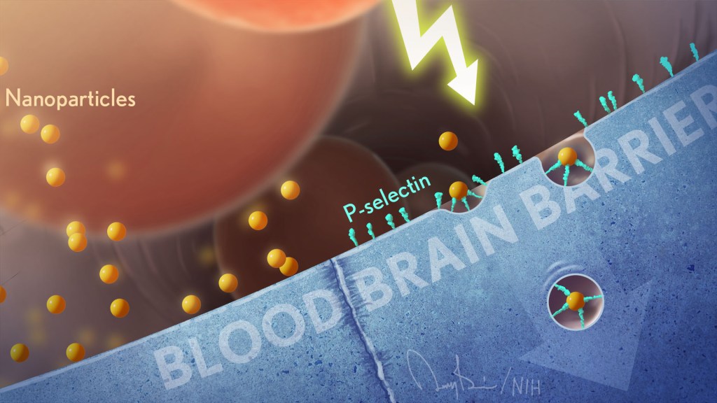
The team, including Daniel Heller, Memorial Sloan Kettering Cancer Center, New York, NY, and Praveen Raju, Icahn School of Medicine at Mount Sinai, New York, NY, wanted to target a protein called P-selectin. The protein is found on blood vessel cells at sites of infection, injury, or inflammation, including cancers. The immune system uses such proteins to direct immune cells to the places where they are needed, allowing them to exit the bloodstream and enter other tissues.

Heller’s team thought they could take advantage of P-selectin and its molecular homing properties as a potential way to deliver cancer drugs to patients. But first they needed to package the drugs in particles tiny enough to stick to P-selectin like an immune cell.

That’s when they turned to a drug-delivery construct called a nanoparticle, which can have diameters a thousand times smaller than that of a human hair. But what’s pretty unique here is the nanoparticles are made from chains of sugar molecules called fucoidan, which are readily extracted from a type of brown seaweed that grows in Japan. It turns out that this unlikely ingredient has a special ability to attract P-selectin.

In the new study, the researchers decided to put their novel fucoidan nanoparticles to the test in the brain, while building on their previous animal work in the lungs [2]. That work showed that when fucoidan nanoparticles bind to P-selectin, they trigger a process that shuttles them across blood vessel walls.

This natural mechanism should also allow nanoparticle-packaged substances in the bloodstream to pass through vessel walls in the BBB and into the surrounding brain tissue. The hope was it would do so without damaging the BBB, a critical step for improving the treatment of brain tumors.

In studies with mouse models of medulloblastoma, the team loaded the nanoparticles with a cancer drug called vismodegib. This drug is approved for certain skin cancers and has been tested for medulloblastoma. The trouble is that the drug on its own comes with significant side effects in children at doses needed to effectively treat this brain cancer.

The researchers found that the vismodegib-loaded nanoparticles circulating in the mice could indeed pass through the intact BBB and into the brain. They further found that the particles accumulated at the site of the medulloblastoma tumors, where P-selectin was most abundant, and not in other healthy parts of the brain. In the mice, the approach allowed the vismodegib treatment to work better against the cancer and at lower doses with fewer side effects.

This raised another possibility. Radiation is a standard therapy for children and adults with brain tumors. The researcher found that radiation boosts P-selectin levels specifically in tumors. The finding suggests that radiation targeting specific parts of the brain prior to nanoparticle treatment could make it even more effective. It also may help to further limit the amount of cancer-fighting drug that reaches healthy brain cells and other parts of the body.

The fucoidan nanoparticles could, in theory, deliver many different drugs to the brain. The researchers note their promise for treating brain tumors of all types, including those that spread to the brain from other parts of the body. While much more work is needed, these seaweed-based nanoparticles may also help in delivering drugs to a wide range of other brain conditions, such as multiple sclerosis, stroke, and focal epilepsy, in which seizures arise from a specific part of the brain. It’s a discovery that brings new meaning to the familiar adage that good things come in small packages.

Endocrine Abnormalities in Aging Survivors of Childhood Cancer: A Report From the Childhood Cancer Survivor Study


Abstract

Purpose The development of endocrinopathies in survivors of childhood cancer as they age remains understudied. We characterized endocrine outcomes in aging survivors from the Childhood Cancer Survivor Study on the basis of therapeutic exposures.

Patients and Methods We analyzed self-reported conditions in 14,290 5-year survivors from the Childhood Cancer Survivor Study, with a median age 6 years (range, < 1 to 20 years) at diagnosis and 32 years (range, 5 to 58 years) at last follow-up. Identification of high-risk therapeutic exposures was adopted from the Children’s Oncology Group Long-Term Follow-Up Guidelines. Cumulative incidence curves and prevalence estimates quantified and regression models compared risks of primary hypothyroidism, hyperthyroidism, thyroid neoplasms, hypopituitarism, obesity, diabetes mellitus, or gonadal dysfunction between survivors and siblings.

Results The cumulative incidence and prevalence of endocrine abnormalities increased across the lifespan of survivors (P < .01 for all). Risk was significantly higher in survivors exposed to high-risk therapies compared with survivors not so exposed for primary hypothyroidism (hazard ratio [HR], 6.6; 95% CI, 5.6 to 7.8), hyperthyroidism (HR, 1.8; 95% CI, 1.2 to 2.8), thyroid nodules (HR, 6.3; 95% CI, 5.2 to 7.5), thyroid cancer (HR, 9.2; 95% CI, 6.2 to 13.7), growth hormone deficiency (HR, 5.3; 95% CI, 4.3 to 6.4), obesity (relative risk, 1.8; 95% CI, 1.7 to 2.0), and diabetes mellitus (relative risk, 1.9; 95% CI, 1.6 to 2.4). Women exposed to high-risk therapies had six-fold increased risk for premature ovarian insufficiency (P < .001), and men demonstrated higher prevalence of testosterone replacement (P < .001) after cyclophosphamide equivalent dose of 20 g/m2 or greater or testicular irradiation with 20 Gy or greater. Survivors demonstrated an increased risk for all thyroid disorders and diabetes mellitus regardless of treatment exposures compared with siblings (P < .001 for all).

Conclusion Endocrinopathies in survivors increased substantially over time, underscoring the need for lifelong subspecialty follow-up of those at risk.

DISCUSSION

In the largest study to date to our knowledge, we examined the evolution of endocrine outcomes in aging survivors of childhood cancer in the context of prior treatment exposures. Our findings indicate that the cumulative risk of developing endocrine disorders steadily increases over time for most outcomes. These risks are particularly great for survivors previously treated with specific high-risk exposures, such as high-dose irradiation of the head, neck, or pelvis, and after exposure to high doses of alkylating agents. For many outcomes, even survivors exposed to non–high-risk therapies were significantly more likely to develop an endocrine disorder compared with siblings.

The magnitude and burden of endocrine abnormalities in childhood cancer survivors, particularly after high-risk cancer therapies, are striking.7,8,16,17 In a study of 310 adult survivors, Brignardello et al7 reported at least one endocrine disorder in more than half of survivors, 16 years after the diagnosis of childhood cancer. Patterson et al16 described a wide range of endocrine health conditions in 519 pediatric-age survivors observed in a survivor program, with many of the identified endocrinopathies associated with radiotherapy or stem-cell transplantation; however, their study did not include survivors of brain tumors, a group typically affected by endocrinopathies. In our study, nearly half of childhood cancer survivors experienced at least one endocrine abnormality, 16.7% at least two, and 6.6% three or more.

Among aging survivors, particularly after cranial irradiation, endocrine conditions with vague or minimal clinical symptoms, such as HP deficits, frequently remain undiagnosed or undertreated because of a lack of formal assessment. In this study, cumulative incidence of GH deficiency in CCSS participants after HP irradiation with 18 Gy or greater reached 17.3% within 15 years of primary cancer diagnosis and subsequently plateaued (Fig 5). These findings reflect an underascertainment of GH-deficient adult CCSS participants without a prior diagnosis of GH deficiency during childhood, resulting from a lack of systematic clinical follow-up. In contrast, Chemaitilly et al8 demonstrated an increasing prevalence of unrecognized adult GH deficiency in a cohort of childhood cancer survivors exposed to cranial irradiation when risk-based screening was applied. Furthermore, they reported a frailty phenotype of 13.1% in their survivors with untreated pituitary deficits at a median age of 33.6 years, a rate observed in the general population among individuals age 65 years or older. These findings underscore the long-term morbidity associated with unrecognized and untreated endocrinopathies in adult childhood cancer survivors.

Similarly, male survivors born in the 1950s were less likely to receive testosterone replacement, despite treatment exposures associated with the development of primary gonadal dysfunction, compared with survivors born after the 1980s. This likely reflects both underdiagnosis and undertreatment of this outcome. Because untreated gonadal insufficiency in men is associated with abdominal obesity, hypertension, dyslipidemia, low bone mineral density, sarcopenia, and frailty,8 timely diagnosis and treatment of gonadal dysfunction in aging male survivors is critical.

The COG-LTFU Guidelines provide consensus-based, exposure-specific recommendations for screening and management of late effects of pediatric cancer therapy and are intended for survivors 2 or more years after completion of therapy.13Both our data and those from the recent St Jude Life studies8,18 validate the utility of the COG-LTFU Guidelines for identifying survivors with exposures that place them at high risk for late adverse endocrine outcomes.19 Nevertheless, as noted in our study, even childhood cancer survivors treated with non–high-risk therapies demonstrated an increased risk for certain endocrine outcomes compared with siblings, highlighting the need for long-term surveillance and individualized screening practices in childhood cancer survivors even in the absence of high-risk treatment exposures.

The CCSS has many unique strengths, including longitudinal characterization of overall health conditions in a large, geographically diverse population of aging childhood cancer survivors, with detailed treatment information through middle adulthood, along with a sibling comparison population. Despite these strengths, a number of limitations must be considered when interpreting our findings. First, the outcomes are self-reported, with external validation only for thyroid malignancies, which could lead to both over- and under-reporting of certain outcomes; however, we used strict criteria, such as reported use of hormone replacement for endocrine outcomes of interest, to overcome some of these limitations. Second, given the lack of hormone data to differentiate between primary versus central endocrine dysfunction in some instances, our findings may have resulted in some misclassification of these outcomes. Third, the role of selection and surveillance bias should be considered when interpreting these results. The latter may explain, at least in part, the elevated risk of thyroid cancer in survivors in the non–high-risk exposure group. Finally, treatments for childhood malignancies have evolved over time, and for some cancers, such as HL and acute lymphoblastic leukemia, in which irradiation has been either eliminated or the dose fields reduced, the risks for several endocrine disorders will likely be lower than those reported in this study. However, the chemotherapy and radiotherapy treatments used in this cohort remain the backbone of therapeutic protocols for many childhood malignancies.20,21 Lastly, our findings are based on the most recent COG-LTFU Guidelines (ie, version 4.0), which provide consensus-based definitions for high-risk exposures for endocrine outcomes of interest. As new information emerges, these definitions may change, which could affect the reported risk estimates.

In summary, the prevalence and cumulative incidence of endocrine outcomes continue to increase as survivors age, particularly after high-risk treatment exposures. These findings underscore the importance of lifelong screening of at-risk childhood cancer survivors for endocrine abnormalities. The National Cancer Policy Board of the Institute of Medicine recommends that survivors receive risk-based care22; thus, our findings provide a compelling rationale for continued risk-based endocrine screening throughout adulthood.


Cranial Radiation Ups Obesity Risk After Childhood Cancer


Childhood cancer survivors are more likely to be obese than other people of the same age, sex, and race without a history of childhood cancer, a new population-based study confirms.

In particular, cranial radiation for childhood cancers appears to have the greatest influence on obesity in adult life, with 47% of those who received this form of treatment becoming obese, vs 29.4% who did not.

The presence of obesity at cancer diagnosis also affects outcomes, as do age at diagnosis and the presence of polymorphisms in genes responsible for neural growth, repair, and connectivity in patients who received cranial radiation.
The study was published online May 11 in Cancer. Lead researcher Carmen Wilson, PhD, from St Jude Children’s Research Hospital, Memphis, Tennessee, told Medscape Medical News, “These results are important because obesity is associated with an increased risk of premature death and a variety of health problems, including cancer and heart disease.”

She also pointed out that the findings stem from “one of the most comprehensive studies yet of obesity in childhood cancer survivors”; of the almost 2000 survivors in the study, “36.2% had a body mass index [BMI] of 30 kg/m2 or more [during 25 years of follow-up], which is defined as obese.”

And she emphasized that the long-term goal of the St Jude research group is to develop effective weight-management strategies for the growing population of childhood cancer survivors in the United States.

Reflecting on the paper, which he found both “fascinating and provocative,” Gerald Denis, PhD, associate professor of pharmacology and medicine, Cancer Research Center, Boston University School of Medicine, Massachusetts, said the results demonstrate the challenges of minimizing the risk of obesity while treating the childhood cancer.

The latter is understandably the priority given the lack of certainty that patients will go on to develop obesity, “but the concern here is partly disciplinary: they [oncologists] are not licensed to treat obesity later in life.”
“Still, in light of these new data, it would be helpful for the professional societies to formally discuss the relative risks and benefits of whether to avoid cranial radiation for any cancer in children unless absolutely necessary,” he commented.

Childhood Cancer Survivors Followed up for 25 Years

Prior studies have identified a high risk of obesity among childhood cancer survivors, particularly those exposed to cranial radiation, and there is also evidence that genetic variation can modify this risk.

In view of these findings, Dr Wilson and colleagues set out to examine factors influencing adult obesity among those included in a register of patients previously treated at St Jude Children’s Research Hospital. In total, 1996 childhood cancer survivors who had received treatment and had survived for over 10 years from diagnosis were included. Their median age at diagnosis was 7.2 years, median age at follow-up was 32.4 years, and median follow-up time was 25 years.

Over a third of individuals (36.2%) were obese at a median follow-up of 25 years after diagnosis; this was 14% higher than the rate in an age-, sex-, and race-matched population drawn from National Health and Nutrition Examination Survey (NHANES) data (31.6%).

The risk of obesity was greatest for those were older at evaluation (≥ 30 years vs < 30 years; P < .001) and those whose pediatric cancer treatment included cranial radiation (P< .001) and chemotherapy with glucocorticoids (P = .004), Dr Wilson and colleagues say.

Patients aged 4 or younger when their cancer was discovered were more likely to be obese than survivors who were older when their cancer was found (40.4% of male and 39.5% of female survivors with a diagnosis before age 5 years were obese).

The researchers also identified genetic polymorphisms in regions near or within the FAM155A, SOX11, CDH18, andGLRA3 genes that may increase the odds of obesity among survivors who received cranial radiation.

Abdominal Radiation Appears to Be Protective

Obesity also differed by cancer type. Over 25% of lymphoma or solid-tumor survivors (excepting females with renal cancer) were obese, compared with 43% of lymphoblastic leukemia survivors.

By gender and cancer type, the highest prevalence of obesity was seen among males who had survived leukemia, at 42.5%, while those who had been treated for other tumors had an obesity prevalence of 38.8%. Among women survivors, the highest prevalence was seen in survivors of neuroblastoma, at 43.6%, and of leukemia, at 43.1%.

But those treated with chest, abdominal, or pelvic radiation were only about half as likely to be obese as other survivors (22.4% vs 40.7%, odds ratio [OR] = 0.48, P < .001); this was regardless of whether they had received cranial radiation or not (P < .0001).

With regard to these findings, Dr Denis said: “It is fascinating that abdominal radiation was inversely associated with obesity. Perhaps visceral adipogenesis was affected because the preadipocytes were ablated due to radiation exposure, whereas subcutaneous preadipocytes elsewhere in the body that were not exposed remained unaffected.

“Additional information about body composition and metabolic parameters would be very helpful to understand the possible mechanisms,” he added.

Cranial Radiation: Does It Damage Hypothalamus?

Dr Wilson hypothesized that the higher prevalence of obesity among childhood cancer survivors who received cranial radiation could be “radiation-induced damage to the hypothalamic-pituitary axis, leading to alterations in leptin sensitivity or growth-hormone production.

“However, this remains to definitively established,” she observed.

And Dr Denis questioned the use of cranial radiation in some patients — specifically those diagnosed with renal cancer or leukemia — and noted that data on whether brain metastases were present or not would have been helpful.

“It would be useful to have more information about the specifics of the cases, because unless it is obvious that the patient requires cranial radiation for a brain tumor, this is clearly a bad idea from the standpoint of obesity risk,” he commented.

He added that it would also be helpful to delve further into the cases of those who survived childhood cancer and were exposed to cranial radiation but who did not become obese later in life.

“Do these patients have protective factors? Could a mechanism be identified that would lead to a novel chemoprotective agent that should be administered to all patients who are about to undergo cranial radiation to reduce their risk for obesity?” he asked.

“These observations will be important for clinical and radiation oncologists to appreciate.”

Genetic Findings May Help Tailor Cancer Treatment in Kids

This study is also one of the first to explore how genetics may affect obesity in childhood cancer survivors, Dr Wilson told Medscape Medical News.

“The four regions of genetic variation identified as being associated with obesity in survivors treated with cranial radiation could lead to better methods of managing obesity risk in young cancer patients during and after treatment,” she observed.

The strongest observed link was near FAM155A, a region previously associated with an increased percentage of fat mass in children of Hispanic descent and with anorexia nervosa.

“Although little is known about the function of FAM155A, it is expressed in the hypothalamus and pituitary, consistent with the hypothesis that [cranial radiation] may modify the risk of obesity among survivors by disrupting the hypothalamic-pituitary axis,” she commented.

Weight Management in High-Risk Groups

In conclusion, Dr Wilson remarked that their results suggest the high prevalence of obesity among survivors of childhood cancer persists many years after cancer therapy and that “the likelihood of a survivor being obese in adulthood may be influenced by the type of therapy they received and whether or not they were obese at diagnosis.”

She emphasized that individuals who care for survivors of childhood cancer “should be aware that the risk of obesity is increased among certain subpopulations of survivors, particularly those treated with cranial radiation and glucocorticoids,” adding that “the findings highlight the need for the development of effective counseling and weight-loss interventions designed to meet the unique needs of childhood cancer survivors.”

And with respect to the genetic associations, “the ability to identify patients at increased risk of obesity on the basis of genetic susceptibility may improve early detection of high-risk subgroups,” she and her colleagues conclude.

Delivery by caesarean section and childhood cancer: a nationwide follow-up study in three countries.


Objective

To investigate the association between delivery by caesarean section and risk of childhood cancer.

Design

A population-based, follow-up study using register data from three countries.

Setting

Denmark, Sweden and Finland.

Population

Children born in Denmark (1973–2007), Sweden (1973–2006) and Finland (randomly selected sample of 90%, 1987–2007;n = 7 029 843).

Methods

Exposure was delivery by caesarean section and the outcome was childhood cancer diagnosis. Follow-up started from birth and ended at the first of the following dates: cancer diagnosis, death, emigration, day before 15th birthday or end of follow-up. Cox regression was used to obtain hazard ratios.

Main outcome measures

Childhood cancer diagnosis.

Results

A total of 882 907 (12.6%) children were delivered by caesarean section. Of these, 30.3% were elective (n = 267 603), 35.9% unplanned (n = 316 536) and 33.8% had no information on planning (n = 298 768). Altogether, 11 181 children received a cancer diagnosis. No evidence of an increased risk of childhood cancer was found for children born by caesarean section (hazard ratio, 1.05; 95% confidence interval, 0.99, 1.11). No association was found for any major type of childhood cancer, or when split by the type of caesarean section (elective/unplanned).

Conclusion

The evidence does not suggest that caesarean section is a risk factor for the overall risk of childhood cancer and possibly not for subtypes of childhood cancer either.

Source: BJOG

Follow-Up Care Crucial for Pediatric Cancer Survivors.


Today 80 percent of children with cancer survive for five years or longer after their diagnosis, and most young survivors grow up to lead long lives. But many deal with after-effects of cancer for their entire lifetimes. Nearly three-quarters of these childhood cancer survivors will later develop a chronic health problem as a delayed effect of treatment, making long-term health monitoring critical to their well-being.

Pictured: Charles Sklar

Pediatric endocrinologist Charles Sklar directs Memorial Sloan-Kettering’s Long-Term Follow-Up Program, which has screened and managed the health of about 2,000 pediatric cancer survivors since its launch in 1990. Dr. Sklar is an active participant in a national research group known as the Childhood Cancer Survivor Study, which monitors the health of pediatric cancer survivors into adulthood to analyze the late effects of cancer treatment and determine how to better detect and treat them.

In a recent interview, Dr. Sklar discussed the Long-Term Follow-Up Program’s role in raising awareness of these lingering effects and why lifelong vigilance is essential.

Are parents of pediatric survivors typically prepared for dealing with late effects of treatment that can impact their child long-term or for the rest of their lives?

Most families that come to us now have heard of these effects, which is somewhat different than when our program started. Oncologists typically discuss most of these potential late effects at the time of diagnosis.

That being said, when you’re the parent of a child with a life-threatening illness, there’s only a limited amount of information you can take in. And often the survivors themselves are not aware because they may have been very young at the time of diagnosis, so these aren’t things that were necessarily discussed with them directly. That’s something we often need to do when we see them in our clinic.

What are the most common delayed effects of cancer treatment such as chemotherapy or radiation on children?

It’s difficult to generalize because treatments are very different for different diseases, and younger children have different vulnerabilities compared to older children. Endocrine, growth, and reproductive problems are very common. Heart and lung problems certainly do occur, but only in select groups of people, and there are very few people who actually suffer from clinically important heart or lung impairments.

How do you diagnose and treat late effects?

Every patient gets a tailored treatment summary that looks at all the therapies they received – including the types and doses of chemotherapy or radiation – as well as the patient’s gender and age at the time of treatment.

We develop a care plan based both on our own experience as well as published guidelines we were instrumental in developing, and we begin a screening program. Some screenings require a yearly blood test; specialized testing like echocardiograms or pulmonary function testing; or sending patients to experts for tests such as neurocognitive testing.

If the tests continue to be normal, there’s obviously nothing to do but continue the screening. Along the way we educate families and survivors about the need to do many of these tests for the rest of their lives.

If we see abnormalities in our screening tests, we treat them or send the patients to specialists who can treat them or perhaps follow them with more sophisticated testing.

How has research on late effects of cancer treatment changed the way that pediatric cancer patients are now treated after their initial cancer diagnosis?

Many changes in treatment have been informed by these types of studies. It’s important to understand that the full scope of late effects and a complete understanding of their prevalence can take 20 to 30 years to come about, so there’s a lot we don’t know yet.

But now we do know, for example, that radiation to the brain – which used to be a standard treatment for almost all children with leukemia – put these children at risk for learning, growth, and endocrine problems. Today, radiation to the brain is only given to a very tiny fraction of children with leukemia, the most common cancer that we see in children.

Radiation to the chest, particularly for young women with Hodgkin’s disease, has now been associated with a very high risk of breast cancer as well as heart problems for both men and women. So the volume, dose, and even the use of radiation has been reduced among these patients over the last 20-plus years.

Are there any new findings from the Childhood Cancer Survivor Study that you find especially compelling?

One study just coming out looked at the interaction between traditional cardiovascular risk factors like high blood pressure, diabetes, and high cholesterol in patients who had cancer treatments that put them at risk for heart problems, such as radiation to the heart area.

While we knew that these children are at risk for certain kinds of heart problems as they age, now we also know that adding in traditional cardiovascular risk factors increases their cardiovascular risks several fold. Their lifetime risk for heart problems and death from heart disease can be significantly reduced if appropriately managed.

What challenges remain in helping childhood cancer survivors?

We need to educate and train physicians and other health care providers to be experts in survivorship. We now have a fellowship here in pediatric survivorship offering specialized, in-depth training to people who want to have a career in taking care of survivors. It’s just now becoming available as a formal area of medical specialization.

Discovery of new gene regulator could precisely target sickle cell disease.


A research team from Dana-Farber/Boston Children’s Cancer and Blood Disorders Center and other institutions has discovered a new genetic target for potential therapy of sickle cell disease (SCD). The target, called an enhancer, controls a molecular switch in red blood cells called BCL11A that, in turn, regulates hemoglobin production.

The researchers — led by Daniel Bauer, MD, PhD, and Stuart Orkin, MD, of Dana-Farber/Boston Children’s — reported their findings today in Science.

Prior work by Orkin and others has shown that when flipped off, BCL11A causes red blood cells to produce fetal hemoglobin that, in SCD patients, is unaffected by the sickle cell mutation and counteracts the deleterious effects of sickle hemoglobin. BCL11A is thus an attractive target for treating SCD.

The disease affects roughly 90,000 to 100,000 people in the United States and millions worldwide.

However, BCL11A plays important roles in other cell types, including the immune system’s antibody-producing B cells, which raises concerns that targeting it directly in sickle cell patients could have unwanted consequences.

The discovery of this enhancer — which regulates BCL11A only in red blood cells — opens the door to targeting BCL11A in a more precise manner. Approaches that disable the enhancer would have the same end result of turning on fetal hemoglobin in red blood cells due to loss of BCL11A, but without off-target effects in other cell types.

The findings were spurred by the observation that some patients with SCD spontaneously produce higher levels of fetal hemoglobin and enjoy an improved prognosis. The researchers found that these individuals possess naturally occurring beneficial mutations that function to weaken the enhancer, turning BCL11A’s activity down and allowing red blood cells to manufacture some fetal hemoglobin.

“This finding gives us a very specific target for sickle cell disease therapies,” said Orkin, a leader of Dana-Farber/Boston Children’s who serves as chairman of pediatric oncology at Dana-Farber Cancer Institute and associate chief of hematology/oncology at Boston Children’s Hospital. “Coupled with recent advances in technologies for gene engineering in intact cells, it could lead to powerful ways of manipulating hemoglobin production and new treatment options for hemoglobin diseases.”

“This is a very exciting study,” said Feng Zhang, PhD, a molecular biologist and specialist in genome engineering at the McGovern Institute for Brain Research at the Massachusetts Institute of Technology (MIT) and the Broad Institute of MIT and Harvard, who was not involved in the study. “The findings suggest a potential new approach to treating sickle cell disease and related diseases, one that relies on nucleases to remove this regulatory region, rather than adding an exogenous gene as in classic gene therapy.”

Source:DFCI


Subsequent neoplasms of the CNS among survivors of childhood cancer: a systematic review.


Childhood cancer survivors are at risk for development of subsequent neoplasms of the CNS. Better understanding of the rates, risk factors, and outcomes of subsequent neoplasms of the CNS among survivors of childhood cancer could lead to more informed screening guidelines. Two investigators independently did a systematic search of Medline and Embase (from January, 1966, through March, 2012) for studies examining subsequent neoplasms of the CNS among survivors of childhood cancer. Articles were selected to answer three questions: what is the risk of CNS tumours after radiation to the cranium for a paediatric cancer, compared with the risk in the general population; what are the outcomes in children with subsequent neoplasms of the CNS who received CNS-directed radiation for a paediatric cancer; and, are outcomes of subsequent neoplasms different from primary neoplasms of the same histology? Our search identified 72 reports, of which 18 were included in this Review. These studies reported that childhood cancer survivors have an 8·1—52·3-times higher incidence of subsequent CNS neoplasms compared with the general population. Nearly all cancer survivors who developed a CNS neoplasm had been exposed to cranial radiation, and some studies showed a correlation between radiation dose and risk of subsequent CNS tumours. 5-year survival ranged from 0—19·5% for subsequent high-grade gliomas and 57·3—100% for meningiomas, which are similar rates to those observed in patients with primary gliomas or meningiomas. The quality of evidence was limited by variation in study design, heterogeneity of details regarding treatment and outcomes, limited follow-up, and small sample sizes. We conclude that survivors of childhood cancer who received cranial radiation therapy have an increased risk for subsequent CNS neoplasms. The current literature is insufficient to comment about the potential harms and benefits of routine screening for subsequent CNS neoplasms.

Source: Lancet


Prenatal Exposure to Diagnostic X-Rays Associated with Nonsignificant Increase in Pediatric Cancers


A BMJ study suggests a slightly — albeit nonsignificant — increased risk for childhood cancer from diagnostic x-rays in utero and in early childhood.

Researchers assessed prenatal and early childhood exposure to radiation and ultrasounds through parental interviews and medical records of some 2700 children with cancer and 4900 age- and sex-matched controls in the U.K.

They found a modestly increased risk for all cancers after prenatal exposure to x-rays, but this result was not statistically significant. Similarly, they found a nonsignificant increase in cancer risk after exposure to diagnostic radiography in early infancy (0 to 100 days). With radiographs in early infancy, the risk for lymphoma reached statistical significance, but it was only based on seven cases. Ultrasound scans were not associated with cancer.

The authors conclude that their results, “which indicate possible risks of cancer from radiation at doses lower than those associated with computed tomography scans, suggest a need for cautious use of diagnostic radiation imaging procedures to the abdomen/pelvis of the mother during pregnancy and in children at very young ages.”

Source:BMJ