Overactive Cell Metabolism Linked to Biological Aging


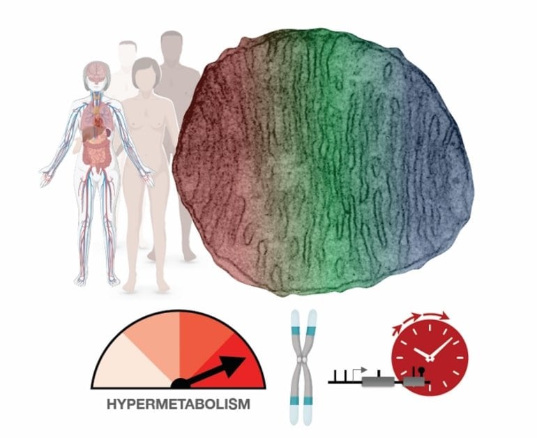
Summary: Human cells with impaired mitochondria expend more energy. While this hypermetabolism enhances a cell’s short-term survival, it also dramatically increases the rate at which the cell ages.

Source: Columbia University

Why do cells, and by extension humans, age? The answer may have a lot to do with mitochondria, the organelles that supply cells with energy. Though that idea is not new, direct evidence in human cells had been lacking. Until now.

In a study published Jan. 12 in Communications Biology, a team led by Columbia University researchers has discovered that human cells with impaired mitochondria respond by kicking into higher gear and expending more energy.

While this adaptation—called hypermetabolism—enhances the cells’ short-term survival, it comes at a high cost: a dramatic increase in the rate at which the cells age.

“The findings were made in cells from patients with rare mitochondrial diseases, yet they may also have relevance for other conditions that affect mitochondria, including neurodegenerative diseases, inflammatory conditions, and infections,” says principal investigator Martin Picard, PhD, associate professor of behavioral medicine (in psychiatry and neurology) at Columbia University Vagelos College of Physicians and Surgeons.

“In addition, hypermetabolism may be a key reason why most cells deteriorate as we get older.”

Hypermetabolic cells age faster

It was generally assumed that mitochondrial defects (which impair the conversion of food sources into usable energy) would force cells to slow their metabolic rate in an effort to conserve energy.

However, by analyzing metabolic activity and energy consumption in cells from patients with mitochondrial diseases, the researchers found that cells with impaired mitochondria double their energy expenditure.

Moreover, re-analyzing data from hundreds of patients with different mitochondrial diseases showed that mitochondrial defects also increase the energetic cost of living at the whole-body level.

Although this energy boost keeps cells running, it also degrades the cell’s telomeres (caps that protect the ends of our chromosomes) and activates stress responses and inflammation. The net effect accelerates biological aging.

“When cells expend more energy to make proteins and other substances essential for short-term survival, they’re likely stealing resources from processes that ensure long-term survival, like maintaining telomeres,” says Gabriel Sturm, a graduate student and lead author on this study.

Hypermetabolism, fatigue, and aging

This hypermetabolic state could explain why people with mitochondrial diseases experience fatigue and exercise intolerance, among other symptoms.

“To make up for the extra energy use in your cells, your body ‘tells’ you not to overexert yourself, to conserve energy. We likely see the same dynamic as people age and their vitality diminishes,” Picard says.

This is a diagram from the study
Mitochondrial defects caused by rare genetic mutations cause human cells to increase their metabolism. Though that helps short-term survival, it comes at a high cost: a dramatic increase in the rate at which the cells age. Hypermetabolism also may be a key reason why most cells deteriorate as everyone gets older. Credit: Martin Picard

The study doesn’t point to any new remedies for patients with mitochondrial diseases, which are currently not treatable, but it does reinforce the current recommendations for patients to move more.

“That may seem counterintuitive, since if you’re more active, you’re going to expend more energy and possibly make your symptoms worse,” Sturm says. 

“But exercise is known to increase the efficiency of an organism. An individual who runs, for example, uses less energy to sustain basic bodily processes than someone who is not physically active.”

Improving organismal efficiency, which would lower energy use in the cells and improve fatigue and other symptoms, may partially explain the health benefits of exercise in patients with mitochondrial diseases and otherwise healthy people.

In their search for new treatments for mitochondrial diseases, researchers should focus on hypermetabolism, Picard says. “Although mitochondrial defects do impair the ability of cells to produce energy, energy deficiency may not be the primary disease initiator. Our study shows these defects increase energy consumption. To move the needle therapeutically, we may need to target hypermetabolism. We need more research to know if that would work.”

Hypermetabolism is also common to other diseases. If increased cellular energy expenditure plays a causal role in driving the aging process, targeting hypermetabolism may be a way to improve fatigue, improve people’s quality of life, or even to slow biological aging.

Abstract

OxPhos defects cause hypermetabolism and reduce lifespan in cells and in patients with mitochondrial diseases

Patients with primary mitochondrial oxidative phosphorylation (OxPhos) defects present with fatigue and multi-system disorders, are often lean, and die prematurely, but the mechanistic basis for this clinical picture remains unclear.

By integrating data from 17 cohorts of patients with mitochondrial diseases (n = 690) we find evidence that these disorders increase resting energy expenditure, a state termed hypermetabolism.

We examine this phenomenon longitudinally in patient-derived fibroblasts from multiple donors. Genetically or pharmacologically disrupting OxPhos approximately doubles cellular energy expenditure.

This cell-autonomous state of hypermetabolism occurs despite near-normal OxPhos coupling efficiency, excluding uncoupling as a general mechanism. Instead, hypermetabolism is associated with mitochondrial DNA instability, activation of the integrated stress response (ISR), and increased extracellular secretion of age-related cytokines and metabokines including GDF15.

In parallel, OxPhos defects accelerate telomere erosion and epigenetic aging per cell division, consistent with evidence that excess energy expenditure accelerates biological aging.

To explore potential mechanisms for these effects, we generate a longitudinal RNASeq and DNA methylation resource dataset, which reveals conserved, energetically demanding, genome-wide recalibrations.

Taken together, these findings highlight the need to understand how OxPhos defects influence the energetic cost of living, and the link between hypermetabolism and aging in cells and patients with mitochondrial diseases.

Plant Hallucinogen Holds Hope for Diabetes Treatment


A potent molecular cocktail containing a compound from ayahuasca spurs rapid growth of insulin-producing cells

Plant Hallucinogen Holds Hope for Diabetes Treatment
Ayahuasca cooking.

For centuries, some indigenous groups in South America have relied on a brew made from the parts of a local vine and a shrub. The effects of this drink, called ayahuasca, would begin with severe vomiting and diarrhea, but the real reason for drinking the tea was the hallucinating that followed. These visions were thought to uncover the secrets of the drinker’s poor health and point the way to a cure.

Modern techniques have revealed that one of the compounds underlying these mystic experiences is the psychoactive drug harmine. What these first users of ayahuasca couldn’t have known was that, one day, this ingredient in their enlightening brew would be positioned as a key to treating diabetes.

Such a cure is a long way off, but researchers took another step toward it when they combined naturally occurring harmine with a compound synthesized from scratch in a lab. Together, the pair can coax the insulin-producing pancreatic cells, called beta cells, into replicating at the fastest rates ever reported, according to findings published December 20 in Cell Metabolism.

Type 1 diabetes arises when the body turns on these cells and destroys them. Type 2 diabetes develops when these same cells wear out and can no longer make insulin. Either effect is a point of no return because the beta cells we make in early life are the only ones we’ll ever have.

If this pair of compounds eventually inches into the treatment toolbox, refreshing a faded cell population could become a reality and a possible treatment for diabetes.  “Looking back 10 years or so, we questioned whether human beta cells could even be coaxed into dividing,’ says Justin Annes, assistant professor of medicine and endocrinology at Stanford University, who also works on beta cell proliferation, with a separate investigator group. “But what began as a fantasy has become aspiration, and perhaps in the coming years, will be a reality.”

One stop on the trip to that reality was a 2015 study showing that harmine treatment of beta cells in a dish promoted their increase at a rate of about 2 percent per day. A promising beginning, says study author Andrew Stewart, scientific director of the Diabetes, Obesity, and Metabolism Institute at the Icahn School of Medicine at Mount Sinai, but a little too slow for someone who needs a replacement population.

In this newest study, Stewart and his colleagues show that combining harmine with a synthetic inhibitor of another molecule kicks up the rate to 5–8 percent on average, and as high as 18 percent using some growth recipes. The one–two punch of this chemical pair isn’t the only possible combination, and other groups also are working on various pairings, Stewart says. Annes and his colleagues have identified several compounds that hold similar promise for pushing insulin-producing cells to reproduce.

“Basically, we’re all competing, but we all know each other so we share reagents and ideas,” says Stewart. “Different people have identified different drugs that make beta cells replicate.” His lab chose harmine because it’s the one they pulled out of their screening of 100,000 compounds in 2015, but “I don’t think harmine is especially better than any other one,” he says.

In 2006, another group of researchers plucked harmine from a molecular haystack in a search for chemicals that interact with a protein associated with Down syndrome. Studies that followed showed harmine’s role in many body systems, including the gut and the brain, explaining in part the effects of ayahuasca on its earliest adopters.

Harmine interferes with an enzyme called dual-specificity tyrosine-regulated kinase 1A, or DYRK1A. Like harmine, DYRK1A operates in a host of tissues.  It helps, for one, in shaping the central nervous system during embryonic development. First identified because of its key involvement in Down syndrome, its routine duty is to add chemical tags to molecules to switch them on or off.

The other molecule in the synergizing pair is an inhibitor of a group of proteins in the transforming growth factor-beta superfamily (TGFβSF). As with DYRK1A, these proteins are active in a large number of body processes, including cell proliferation.

Stewart and his team homed in on TGFβSF and DYRK1A after probing the secrets of cells from benign pancreatic tumors called insulinomas. They reasoned that if they could pinpoint what made these tumors grow, they could co-opt that information to encourage growth of normal beta cells. Their exploration uncovered DYRK1A and TGFβSF-related targets.

Inhibiting these molecules in human beta cells in a dish shuts down the cell regulators that usually keep the brakes on cancer’s out-of-control cell growth. Because harmine and TGFβSF inhibitor release this brake and DYRK1A and TGFβSF are active in many tissues, any treatment involving the pair of inhibitors must be closely targeted. “Certainly, we have a long way to go before these medications can be used in humans,” says Annes, calling the concern about cancer risk “reasonable.”

Adding to that concern is that harmine affects other cell types, says Klaus Kaestner, professor of genetics and associate director of the Penn Diabetes Research Center at the University of Pennsylvania, who was not involved in the study. In 2016, his group reported that harmine triggers many types of hormone-producing cells to divide, including other cells in the pancreas.

Stewart and his colleagues are sorting through a number of potential chemical tags that might help guide the inhibitors to the right location. But for now, says Stewart, “we are Amazon and have a bunch of parcels, and we know that they’re for you, but we don’t know the address.”

Type 1 diabetes poses another hurdle. Although the immune system targets and destroys these cells in this form of diabetes, a small pool of beta cells often remains, Stewart says. What’s unknown is if a new population grown from these cells would simply attract further immune destruction. Stewart says that if the harmine-TGFβSF inhibitor combination ever makes it to trials, the population it might initially suit best are those who have type 2 diabetes. Then the journey from a South American rainforest to a clinical treatment would be complete.

Obscure Asthma Drug Shows Promise for Treating Diabetes


A little-used asthma drug called amlexanox may potentially be repurposed to treat type 2 diabetes, according to findings from a small proof-of-concept study published in the July issue of Cell Metabolism.

Results showed that using the drug for 12 weeks was linked to significantly reduced HbA1c in some patients with obesity, type 2 diabetes, and nonalcoholic fatty-liver disease (NAFLD).

“The overall significant reduction in HbA1c over this relatively short trial indicates that amlexanox can benefit some patients with type 2 diabetes. The reduction in HbA1c is on the order of a [dipeptidyl peptidase-4] DPP-4 inhibitor when given alone over the same time period,” commented first author Elif Oral, MD, director of the MEND Obesity and Metabolic Disorder Program at the University of Michigan, Ann Arbor.

Researchers also looked at baseline inflammation, which revealed an interesting finding: people with higher levels of inflammation responded better.

“Among drug-treated patients, there seemed to be a greater degree of inflammation in responders compared with nonresponders. This is interesting, since we know that inflammatory pathways drive up expression of the targets of amlexanox,” Dr Oral said.

Amlexanox inhibits two enzymes: IKKƐ and TBK1. Studies in mice have shown that inhibiting these enzymes improves weight, insulin resistance, fatty liver, and inflammation.

Another intriguing result: responders showed over 1100 gene changes, and these changes were found only in this group.

“The drug response was characterized by a unique and dramatic molecular signature of gene-expression changes, consistent with what was seen in mouse models, in which expression of energy-expenditure genes were increased. We’re still investigating the importance and significance of these gene-expression changes,” Dr Oral added.

Amlexanox Developed in Japan to Treat Allergies and Asthma

Amlexanox was developed in Japan in the 1980s to treat asthma and allergic rhinitis. However, it requires thrice-daily dosing and was never introduced to the United States, because of heavy competition from more popular medications like montelukast, which can be taken once a day. Even in Japan, the prescription rate was very low, and therefore amlexanox was discontinued this year for commercial reasons.

However, its exact mechanism of action has never been fully investigated. It was not until Dr Oral and colleagues screened 150,000 chemicals, looking for inhibitors of IKKƐ and TBK1, that they hit upon amlexanox as a potential antidiabetes drug.

They first tested amlexanox in mice and did an open-label safety study in humans. Both the animal and human trials pointed to fat tissue as an important target for amlexanox.

So researchers next tested amlexanox in a randomized double-blind placebo controlled study that included 42 obese individuals with type 2 diabetes and NAFLD. Participants were randomized to 12 weeks of 50-mg amlexanox three times daily or placebo.

About one-third of participants showed a robust response to amlexanox, with reductions in HbA1c of ≥ 0.5% percentage points or more, which was significantly different from placebo (= .05). Responders also showed significant decreases in fructosamine, a marker for shorter-term glucose control (= .024).

Similar to results in mice, at 2 to 4 weeks responders showed a transient increase in IL-6, followed by decreased fasting glucose and improved insulin sensitivity. A subgroup of responders with NAFLD showed improvement in fatty liver.

Responders also had higher levels of baseline inflammation than nonresponders or placebo patients, including higher levels of CRP, which correlated with the amount of reduction in HbA1c. And analyses of fat biopsies showed they also had higher baseline activation of genes involved in inflammation.

Fat biopsies also replicated findings from the open-label study in humans, showing responders treated with amlexanox had higher expression of genes involved in energy expenditure and “browning” of fat.

Seven patients in the amlexanox group developed a rash at 4 weeks, which resolved within 2 weeks using local treatment. No other adverse events attributable to amlexanox occurred. This is consistent with the long-term safety profile in Japan, in which about 5% of patients developed rash, Dr Oral pointed out.

“We don’t understand the mechanism for why participants with more underlying inflammation responded better. However, previous work has shown that TBKI and IKKƐ are upregulated in the setting of more inflammation. So it is possible that inflammation oversensitizes the pathway that the drug targets,” she explained.

More Studies Planned

The team is now planning a longer 6-month prospective, randomized study in humans that will test whether individuals with elevated CRP and higher levels of fat inflammation at baseline have better responses to amlexanox.

They also plan another trial in humans that will test amlexanox in combination with mirabegron (Myrbetriq, Astellas Pharma), a pure beta agonist used to treat overactive bladder. The idea is to see whether amlexanox can restore catecholamine sensitivity.

Future studies will also determine the optimal safe dose and dosing regimen for amlexanox.

“If we can really prove that those patients with higher inflammation will respond better with this drug, it will be the first time that such an observation will be made, which is exciting. It’s another way of customizing therapy for patients,” Dr Oral stressed.

The group is currently looking for ways to partner with companies and investors, but currently none are involved.

A brand new type of insulin-producing cell has been discovered hiding in the pancreas.


A missing piece of the diabetes puzzle.


Researchers have found a brand new type of insulin-producing cell hiding in plain sight within the pancreas, and they offer new hope for better understanding – and one day even treating – type 1 diabetes.

Type 1 diabetes occurs when a person’s own immune system kills off most of their insulin-producing beta cells. And seeing as insulin is the hormone that regulates our blood sugar, type 1 diabetics are left reliant on injecting themselves with insulin regularly.

 While the condition can usually be managed effectively, in order to properly treat it, researchers would need to find a way to regenerate a patient’s beta cells and prevent them from being attacked in future – something we’re getting better at, but ultimately has eluded scientists so far.

The discovery of these previously unnoticed cells in the pancreas – which the team are calling ‘virgin beta cells‘ – could offer a new route for regrowing healthy, mature beta cells – and also provides insight into the basic mechanisms behind the disease.

“We’ve seen phenomenal advances in the management of diabetes, but we cannot cure it,” said lead researcher Mark Huising from the University of California, Davis.

“If you want to cure the disease, you have to understand how it works in the normal situation.”

To get a better insight into exactly what happens in type 1 diabetes, the researchers studied both mice and human tissue.

Huising and his team were looking at regions inside the pancreas known as the islets of Langerhans, which in healthy humans and mice are the regions that contain the beta cells that detect blood sugar levels around the body and produce insulin in response.

 Researchers also know that the islets contain cells called alpha cells, which produce glucagon, a hormone that raises blood sugar. These alpha cells, combined with the blood sugar-lowering beta cells, are how the body regulates blood sugar levels.

But in patients with type 1 diabetes, two things go wrong: the beta cells are killed off by the body’s own immune system, and then they fail to regenerate.

In order to treat the disease, we need to find a way to overcome both of those problems.

For decades, scientists have been trying to do this, and they had long assumed that there was one main way for beta cells to be produced – through other adult beta cells dividing.

But after using new microscope techniques to study islet tissue in the lab, Huising and his team found a new type of cell scattered around the edges of the islets that no one had noticed before – and they looked a lot like immature beta cells, suggesting that maybe there was actually another way beta cells were being made.

Further study revealed that these new virgin beta cells could make insulin, but they didn’t have the receptors to detect glucose, so couldn’t function as mature beta cells.

But that wasn’t all the researchers found. They also observed some mature beta cells in the islets transitioning into alpha cells – representing a completely unexpected alpha cell generation pathway.

“There’s much more plasticity in the system than was thought,” said Huising.

It’s still very early days, and these new cells now need to be confirmed in live humans and animals – not just tissue in the lab. But the fact that we now have evidence that they exist opens up a whole avenue of research on type 1 diabetes and potential treatments.

According to Huising, there are three main reasons to get excited about the result: firstly, it represents a new beta cell population in both humans and mice that we had no idea about before, and secondly, it also provides a potential new source of beta cells that could be used to treat diabetics.

“Finally, understanding how these cells mature into functioning beta cells could help in developing stem cell therapies for diabetes,” a press release explains.

The research could also have benefits for type 2 diabetes, which occurs when beta cells become inactive and stop releasing or secreting insulin.

Source: Cell Metabolism.

Diets That Mimic Fasting: How To Lose Belly Fat, Improve Memory, And Increase Lifespan The Safe Way


“Strict fasting is hard for people to stick to, and it can also be dangerous, so we developed a complex diet that triggers the same effects in the body,” said the study’s lead researcher Valter Longo, director of the USC Longevity Institute, in a press release. “I’ve personally tried both, and the fasting mimicking diet is a lot easier and also a lot safer.”

The human trial involved 19 participants and was designed to replicate Longo’s yeast andmouse trials. Once a month for five days, participants limited their caloric intake by 34 to 54 percent — just low enough to mimic the effects of fasting. For the other 25 days of the month, participants returned to their normal eating habits.

After three months, Longo and his research team measured the participants’ biomarkers and found they were at a decreased risk of aging, diabetes, heart disease, and cancer. They’re calling it the “fasting mimic diet (FMD),” and it was shown to cut belly fat, improve learning and memory skills, and increase the number of stem cells ultimately leading to a longer lifespan.

It turns out that when there are a certain amount of proteins, carbohydrates, fats, and micronutrients, the body lowers the amount of hormone IGF-I it produces. Not only is this hormone responsible for promoting aging but it has also been linked to cancer susceptibility, which means less of it is better. Longo proved this theory before when he demonstrated how to starve cancer cells while protecting other cells from harm.

“It’s about reprogramming the body so it enters a slower aging mode, but also rejuvenating it through stem cell-based regeneration,” Longo said. “Not everyone is healthy enough to fast for five days, and the health consequences can be severe for a few who do it improperly.”

Fasting can hurt the body if it’s not done right. Women who try water-only diets, for example, put themselves at risk of developing gallstones if they aren’t properly supervised, Longo explained. Fasting isn’t for everyone, either. People with a body mass index below 18 — considered a normal weight — should not engage in fasting of any sort.

Diabetics also shouldn’t partake in fasting or fasting mimic diets while they receive insulin or other drugs because the body uses up glucose energy supplies before it begins to burn fat. The process of burning fat to convert into fuel, also known as “ketosis,” makes the blood become more acidic, leading to bad breath, fatigue, and eventual kidney and liver damage. The FMD diet, however, is unique in that it allows the person to return to normal caloric intake for a majority of the month. Some fitness experts like Jillian Michaels, believe fasting can turn into an unhealthy “yo-yo” effect, and cause a person to cyclically fast and binge. The trick to avoiding the dreaded yo-yo effect is to not cut calories altogether but instead limit calories for one week a month, and gradually return to your normal caloric intake for the other three weeks.

The research team is set to meet with Food and Drug Administration officers soon, Longo said. They’ll work out the details on how to implement the diet safely in order to prevent and treat obesity. In the meantime, patients shouldn’t try it at home until Longo and his team finish testing through a randomized clinical trial, which will involve 70 patients over the span of six months.

“It’s not a typical diet because it isn’t something you need to stay on,” Longo said. “If the results remain as positive as the current ones, I believe this FMD will represent the first safe and effective intervention to promote positive changes associated with longevity and health span, which can be recommended by a physician.”

Source:  Longo V. Cell Metabolism. 2015.