Study shows that ‘transcendent’ thinking may grow teens’ brains over time


Landmark study shows that 'transcendent' thinking may grow teens' brains over time
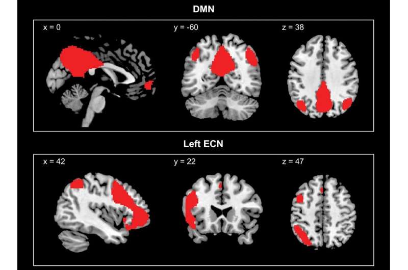
Depiction of coronal, sagittal, and axial views of the group-level default mode network (DMN; top) and left executive control network (left ECN; bottom) maps, derived from a 20-component group independent component analysis of the resting-state data (concatenating data from all participants from the two data collections), transformed into z-score maps and thresholded at z = 2. Credit: Scientific Reports (2024). DOI: 10.1038/s41598-024-56800-0

Scientists at the USC Rossier School of Education’s Center for Affective Neuroscience, Development, Learning, and Education (CANDLE) have shown for the first time that a type of thinking that has been described for over a century as a developmental milestone of adolescence may grow teenagers’ brains over time.

This kind of thinking, which the study’s authors call “transcendent,” moves beyond reacting to the concrete specifics of social situations also to consider the broader ethical, systems-level and personal implications at play. Engaging in this type of thinking involves analyzing situations for their deeper meaning, historical contexts, civic significance, and/or underlying ideas.

The research team, led by USC Rossier Professor Mary Helen Immordino-Yang, includes Rebecca J.M. Gotlieb, a research scientist at UCLA, and Xiao-Fei Yang, an assistant research professor at USC Rossier. The published study “Diverse adolescents’ transcendent thinking predicts young adult psychosocial outcomes via brain network development” is published in Scientific Reports.

In previous studies, the authors had shown that when teens and adults think about issues and situations in a transcendent way, many brain systems coordinate their activity, among them two major networks important for psychological functioning: the executive control network and the default mode network.

The executive control network is involved in managing focused and goal-directed thinking, while the default mode network is active during all kinds of thinking that transcends the “here and now,” such as when recalling personal experiences, imagining the future, feeling enduring emotions such as compassion, gratitude, and admiration for virtue, daydreaming or thinking creatively.

The researchers privately interviewed 65 14–18-year-old high school students about true stories of other teens from around the world and asked the students to explain how each story made them feel. The students then underwent fMRI brain scans that day and again two years later. The researchers followed up with the participants twice more over the next three years as they moved into their early twenties.

What the researchers found is that all teens in the experiment talked at least some about the bigger picture—what lessons they took from a particularly poignant story or how a story may have changed their perspective on something in their own life or the lives and futures of others. However, they found that while all of the participating teens could think transcendently, some did it far more than others.

And that was what made the difference. The more a teen grappled with the bigger picture and tried to learn from the stories, the more that teen increased the coordination between brain networks over the next two years, regardless of their IQ or their socioeconomic status.

This brain growth—not how a teen’s brain compared to other teens’ brains but how a teen’s brain compared to their own brain two years earlier—in turn predicted important developmental milestones, like identity development in the late teen years and life satisfaction in young adulthood, about five years later.

The findings reveal a novel predictor of brain development—transcendent thinking. The researchers believe transcendent thinking may grow the brain because it requires coordinating brain networks involved in effortful, focused thinking, like the executive control network, with those involved in internal reflection and free-form thinking, like the default mode network.

These findings “have important implications for the design of middle and high schools, and potentially also for adolescent mental health,” senior researcher Immordino-Yang says. The findings suggest “the importance of attending to adolescents’ needs to engage with complex perspectives and emotions on the social and personal relevance of issues, such as through civically minded educational approaches,”‘

Immordino-Yang explains. Overall, Immordino-Yang underscores “the important role teens play in their own brain development through the meaning they make of the social world.”
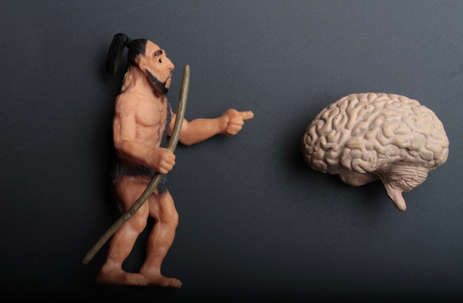
Brutes and Brains: What We Know About Neanderthal Brain Size.


The Neanderthals were smart. But, what, specifically, differentiated their brains from those of our own ancestors?

Neanderthal Brain

Specialists say that the brains of Homo neanderthalensis and Homo sapiens were similar. But slight differences in the structure and development of the two species’ brains could’ve changed the ways they thought about the world.

Tradition says that Homo neanderthalensis and Homo sapiens were intellectually distinct. But archaeologists and anthropologists increasingly insist that the intellectual divide between the two species is shrinking.

In fact, the traces of their ancient activities show more and more that the two species followed similar survival strategies. Residing in similar societies, they made similar sounds and manipulated similar tools. Apart from that, the most recent research shows that the two created similar art, too, signifying their shared taste for abstraction.

But what about their brains? Was there anything in their anatomy that differentiated their thinking?

Some specialists say yes. Working in the fields of paleontology, paleoneurology and paleogenetics, experts suspect that the slightest distinctions in the structure and development of the two species’ brains could’ve set their cognition apart in complex and consequential ways.

The Brutish Beginning of the Neanderthal

When academics found the first Neanderthal fossils around two centuries ago, they weren’t sure what they were working with. While some supposed that they stumbled across their own ancient ancestors, others thought that the specimens signified something else entirely.

At the time, there was only one conclusion for which they felt confident: Whatever the fossils’ formal classification, the creature that they found was far from intelligent, they thought, when it wandered the world thousands of years ago


Neanderthal Thinking

“Darkness characterized the being to which the fossil belonged,” asserts an analysis of one set of specimens from the 1860s. “The thoughts and desires which once dwelt within it never soared beyond those of a brute.”

Articulated around the same time that the Neanderthals secured their status as a separate species, this now out-of-favor sentiment stemmed from the fact that the Neanderthals displayed a “singularly different” skull structure from that of our own species.

Because of this difference, the Neanderthals were swiftly branded as a species of brutes, who were trapped in ignorant “benightedness” until our own intellectually superior species beat them around 40,000 years ago.

Neanderthal Intelligence

In the years since then, archaeologists and anthropologists have found an abundance of hints that the Neanderthals acted with a similar sophistication to our own species. In addition to their similar survival strategies and tools, recent research also suggests that the Neanderthals communicated and created cultures of art and adornment comparable to those of our ancestors.

The complexity of these activities suggest that the Neanderthals’ minds mirrored our own. But what, specifically, do we know about the structure and development of their brains?


Did Neanderthals Have Bigger Brains?

Specialists are still searching for their first bits of brain from the Neanderthals, since these tissues tend to be the first part of the body to decompose after death. That said, they’ve found an abundance of preserved skulls from the species, which feature the casts or inner surfaces of the brains they once contained.

Neanderthal Brain Size

Overall, these skulls indicate that the brains of H. neanderthalensis and H. sapiens were similarly sized, with H. neanderthalensis brains beating H. sapiens brains only slightly in terms of their total volume.

Neanderthal Cranial Capacity

Though the measurements aren’t always consistent and change over time, specialists say that the typical Neanderthal skull contained around 1500 cubic centimeters (or 51 ounces) of cerebral tissue, though the skulls of their more modern counterparts showed a smaller cranial capacity of only 1350 cubic centimeters (or 46 ounces).

A Stretched Brain

In addition to their size, Neanderthal brains and braincases were also slightly stretched, producing a strange, semi-spherical skull that terminated in a big bump toward the back. Termed the “occipital bun,” this bump was one of the first features of Neanderthal anatomy that experts discovered and described.


The Structure of the Neanderthal Brain

Paleontologists and paleoneurologists suspect that the strange traits of Neanderthals impacted the size, shape and arrangement of the separate structures inside their brain, too, transforming their particular patterns of thinking.

Some studies suppose, for instance, that the structure of the Neanderthal skull meant that the H. neanderthalensis cerebellum was smaller than the H. sapiens cerebellum. A small cerebellum, these studies say, can cut a species’ capacity to learn and think logically, to process language, and to interact socially, which would all substantially impact its survival.

Adding to this is an assortment of similar proposals about the peculiarities inside the Neanderthal mind. For instance, some paleontologists and paleoneurologists say that much more of the species’ brain was concentrated on controlling basic body movements, all thanks to the species’ bigger, bulkier bodies, which were more difficult to move than our own.

The Development of the Neanderthal Brain

Beyond its simple structure, specialists are also learning a lot about the development of the Neanderthal brain from birth to adulthood.

For instance, the shape and size of juvenile, adolescent and adult skulls suggest that H. neanderthalensis minds matured much more slowly than our own minds mature. They were below 90 percent of their average adult volume at 8 years old. And though that may not seem sluggish, specialists say that that’s basically the same age that H. sapiens brains become full-fledged today, in terms of their overall volume.

Building Neanderthal Brains

Genetic analyses add additional support to the idea that the development of the Neanderthal brain differed from our own. In fact, though specialists traditionally stick to skulls in their attempts to study Neanderthal smarts, the recent reconstruction of the Neanderthal genome is inspiring some to turn to the ancient genes that guide brain growth, instead, as a way to differentiate between H. neanderthalensis and H. sapiens thinking


NOVA1

In 2021, for instance, a team of geneticists investigated a gene called NOVA1, which directs the development of brain tissues in H. neanderthalensis and H. sapiens. Inserting two distinct forms of the gene into clusters of uncultured cells, the team discovered that the form of NOVA1 found in H. neanderthalensis created bumpier blobs of brain tissue when cultured, while the form of NOVA1 found in H. sapiens created smooth, spherical clumps.


TKTL1

The following year, in 2022, a second team of geneticists followed a similar approach with a gene called TKTL1, which prompts neuron production. While the H. neanderthalensis form of TKTL1 fostered some neurons, the H. sapiens form fostered many more.

Though these findings do demonstrate that the brains of H. neanderthalensis and H. sapiens developed differently, they don’t disclose all the details of this difference. Individual genes are only tiny threads in a greater genetic tapestry, specialists say, suggesting that a true appreciation of the influence of NOVA1 and TKTL1 on something as complex as cognition can only come in the context of a more complete genome.


Though it will take time, specialists say that future work within this field will untangle the influence of an assortment of other genes on the growth of the Neanderthal brain. Combined with the insights taken from the structure of skulls, this work will ultimately create a clearer picture of the differences between our own cognition and that of our closest cousins.

How Air Pollution Affects Our Brains


An expert Harvard panel discusses the links between air pollution and dementia, learning, mental health, and mood.

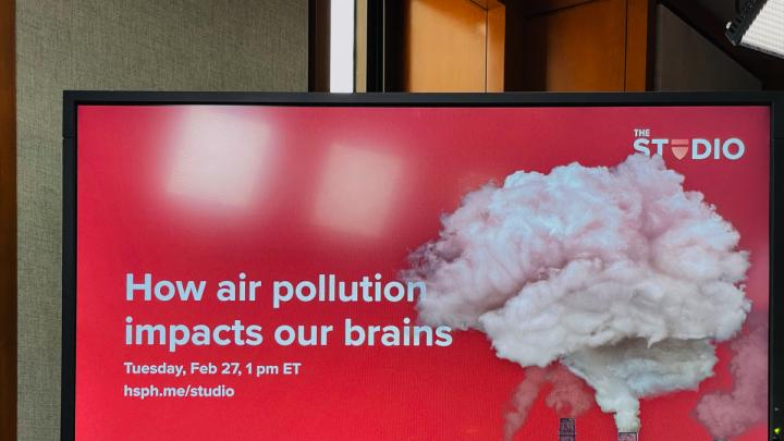
How air pollution impacts our brains
At Harvard, an expert panel discussed how air pollution affects dementia, learning, mental health and mood.

Emerging evidence shows that exposure to air pollution increases the incidence and progression of Alzheimer’s, Parkinson’s, and other neurocognitive diseases, according to Francesca Dominici. She spoke as part of a panel on how air pollution affects the human brain, held February 27 at the Harvard T. H. Chan School of Public Health (HSPH). Dominici, one of world’s leading data scientists studying the health effects of fine particles, was joined on the panel by Joseph Allen, an expert on indoor air pollution; Maite Arce, president and CEO of the Hispanic Access Foundation; and Marc Weisskopf, who studies the biological mechanisms that make PM 2.5 (particles that are 2.5 microns or smaller in diameter) deadly. The group discussed both outdoor and indoor air pollution, the physiological pathways particles travel within the human body, their mental health effects, the elevated impacts on underprivileged groups in the United States, and the Environmental Protection Agency’s (EPA) new fine particle regulations.

Dominici, the Gamble professor of biostatistics, population, and data science, has been working with HSPH colleagues to analyze the healthcare records of millions of Americans in the Medicare and Medicaid programs, and linking them to each person’s long-term exposure to fine particle pollution. “What we are finding, with enormous statistical power,” she said, “is that long-term exposure to fine particulate matter increased the incidence of hospitalization for Alzheimer’s disease.” It also accelerates the progression of the disease, as well as the rate of hospitalization and mortality from all causes.

Why are fine particles so biologically dangerous? “The exploration of air pollution and the brain is relatively new,” Weisskopf said. The “wealth of data” and research documenting the cardiovascular effects of inhaled fine particles, much of it conducted at HSPH, sparked interest in the possibility that the brain, which is “hugely dependent” on the blood supply, might also be affected, he continued. Particles can generate inflammatory immune responses in the systemic circulation, according to Weisskopf, Drinker professor of environmental epidemiology and physiology, which can then affect the brain. But fine particles may be able to reach the brain directly, he continued: “When you breathe in through your nose, you’re smelling things because you have neurons that are kind of sticking out in the world, exploring it that way. And we now understand that some particles, or aspects of particles, can actually get transported directly back into the brain—skipping the lungs and the cardiovascular system.” The precise biological pathways, he said, are an active area of research.

Air pollution also affects mental health, explained associate professor of exposure assessment science Joseph Allen. An HSPH program studying the research into this link, largely in children, he said, has found that “air pollution is linked to anxiety and suicide ideation, as well as pediatric hospitalizations for pediatric disorders.”

Allen’s own work focuses on indoor air quality. One of his double-blind studies of office workers introduced chemicals off-gassed by dry cleaning and dry-erase markers into environmentally controlled workspace. During such exposure, office workers performed worse on tests of cognitive function, such as seeking and utilizing information, as well as on tests of strategic decision-making, and response to a crisis. A second study that assessed the impact of air pollution penetrating a building from outside and affecting workers at their desks demonstrated similar outcomes. As Allen summarized the research, “In all these dimensions—in office workers, university workers, kids in elementary school, high school students—we see over and over the impacts of air pollution on the whole range of mental health, from anxiety all the way through higher order cognitive function.”

Sabrina Shankman, a Boston Globe reporter who moderated the event, then asked about the disparate impact of air pollution on communities of color. Dominici recalled that in one of her studies of air pollution by U.S. census tract, published three years ago in Nature, she and her colleagues found higher percentages of underrepresented minorities in the most polluted areas. Such pollution has decreased substantially due to the Clean Air Act, she noted—but strikingly, the racial disparities crossed socioeconomic lines. “Very surprising to me,” Dominici said, “was that high socioeconomic status black Americans” were exposed to higher levels of air pollution than the “lowest socioeconomic status white Americans.” She continued, “That tells you a lot about systemic bias and racism.” The reason, she believes, is less pressure from local communities on siting decisions and less attention to enforcement of EPA fine particle regulations. Such disparities are not just artifacts of historic practices such as redlining. “A study that we are conducting right now on exposure to air pollution from cryptocurrency data mining, which is not regulated in the United States,” Dominici said, indicates that the buildings that house these energy-intensive operations tend to be located in lower socioeconomic status and underrepresented minority neighborhoods, where there is less knowledge and pressure from citizens and local governments.

Maite Arce, of the Hispanic Access Foundation, has been trying to change that dynamic in Latino communities through a program called “The Air That We Breathe.” The effort has recruited citizen scientists from 12 communities who are talking about air pollution and learning from experts in air quality. The program is deploying sensors in churches to monitor and report local air quality; the data gathered are then entered into an online map. Because “we’re starting at zero in terms of the knowledge” about PM 2.5, Arce said, local leaders are developing materials tailored to the needs of their primarily Spanish-speaking or immigrant communities. They’re disseminating that information through “sermons…, film screenings, roundtables, community workshops, radio and social media.” And they help residents prepare to react on days of poor air quality through masking or staying indoors. Parents understand that airborne pollution “may increase respiratory infections in children, which can lead to symptoms like asthma, and that can lead to school absences,” she reported. But the understanding that proceeds from this public health education is “eye-opening for them, and makes them feel that urgency to take action,” Arce said, “…to really urge for policy change.”

The conversation then turned to the new EPA fine particle regulations, which reduce the standard for U.S. air pollution from 12 micrograms of particles per cubic meter of air to nine. Dominici called this “the biggest public health victory in a long time” and expressed pride in HPSH’s instrumental role in the decision, stretching from the early “Six Cities” study 30 years ago to the most recent research. States will have to develop action plans and examine the inventory of local sources of particle pollutants, from traffic, industry, or power plants, she explained. The result will be “longer life for everybody, so it’s really good, a fantastic victory.”

On the other hand, there is no safe level of particle pollution—the cleaner the air, the healthier are the people who breathe it, the data show—so the standard could have been lower. “One might ask, well, why not zero?” she asked. “But that is nearly impossible because there is always going to be some background fine particulate matter.” Dominici characterized the new standard—nine micrograms per cubic meter—as “a compromise between the science and the political landscape.” Weisskopf, who served on the panel that recommended the new standard, provided some insight into “how the sausage gets made.” Some of the decision-makers on the panel argued for a higher number, while he and other public health advocates pushed for lower exposures. Paradoxically, a lot of the best data showing the health benefits of even lower exposures come from Canada, which has cleaner air than the United States. But the panel favored U.S. evidence, so demonstrating the value of an even better standard was difficult. The World Health Organization, he noted, suggests a target of five micrograms of fine particle pollutants per cubic meter.

Given that context, Allen segued to the “major disconnect” between the legally enforceable limits on particles outdoors, and those applying indoors. The Occupational Safety and Health Administration (OSHA) standard for workplaces, currently 5,000 micrograms per cubic meter, is more than 500 times the outdoor standard. That limit is “grossly out of date,” he said, a fact that OSHA acknowledges. The agency does enforce lower standards for chemicals, silica, asbestos, and other hazardous materials, but lacks the legal authority to set standards outside the workplace. In other words, Allen said, there is no national indoor air quality standard for all the places where OSHA doesn’t have oversight: classrooms, offices, and so on. Nor are there sufficient data about the long-term effects of indoor air pollution—even though that is where most exposures occur, because people spend most of their time indoors. “A loose collection of researchers” is trying to raise funds for such studies, he reported, but with no encompassing regulatory authority to enforce standards it’s unclear how such data might be used.

Well-designed buildings can cut indoor exposure to dangerous levels of outdoor air pollution to well below the EPA standard, as occurred during last summer’s wildfires. Allen said that even retroactively fitted high quality air filters can dramatically improve indoor air in schools, offices, and homes during such events, which are expected to increase in frequency with climate change. Weisskopf added that more research is also needed on the toxicity of various indoor air pollutants; a recent study implicated wildfire smoke, which can penetrate indoors, in dementia. More work needs to be done to assess the toxicity of other types of particles, he emphasized, because their chemical composition varies depending on their source (a gas stove, an oil-filled cooking skillet, a farm engaged in agricultural burning, or something else entirely), as may the physiological effects.

Shankman brought the event to a close with a final question for each of the panelists: “If you could change one thing today to improve air quality what would it be?”

  • “Expand the number of citizen scientists in the most affected communities,” Arce said.
  • “Get the standards changed for how we design and operate buildings,” Allen said.
  • “Shut down all the coal-fired power plants,” Dominici said.
  • “Lower the [EPA] standard further,” Weisskopf said.

And with that, the audience dispersed, trading the well-ventilated environs of HSPH for the ambient air of Huntington Avenue and the nearby Mission Hill neighborhood.

Ancient Viruses Shaped Our Brains


Summary: Ancient viruses played a pivotal role in the development of myelin, crucial for complex vertebrate brains. The discovery of “RetroMyelin,” a retrovirus-derived element essential for myelin production across mammals, amphibians, and fish, underscores the impact of viral genes on vertebrate evolution.

The study demonstrates that myelination, a key factor in nerve impulse conduction and vertebrate diversity, owes its existence to ancient viral insertions, challenging previous understandings of evolutionary biology. This convergence of virology and neurobiology opens new avenues for exploring the molecular mechanisms behind evolution and the intricate relationship between viruses and vertebrate development.

Key Facts:

  1. “RetroMyelin,” a gene sequence derived from ancient retroviruses, is vital for the production of myelin in vertebrates.
  2. The presence of RetroMyelin in diverse vertebrate groups suggests separate viral genome integration events, highlighting its role in convergent evolution.
  3. Experimental disruption of RetroMyelin in zebrafish and frogs led to significantly reduced myelin production, proving its functional role in myelination.

Source: Cell Press

Researchers report February 15 in the journal Cell that ancient viruses may be to thank for myelin—and, by extension, our large, complex brains.

The team found that a retrovirus-derived genetic element or “retrotransposon” is essential for myelin production in mammals, amphibians, and fish. The gene sequence, which they dubbed “RetroMyelin,” is likely a result of ancient viral infection, and comparisons of RetroMyelin in mammals, amphibians, and fish suggest that retroviral infection and genome-invasion events occurred separately in each of these groups.

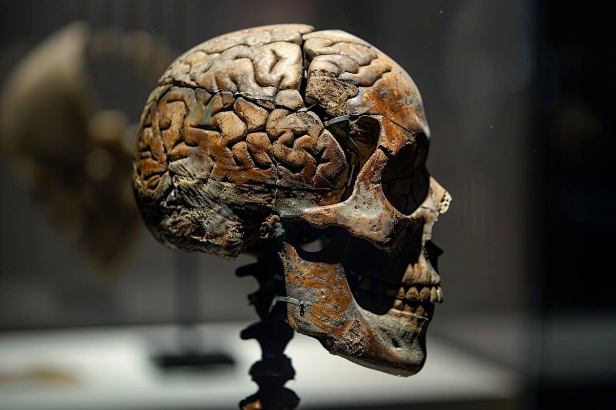
This shows a statue of a skull.
When they experimentally disrupted the RetroMyelin gene sequence in the fertilized eggs of zebrafish and frogs, they found that the developing fish and tadpoles produced significantly less myelin than usual.

“Retroviruses were required for vertebrate evolution to take off,” says senior author and neuroscientist Robin Franklin of Altos Labs-Cambridge Institute of Science.

“If we didn’t have retroviruses sticking their sequences into the vertebrate genome, then myelination wouldn’t have happened, and without myelination, the whole diversity of vertebrates as we know it would never have happened.”

Myelin is a complex, fatty tissue that ensheathes vertebrate nerve axons. It enables rapid impulse conduction without needing to increase axonal diameter, which means nerves can be packed closer together. It also provides metabolic support to nerves, which means nerves can be longer.

Myelin first appeared in the tree of life around the same time as jaws, and its importance in vertebrate evolution has long been recognized, but until now, it was unclear what molecular mechanisms triggered its appearance.

The researchers noticed RetroMyelin’s role in myelin production when they were examining the gene networks utilized by oligodendrocytes, the cells that produce myelin in the central nervous system.

Specifically, the team was investigating the role of noncoding regions including retrotransposons in these gene networks—something that hasn’t previously been explored in the context of myelin biology.

“Retrotransposons compose about 40% of our genomes, but nothing is known about how they might have helped animals acquire specific characteristics during evolution,” says first author Tanay Ghosh, a computational biologist at Altos Labs-Cambridge Institute of Science.

“Our motivation was to know how these molecules are helping evolutionary processes, specifically in the context of myelination.”

In rodents, the researchers found that the RNA transcript of RetroMyelin regulates the expression of myelin basic protein, one of the key components of myelin. When they experimentally inhibited RetroMyelin in oligodendrocytes and oligodendrocyte progenitor cells (the stem cells from which oligodendrocytes are derived), the cells could no longer produce myelin basic protein.

To examine whether RetroMyelin is present in other vertebrate species, the team searched for similar sequences within the genomes of jawed vertebrates, jawless vertebrates, and several invertebrate species.

They identified analogous sequences in all other classes of jawed vertebrates (birds, fish, reptiles, and amphibians) but did not find a similar sequence in jawless vertebrates or invertebrates.

“There’s been an evolutionary drive to make impulse conduction of our axons quicker because having quicker impulse conduction means you can catch things or flee from things more rapidly,” says Franklin.

Next, the researchers wanted to know whether RetroMyelin was incorporated once into the ancestor of all jawed vertebrates or whether there were separate retroviral invasions in the different branches.

To answer these questions, they constructed a phylogenetic tree from 22 jawed vertebrate species and compared their RetroMyelin sequences. The analysis revealed that RetroMyelin sequences were more similar within than between species, which suggests that RetroMyelin was acquired multiple times through the process of convergent evolution.

The team also showed that RetroMyelin plays a functional role in myelination in fish and amphibians. When they experimentally disrupted the RetroMyelin gene sequence in the fertilized eggs of zebrafish and frogs, they found that the developing fish and tadpoles produced significantly less myelin than usual.

The study highlights the importance of non-coding regions of the genome for physiology and evolution, the researchers say. “Our findings open up a new avenue of research to explore how retroviruses are more generally involved in directing evolution,” says Ghosh.

Funding:

This research was supported by the Adelson Medical Research Foundation, the UK Multiple Sclerosis Society, the Wellcome Trust, and the Altos Labs-Cambridge Institute of Science.


Abstract

A retroviral link to vertebrate myelination through retrotransposon RNA-mediated control of myelin gene expression

Highlights

  • RNA expression of retroviral element RNLTR12-int is crucial for myelination
  • RNLTR12-int binds to SOX10 to regulate Mbp expression
  • RNLTR12-int-like sequences (RetroMyelin) were identified in all jawed vertebrates
  • Convergent evolution likely led to RetroMyelin acquisition, adapted for myelination

Summary

Myelin, the insulating sheath that surrounds neuronal axons, is produced by oligodendrocytes in the central nervous system (CNS). This evolutionary innovation, which first appears in jawed vertebrates, enabled rapid transmission of nerve impulses, more complex brains, and greater morphological diversity.

Here, we report that RNA-level expression of RNLTR12-int, a retrotransposon of retroviral origin, is essential for myelination. We show that RNLTR12-int-encoded RNA binds to the transcription factor SOX10 to regulate transcription of myelin basic protein (Mbp, the major constituent of myelin) in rodents. RNLTR12-int-like sequences (which we name RetroMyelin) are found in all jawed vertebrates, and we further demonstrate their function in regulating myelination in two different vertebrate classes (zebrafish and frogs).

Our study therefore suggests that retroviral endogenization played a prominent role in the emergence of vertebrate myelin.

When Do Brains Grow Up? New Findings Shock Neuroscientists


Recent research has revealed that mice and primates, despite their differing lifespans, develop brain synapses at the same rate. This surprising discovery challenges previous assumptions in neuroscience about aging and disease, and it opens new avenues for understanding human brain development and improving neurological disorder treatments.

Recent research indicates that mouse and primate brains mature at a similar rate.

A study by Argonne National Laboratory finds that both short-lived mice and longer-living primates develop brain synapses on the exact same timeline, challenging assumptions about disease and aging. But what does this mean for humans — and past research?

Mice typically live two years and monkeys live 25 years, but the brains of both appear to develop their synapses at the same time. This finding, published in a recent study led by neuroscientist Bobby Kasthuri of the U.S. Department of Energy’s (DOE) Argonne National Laboratory and his colleagues at the University of Chicago, is a shock for neuroscientists.

Shattering Previous Assumptions in Neuroscience

Until now, brain development was understood as happening faster in mice than in other, longer-living mammals such as primates and humans. Those studying the brain of a 2-month-old mouse, for example, assumed the brain was already finished developing because it had a shorter overall lifespan in which to develop. In contrast, the brain of a 2-month-old primate was still considered going through developmental changes. Accordingly, the 2-month-old mouse brain was not considered a good comparison model to that of a 2-month-old primate.

That assumption appears to be completely wrong, which the authors think will call into question many results using young mouse brain data as the basis for research into various human conditions, including autism and other neurodevelopmental disorders.

“A fundamental question in neuroscience, especially in mammalian brains, is how do brains grow up?” said Kasthuri. ​“It turns out that mammalian brains mature at the same rate, at every absolute stage. We are going to have to rethink aging and development now that we find it’s the same clock.”

Study Methodology and Astonishing Findings

Gregg Wildenberg is a staff scientist at The University of Chicago and the lead author of the study along with Kasthuri and graduate students Hanyu Li, Vandana Sampathkumar, and Anastasia Sorokina. He looked closely at the neurons and synapses firing in the brains of very young mice. He marveled that the baby mouse crawled, ate, and behaved just as one would expect despite having next to no measurable connections in its brain circuitry.

“I think I found one synapse along an entire neuron, and that is shocking,” said Wildenberg. ​“This living baby animal existed outside of the womb six days after birth, behaving and experiencing the world without any of its brain’s neurons actually connected to each other. We have to be careful about overinterpreting our results, but it’s fascinating.”

Brain neurons are different than every other organ’s cells’ neurons because brain cells are post-mitotic, meaning they never divide. All other cells in the body — liver, stomach, heart, skin, and so on — divide, get replaced, and deteriorate over the course of a lifetime. This process begins at development and ultimately transitions into aging. The brain, however, is the only mammalian organ that has essentially the same cells on the first and the last day of life.

Exploring Developmental Mysteries and Technological Advances

Complicating matters, early embryonic cells of every species appear identical. If fish, mouse, primate, and human embryos were all together in a petri dish, it would be virtually impossible to figure out which embryo would develop into what species. At some mysterious point, a developmental programming change happens within an embryo and only one specific species emerges. Scientists would like to understand the role of brain cells in brain development as well as in the physical development within species.

Kasthuri and his team were able to advance their recent discovery thanks to the Argonne Leadership Computing Facility (ALCF), a DOE Office of Science user facility. The ALCF is able to handle enormous datasets — ​“terabytes, terabytes and more terabytes of data,” said Kasthuri — to look at brain cells at the nanoscale. The researchers used the supercomputer facility to look at every neuron and count every synapse across multiple brain samples at multiple ages of the two species. Collecting and analyzing that level of data would have been impossible, said Kasthuri, without the ALCF.

Reevaluating Past Research and Looking Forward

Kasthuri knows many scientists will want more data to confirm the findings of the recent study. He himself is reconsidering past research results in the context of the new information.

“One of the previous studies we did was comparing an adult mouse brain to an adult primate brain. We thought primates are smarter than mice so every neuron should have more connections, be more flexible, have more routes, and so on,” he explained. ​“We found that the exact opposite is true. Primate neurons have way fewer connections than mouse neurons. Now, looking back, we thought we were comparing similar species but we were not. We were comparing a 3-month-old mouse to a 5-year-old primate.”

The research’s implications for humans is blurry. For one thing, behaviorally, humans develop more slowly than other species. For example, many four-legged mammals can walk within the first hour of life whereas humans frequently take more than a year before walking first steps. Are the rules and pace of synaptic development different in human brains compared to other mammalian brains?

“We believe something remarkable, something magical, will be revealed when we are able to look at human tissues,” said Kasthuri, who suspects humans may be on a different schedule altogether. ​“That’s where the clock that is the same for all these other mammalian species may get broken.”

Wildenberg hopes the information gathered during the study will lead to the development of pharmaceuticals that better target human neurological disorders and diseases.

“Mouse models may be great for developing cardiovascular medicines because hearts, which are basically pumps, work similarly across species,” he said. ​“However, developing drugs for neurological conditions is extremely hard. It’s important to understand how different species’ brains evolve so that scientists can tailor approaches based on the brain’s innovations and adaptations.”

Octopus Intelligence Sheds Light on Evolution of Complex Brains


Octopuses have both a central brain and a peripheral nervous system–one that is capable of acting independently.

Meeting an octopus is, in many respects, the closest we can come to meeting interplanetary aliens. Yet, new research shows their brains have some startling commonalities to those of humans—perhaps most notably in terms of microRNA (miRNA) and the role it plays in brain development. miRNA may be crucial for the development of complex brains.

“Complex brains with higher cognitive features have only evolved in vertebrates…with one exception: soft body cephalopods, for example octopuses,” notes Nikolaus Rajewsky, PhD, scientific director of the Berlin Institute for Medical Systems Biology of the Max Delbrück Center (MDC-BIMSB), and head of the systems biology of gene regulatory elements lab. “This is important because the octopus brain has evolved completely independently from the complex mammalian brain.”

Rajewsky began his research after reading scientific literature noting that octopuses are adept at RNA editing. He hypothesized that perhaps the octopus is therefore an “RNA-artist” and has evolved other RNA-based mechanisms. “Those mechanisms would be interesting to identify—not only to understand octopus evolution better but also to potentially harness new tools for RNA applications in humans,” Rajewsky says.

He profiled messenger RNAs, noncoding RNAs and, specifically, small RNAs in18 different tissue types from deceased octopuses. While RNA editing turned out to be less interesting because editing sites did not map to important sites, the researchers discovered 42 novel miRNA families in neural tissue—primarily in the brain.

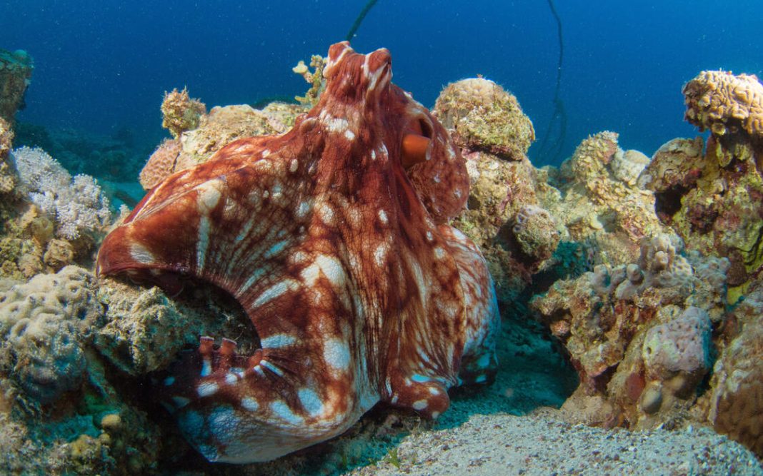
Octopuses have complex “camera” eyes, as seen here in a juvenile animal. [Nir Friedman]

“This is the third-largest expansion of microRNA families in the animal world, and the largest outside of vertebrates,” Grygoriy Zolotarov, MD, lead author of a paper in Science Advances, said when the announcement was made. Therefore, miRNA appears to be closely linked to the evolution of complex animal brains.

The 42 miRNA families Rajewsky and his team identified are not shared with humans. In fact, the most recent common ancestor of octopuses and humans was a primitive flatworm that lived approximately 750 million years ago. These genes were conserved during the evolution of the octopus, so they are likely to be beneficial. Now the question is exactly what benefits they provide.

Another style of intelligence

Octopuses are curious creatures possessing an inventive mindset exemplified by their ability to disguise and protect themselves using opened shells for protection or as projectiles and to gather and store them for later use, as well as their well-known camouflage abilities. They also remember people and things and have distinct preferences. A team of Brazilian researchers thinks they may even dream.

Some researchers have reported they aren’t motivated by snacks (like other animals). As Rajewsky points out, “They do have personalities, so—just maybe—they realize you’re trying to reward them with food and don’t like to be manipulated. I’m not a behavioral scientist. I’m just speculating,” he stresses, “but it does account for the fact that there’s intelligence there that you cannot right away compare with our concepts.

“The octopus is a special invertebrate. By studying how the brain functions in octopuses, we can maybe learn new tools to interfere with our nervous systems or to understand our nervous system better,” Rajewsky tells GEN. While researchers are clearly interested in studying this animal, it’s not in danger of becoming the next lab rat. Its brain is “the most distant brain of all other animal brains.”

Octopuses also are difficult to study, needing aquariums. Rajewsky is a systems biologist working in molecular biology to understand the functioning of cells in tissues, as well as diseases, by studying molecular interactions. “To do this in the octopus, I can only do descriptive studies. I cannot do molecular experiments in octopuses because I don’t have tanks with animals.” It also would require adjusting the existing tools to octopus biochemistry. Therefore, he says, “I’m not intending to experiment on octopuses.” Instead, he analyzes frozen tissue samples collected from a marine station in Naples.

Finding the evolutionary departure

Rajewsky and his team began by quantifying major modes of posttranscriptional regulation across the 18 octopus tissues. They found that A-to-I editing was separate from the miRNA function, and therefore did not modulate it in any functionally important regard. Specifically, “…most of that editing occurred in the introns and 3 prime UTRs of mRNAs,” they wrote, with alternative splicing highest in neural tissues, as expected. Editing rarely altered the splice sites. The transcriptome, in fact, generally resembled that of other invertebrates.

The major evolutionary departure of the octopus from other invertebrates became evident when the researchers investigated miRNAs, they knew bound to the 3’ untranslated regions (3’UTRs).

The octopus genome has 138 miRNA families. When miRNA from Octopus vulgaris was compared with that of another species, O. bimaculoides, which last shared a common ancestor approximately 50 million years ago, the researchers found 43 novel miRNA families were shared among the two octopus species and squid. (Research also shows that 12 miRNAs are shared between squid and nautilus, and another 35 miRNAs are restricted to the octopus lineage.)

The research team is now planning to apply a technique, which will make the cells in octopus tissue visible at a molecular level. [Nir Friedman]

Of the 43 miRNAs unique to the octopus tissue, 34 were expressed in greatest quantity in neural tissues—typically at rates 13 times higher than when expressed in non-neuronal tissue. Levels were particularly high in the visceral ganglion, vertical lobe, and optic gland. Notably, these novel miRNAs were present in late-stage embryos and in just-hatched octopuses as well as in adult tissue, thus underscoring their likely involvement in brain development.

The paper notes that these miRNA sites are conserved throughout evolution and the researchers suggest conservation is the result of functional interaction between the miRNA and miRNA response elements in specific tissues.

Next research goals

There are three things Rajewsky says he wants to learn from this research:

  • In which specific cell types the newly discovered miRNAs are active. To do this, “We’ve developed a method to quantify miRNAs’ single cells,” he says, making them visible at a molecular level.
  • How the various type of neurons and other cells in the brain communicate with each other at a molecular level and how they’re organized. “For this we will perform spatial transcriptomics.”
  • How organism-specific miRNAs relate to brain evolution and either differ or correlate function between the octopi and human brains. “We can do experiments in human organoids to study human-specific miRNAs,” to determine this, he says.

Going further, Rajewsky plans to form a network of other octopus scientists to exchange insights.

Rajewsky became fascinated with octopuses after a trip to California’s Monterey Bay Aquarium, becoming “mesmerized” by an octopus in one of the tanks. “I have the feeling that this is a special and intelligent mind…so I read about octopuses as a hobby. Then, three years ago, I read a paper saying octopuses have elevated levels of RNA editing—a mechanism exists in humans in which RNA molecules can be reprogrammed, thus changing the nucleotide sequence in the RNA.

“Suddenly, I thought, maybe the octopus is an RNA extremophile. If they do this editing, maybe they do other things at the nuclear RNA level, as well.” He then embarked on a project to better understand how “this amazing animal has evolved, maybe learn something fundamental about how complex brains evolve and also something cool about RNA.”

What he has learned opens the door to a deeper understanding of miRNA’s role in the development of complex brains. “That is something that has been hypothesized for a long time,” Rajewsky says, and now it is becoming clearer. “It probably says, also, that miRNA is doing something in the brain that we still don’t understand.”

Brains with more vitamin D may work better


“We wanted to know if vitamin D is even present in the brain, and if it is, how those concentrations are linked to cognitive decline,” says Kyla Shea.

The brains of people with cognitive decline fare better with higher levels of vitamin D, research finds.

The study appears in in Alzheimer’s & Dementia: The Journal of the Alzheimer’s Association.

“This research reinforces the importance of studying how food and nutrients create resilience to protect the aging brain against diseases such as Alzheimer’s disease and other related dementias,” says senior and corresponding author Sarah Booth, director of the Jean Mayer USDA Human Nutrition Research Center on Aging (HNRCA) at Tufts and lead scientist of the HNRCA’s Vitamin K Team.

Vitamin D supports many functions in the body, including immune responses and maintaining healthy bones. Dietary sources include fatty fish and fortified beverages (such as milk or orange juice); brief exposure to sunlight also provides a dose of vitamin D.

“Many studies have implicated dietary or nutritional factors in cognitive performance or function in older adults, including many studies of vitamin D, but all of them are based on either dietary intakes or blood measures of vitamin D,” says lead author Kyla Shea, a scientist on the Vitamin K Team and an associate professor at the Friedman School of Nutrition Science and Policy at Tufts. “We wanted to know if vitamin D is even present in the brain, and if it is, how those concentrations are linked to cognitive decline.”

Booth, Shea, and their team examined samples of brain tissue from 209 participants in the Rush Memory and Aging Project, a long-term study of Alzheimer’s disease that began in 1997. Researchers at Rush University assessed the cognitive function of the participants, older people with no signs of cognitive impairment, as they aged, and analyzed irregularities in their brain tissue after death.

In the study, researchers looked for vitamin D in four regions of the brain—two associated with changes linked to Alzheimer’s disease, one associated with forms of dementia linked to blood flow, and one region without any known associations with cognitive decline related to Alzheimer’s disease or vascular disease. They found that vitamin D was indeed present in brain tissue, and high vitamin D levels in all four regions of the brain correlated with better cognitive function.

However, the levels of vitamin D in the brain didn’t associate with any of the physiological markers associated with Alzheimer’s disease in the brain studied, including amyloid plaque buildup, Lewy body disease, or evidence of chronic or microscopic strokes. This means it’s still unclear exactly how vitamin D might affect brain function.

“Dementia is multifactorial, and lots of the pathological mechanisms underlying it have not been well characterized,” Shea says. “Vitamin D could be related to outcomes that we didn’t look at yet, but plan to study in the future.”

Vitamin D is also known to vary between racial and ethnic populations, and most of the participants in the original Rush cohort were white. The researchers are planning follow-up studies using a more diverse group of subjects to look at other brain changes associated with cognitive decline. They hope their work leads to a better understanding of the role vitamin D may play in staving off dementia.

However, experts caution people not to use large doses of vitamin D supplements as a preventive measure. The recommended dose of vitamin D is 600 IU for people 1-70 years old, and 800 IU for those older—excessive amounts can cause harm and have been linked to the risk of falling.

“We now know that vitamin D is present in reasonable amounts in human brains, and it seems to be correlated with less decline in cognitive function,” Shea says. “But we need to do more research to identify the neuropathology that vitamin D is linked to in the brain before we start designing future interventions.”

Support for the work came from the National Institutes of Health’s National Institute on Aging, as well as the US Department of Agriculture’s Agricultural Research Service. Complete information on authors, funders, and conflicts of interest is available in the published paper. The content is solely the responsibility of the authors and does not necessarily represent the official views of the National Institutes of Health or the US Department of Agriculture.

Source: Tufts University

Axolotls Can Regenerate Their Brains


Summary: Axolotls have the ability to regenerate brain areas following an injury. Researchers have mapped cell types and genes associated with neurodegeneration in the axolotl brain, discovering some similarities in the human brain. The findings could pave the way for new neurodegenerative therapies.

Source: The Conversation

The axolotl (Ambystoma mexicanum) is an aquatic salamander renowned for its ability to regenerate its spinal cord, heart and limbs. These amphibians also readily make new neurons throughout their lives. In 1964, researchers observed that adult axolotls could regenerate parts of their brains, even if a large section was completely removed. But one study found that axolotl brain regeneration has a limited ability to rebuild original tissue structure.

So how perfectly can axolotl’s regenerate their brains after injury?

As a researcher studying regeneration at the cellular level, I and my colleagues in the Treutlein Lab at ETH Zurich and the Tanaka Lab at the Institute of Molecular Pathology in Vienna wondered whether axolotls are able to regenerate all the different cell types in their brain, including the connections linking one brain region to another.

In our recently published study, we created an atlas of the cells that make up a part of the axolotl brain, shedding light on both the way it regenerates and brain evolution across species.

Why look at cells?

Different cell types have different functions. They are able to specialize in certain roles because they each express different genes. Understanding what types of cells are in the brain and what they do helps clarify the overall picture of how the brain works. It also allows researchers to make comparisons across evolution and try to find biological trends across species.

One way to understand which cells are expressing which genes is by using a technique called single-cell RNA sequencing (scRNA-seq). This tool allows researchers to count the number of active genes within each cell of a particular sample. This provides a “snapshot” of the activities each cell was doing when it was collected.

https://www.youtube.com/embed/k9VFNLLQP8c?feature=oembedCredit: UCSF

This tool has been instrumental in understanding the types of cells that exist in the brains of animals. Scientists have used scRNA-seq in fishreptilesmice and even humans. But one major piece of the brain evolution puzzle has been missing: amphibians.

Mapping the axolotl brain

Our team decided to focus on the telencephalon of the axolotl. In humans, the telencephalon is the largest division of the brain and contains a region called the neocortex, which plays a key role in animal behavior and cognition.

Throughout recent evolution, the neocortex has massively grown in size compared with other brain regions. Similarly, the types of cells that make up the telencephalon overall have highly diversified and grown in complexity over time, making this region an intriguing area to study.

We used scRNA-seq to identify the different types of cells that make up the axolotl telencephalon, including different types of neurons and progenitor cells, or cells that can divide into more of themselves or turn into other cell types.

We identified what genes are active when progenitor cells become neurons, and found that many pass through an intermediate cell type called neuroblasts – previously unknown to exist in axolotls – before becoming mature neurons.

https://www.youtube.com/embed/uooR4293p_4?feature=oembedCredit: TED Ed

We then put axolotl regeneration to the test by removing one section of their telencephalon. Using a specialized method of scRNA-seq, we were able to capture and sequence all the new cells at different stages of regeneration, from one to 12 weeks after injury. Ultimately, we found that all cell types that were removed had been completely restored.

We observed that brain regeneration happens in three main phases. The first phase starts with a rapid increase in the number of progenitor cells, and a small fraction of these cells activate a wound-healing process. In phase two, progenitor cells begin to differentiate into neuroblasts. Finally, in phase three, the neuroblasts differentiate into the same types of neurons that were originally lost.

Astonishingly, we also observed that the severed neuronal connections between the removed area and other areas of the brain had been reconnected. This rewiring indicates that the regenerated area had also regained its original function.

See also

This shows a pregnant woman's belly

FeaturedNeurologyNeuroscience

May 3, 2022

Brain Folds Formed During Fetal Stage Could Affect Onset of Dementia

Amphibians and human brains

Adding amphibians to the evolutionary puzzle allows researchers to infer how the brain and its cell types has changed over time, as well as the mechanisms behind regeneration.

When we compared our axolotl data with other species, we found that cells in their telencephalon show strong similarity to the mammalian hippocampus, the region of the brain involved in memory formation, and the olfactory cortex, the region of the brain involved in the sense of smell. We even found some similarities in one axolotl cell type to the neocortex, the area of the brain known for perception, thought and spatial reasoning in humans.

These similarities indicate that these areas of the brain may be evolutionarily conserved, or stayed comparable over the course of evolution, and that the neocortex of mammals may have an ancestor cell type in the telencephalon of amphibians.

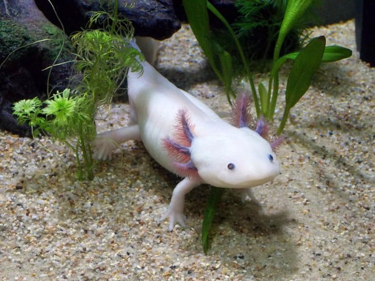
This shows an axolotl.
Axolotls are a model organism researchers use to study a variety of topics in biology. Image is in the public domain

While our study sheds light on the process of brain regeneration, including which genes are involved and how cells ultimately become neurons, we still don’t know what external signals initiate this process. Moreover, we don’t know if the processes we identified are still accessible to animals that evolved later in time, such as mice or humans.

But we’re not solving the brain evolution puzzle alone. The Tosches Lab at Columbia University explored the diversity of cell types in another species of salamander, Pleurodeles waltl, while the Fei lab at the Guangdong Academy of Medical Sciences in China and collaborators at life sciences company BGI explored how cell types are spatially arranged in the axolotl forebrain.

Identifying all the cell types in the axolotl brain also helps pave the way for innovative research in regenerative medicine. The brains of mice and humans have largely lost their capacity to repair or regenerate themselves. Medical interventions for severe brain injury currently focus on drug and stem cell therapies to boost or promote repair.

Examining the genes and cell types that allow axolotls to accomplish nearly perfect regeneration may be the key to improve treatments for severe injuries and unlock regeneration potential in humans.

What Can Wordle Do for Our Brains?


Forget morning coffee, stretches, or meditation. There’s a new way to kick-start your brain.

Jeanenne Ray, a book editor in Marin County, CA, tackles it first thing in the morning, while still lying in bed. It’s also the first on the to-do list of Shelly Groves, who owns a dog walking and pet sitting service in Avondale Estates, GA. That’s also the pattern of Todd Siesky, an Atlanta communications professional, but he knows to walk away if it gets too frustrating.

The three are among the millions playing Wordle, the “it” puzzle/brain teaser of the moment. Created by software engineer Josh Wardle of Brooklyn, NY, for his partner during the pandemic, it’s now been sold to The New York Times, and initially will remain free.

For those who’ve never tested their brain power on Wordle, it’s simple but challenging. Players get six attempts to guess the five-letter word of the day. After plugging in a word as their first guess, they get feedback, with color coded blocks telling them if their chosen letters are correct and in the right position.

Can It Help Brain Power?

Besides providing us with fresh fodder for bragging rights on social media, where players obsessively post their scores, can playing Wordle daily improve our memory and overall brain power?

Probably, say two neuroscientists who study the workings of the human brain, as long as frustration doesn’t undo the benefits.

Michael Yassa, PhD, professor and director of the center for the Neurobiology of Learning and Memory at the University of California, Irvine, began playing Wordle in January.

“It activates our dopamine,” he says.

That’s the neurotransmitter linked with feeling pleasure, satisfaction, and motivation. “That can color your day in a positive way,” he says.

Playing the game also gets your problem-solving skills going, Yassa says.

Wordle 236 2/6

⬜⬜🟨⬜🟩
🟩🟩🟩🟩🟩— Kiernan Shipka (@kiernanshipka) February 10, 2022

Another benefit, he says, is the social interaction that naturally follows for most. When a player gets the answer in two or three tries, boasting on social media is common.

“We know that social interactions are good for our brain,” Yassa says.

When you interact with others, he says, there’s more release of dopamine, along with oxytocin, the so-called love hormone that rises during hugging and is linked with empathy, trust, and relationship-building.

Sharing results is usually a healthy competition, Yassa says. He compares results with his brother, who lives on the East Coast.

“I feel like I’ve bonded with my brother a lot more,” he says. As for wins, “we go back and forth,” with one winning one day, the other the next.

What about the claim from some experts that Wordle will create new brain synapses, needed for communication between cells, or will strengthen existing ones? There’s no study on Wordle and synapse-building that Yassa is aware of, but he says it makes sense that it would build or strengthen them.

“When you are engaging in a novel activity, you can create new synapses,” he says, and scientists know that’s part of the brain’s ongoing plasticity, the ability of the nervous system to change in response to stimuli, either internal or external.

But it’s not possible at this point so say how much synapse-building Wordle might do, Yassa says.

“Anything that causes a high level of engagement — something that engages memory, problem solving — is good for your brain, and will strengthen those processes in your brain,” says Earl Miller, PhD, professor of neuroscience at the Massachusetts Institute of Technology. “Your brain is like a muscle, and the more you use it the better it gets at doing things.”

But Yassa cautions it may take some time to see effects on memory. And occasional players may not see the same benefits as daily fans.

Word Puzzle Research

In a previous study, conducted well before Wordle debuted, researchers studied the links between word puzzle habits and 14 cognitive measures, such as memory and attention, in more than 19,000 adults, ages 50 to 93. Some never played word puzzles, while others did occasionally, frequently, or even more than once a day.

For each measure tested, those who never did word puzzles or did them only occasionally performed more poorly than virtually every other group, the researchers found.

Players’ Experiences

Many players say Wordle is just plain fun. “Having a puzzle that is rooted in words is both fascinating and enjoyable,” Siesky says. There is a logic to all puzzles, he says, including Wordle’s. That’s part of the attraction for him.

“I feel like it’s good for my 58-year-old brain,” Groves says, although she doesn’t think she’s been doing it long enough to see improvements in memory. It hasn’t changed her social media use one way or the other. She sees sharing results there as ”a humble brag or perhaps a humbling moment” for those times when it takes all six guesses to get the word, or, shudder, if you don’t get it at all.

Ray doesn’t compete with anyone, but gives feedback when she sees results on social media. A former high school classmate got the answer in two tries the other day, she says, and that led to some congratulations and pleasant conversation.

Wordle 235 3/6

⬛🟨⬛🟨⬛
⬛🟩⬛🟩🟩
🟩🟩🟩🟩🟩— Tom Joseph (@TomJChicago) February 9, 2022

Players often trade tips, with a little good-natured ribbing as well as advice. For instance, while “adieu” is a favorite start word for some, due to all the vowels, it has been scorned by others.

In January, British players were not pleased, pointing out that “favor,” the word of the day, was ”Americanized” and is actually spelled “favour.”

Sharing the best tips is apparently expected. Tweeted one player recently: “Just told my bf that I always start with GRAVY on wordle and he is absolutely furious with me.”

Frustration Factor

Some days are more difficult than others, of course. “If I get really frustrated, I force myself to think about patterns and language,” Siesky says. If that doesn’t work, he takes a break.

Yassa acknowledges that frustration factor, as he’s experienced it firsthand. He says he has never solved the puzzle in one try. “I’ve gotten it in two tries twice, and a lot more in four tries. One took six,” he laughs, ”and that one almost gave me a heart attack.”

If it’s too stressful, it might not be your game, Yassa and Miller agree. “Stress is counterproductive to your health,” Miller says. Momentary frustration with Wordle is OK, but if it’s really stressing you out, ”find something you are better at,” he suggests.

“It’s trial and effort,” Yassa says about the best choices for people. If Wordle isn’t your thing, maybe you’re better at numbers than words, Yassa says, and should try a numbers-based puzzle like Sudoku. That is one, Yassa admits, that he avoids.