Viruses Can Exploit Built-In Antibody Bias to Reinfect Hosts


A massive crowd of people shouts as one, “WE ARE ALL INDIVIDUALS!” Then, a lone person in the crowd sheepishly mutters, “I’m not.”

The scene above, one of the best from the Monty Python troupe, brings to mind a phenomenon known as the immunodominant public antibody response. It is a widely shared immune response to a pathogen. In different people, the same epitopes on viral proteins end up being targeted by the same antibodies—public antibodies.

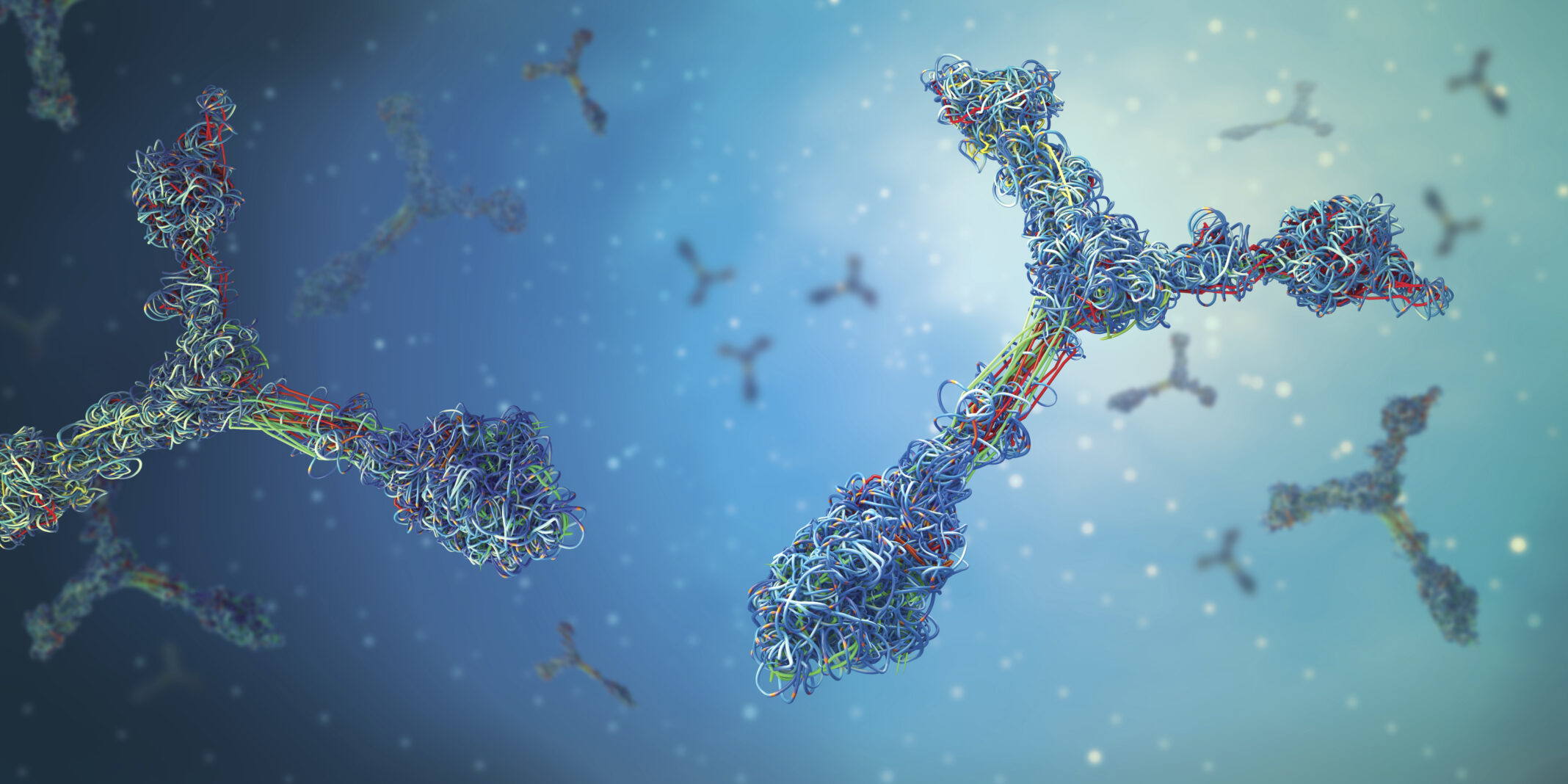
The phenomenon, which appears to reflect our immune systems’ tendency to be efficient, can sometimes leave us vulnerable. That is, it may cause us to produce antibodies that repeatedly target the same epitopes, even if the antibodies are not protective. Or, it can give viruses, such as SARS-CoV-2, an easy way to evade the immune response. For example, a virus could mutate just a few target residues and gain the ability to outmaneuver antibodies that are widely shared by many people.

A better understanding of the phenomenon is now possible thanks to a new study led by investigators from Brigham and Women’s Hospital. According to this study, the generation of antibodies is far from random because of germline-encoded amino acid–binding (GRAB) motifs. GRAB motifs represent a germline-encoded component of the architecture of the antibody repertoire that predisposes antibodies to recognize particular structures and thus influences epitope selection and composition.

The study—”Germline-encoded amino acid–binding motifs drive immunodominant public antibody responses”—was published in Science. “Our research may help explain a lot of the patterns we’ve seen during the COVID-19 pandemic, especially in terms of re-infection,” said corresponding author Stephen J. Elledge, PhD, the Gregor Mendel professor of genetics at the Brigham and Harvard Medical School. “Our findings could help inform immune predictions and may change the way people think about immune strategies.”

Before the study, there were hints, but no clear evidence, that people’s immune systems didn’t target sites on a viral protein at random. In isolated examples, investigators had seen recurrent antibody responses across individuals—people recreating antibodies to home in on the same viral protein location (known as an epitope). But the study by Elledge and colleagues helps explain the extent and underlying mechanisms of this phenomenon.

The team used a tool the Elledge lab developed in 2015 called VirScan, which can detect thousands of viral epitopes—sites on viruses that antibodies recognize and bind to—and give a snapshot of a person’s immunological history from a single drop of blood. For the new study, the researchers used VirScan to analyze 569 blood samples from participants in the United States, Peru, and France. They found that recognition of public epitopes—viral regions recurrently targeted by antibodies—was a general feature of the human antibody response.

“By mapping 376 immunodominant ‘public epitopes’ at high resolution and characterizing several of their cognate antibodies, we concluded that germline-encoded sequences in antibodies drive recurrent recognition,” the article’s authors wrote. “Systematic analysis of antibody-antigen structures uncovered 18 human and 21 partially overlapping mouse GRAB motifs within heavy and light V gene segments that in case studies proved critical for public epitope recognition.”

GRAB motifs correspond to antibody regions that are particularly good at picking out one specific amino acid. They help explain why human antibodies tend to focus on regions where these amino acids are available for binding, and thus repeatedly bind the same spots. A small number of mutations can help a virus avoid detection by these shared antibodies, allowing the virus to reinfect populations that were previously immune.

“We find an underlying architecture in the immune system that causes people, no matter where in the world they live, to make essentially the same antibodies that give the virus a very small number of targets to evade in order to reinfect people and continue to expand and further evolve,” said lead author Ellen L. Shrock, PhD, of the Elledge lab.

Interestingly, the team noted that nonhuman species produce antibodies that recognize different public epitopes from those that humans recognize. And, while it is more likely for a person to produce antibodies against a public epitope, some people do produce rarer antibodies, which may more effectively protect them from reinfection. These insights could have important implications for treatments developed against COVID-19, such as monoclonal antibodies, as well as for vaccine design.

“The more unique antibodies may be a lot harder to evade,” Elledge said. “[This] is important to consider as we think about the design of better therapies and vaccines.”

FDA OKs First Rapid Test for HIV-1/2 Antibodies, HIV-1 Antigen.


The US Food and Drug Administration (FDA) today approved the first rapid diagnostic test to spot antibodies to both HIV types 1 and 2 as well as the HIV-1 p24 antigen. The new test will enable HIV testing to expand to regions without traditional healthcare facilities and speed up treatment, according to the agency.

The Alere Determine HIV-1/2 Ag/Ab Combo test, manufactured by Orgenics of Yavne, Israel, can detect HIV antibodies and the HIV-1 p24 antigen in human serum, plasma, and venous or fingerstick whole-blood specimens. The HIV-1 antigen is an earlier tip-off to HIV-1 infection than HIV-1 antibodies by themselves. The new test can tell the difference between acute vs established HIV-1 infections when the blood specimen tests positive for the p24 antigen, but negative for HIV-1 and HIV-2 antibodies.

Most HIV infections worldwide stem from HIV-1. HIV-2 infections occur mostly in West Africa.

“This test helps diagnose HIV infection at an earlier time in outreach settings, allowing individuals to seek medical care sooner,” said Karen Midthun, MD, director of the FDA’s Center for Biologics Evaluation and Research in a news release. “Earlier diagnosis may also help to reduce additional HIV transmission.”

The Alere Determine HIV-1/2 Ag/Ab Combo test is not intended to screen blood donors.

Source: Medscape.com


;<�? n<� 0� http://www.medscape.com/viewarticle/779445&#8243; target=”_blank”>priority review status to dolutegravir. Its approval comes 4 days after the FDA okayed the first rapid diagnostic test to spot antibodies to both HIV types 1 and 2 as well as the HIV-1 p24 antigen.


The FDA determined that dolutegravir is safe and effective on the basis of 4 clinical trials involving 2539 participants with HIV. Drug regimens for treating HIV that included dolutegravir proved effective in reducing viral loads, according to the agency. The pharmacokinetics, safety, and activity of the drug with regard to children defined in the pediatric indication were established in a fifth clinical trial.

Insomnia and headache were common adverse events observed in the clinical trials. Among the more serious adverse events were hypersensitivity reactions and abnormal liver functions among patients who also were infected with hepatitis B or C, or both. The drug’s label contains instructions on how to monitor patients for such serious adverse events.

Source: Medscape.com


VariZIG for Varicella Postexposure Prophylaxis: Updated CDC Recommendations.


The new recommendations extend the treatment window from 4 to 10 days postexposure.


In the 1960s, zoster immune globulin (immune globulin prepared from patients recovering from zoster) was found to reduce severity of primary varicella as well as attack rates. The development of varicella zoster immune globulin (VZIG) followed in the late 1970s for high-risk patients with varicella exposure. In 2006, VZIG was replaced by a similar product — VariZIG. VariZIG was available as an investigational new drug expanded access protocol until late 2012 and administration was recommended up to 4 days after exposure.

In December 2012, the FDA approved VariZIG for postexposure prophylaxis of varicella in high-risk susceptible individuals. Now, the CDC recommendations extend the treatment window to 10 days postexposure. This recommendation concurs with the current American Academy of Pediatrics recommendations for VariZIG use for the following exposed groups:

  • Immunocompromised patients without evidence of immunity
  • Newborn infants whose mothers have signs of varicella 5 days before to 2 days after delivery
  • Hospitalized premature infants ≥28 weeks’ gestation born to susceptible mothers
  • Hospitalized premature infants <28 weeks’ gestation and birth weight ≤1000 g irrespective of maternal immunity
  • Pregnant women without evidence of immunity

COMMENT

These new recommendations outline the use of passive immunoglobulin (VariZIG) for postexposure prophylaxis of susceptible patients with varicella exposure. The patient groups for which prophylaxis is recommended have not changed from the original VZIG recommendations, but the time for administration of postexposure prophylaxis has been extended to 10 days following exposure. Efficacy is increased if prophylaxis is given as soon as possible after exposure. Vaccination against varicella provides the best protection; however, in patients for whom a live viral vaccine is contraindicated, passive immunity with VariZIG provides protection. VariZIG is available in the U.S. through FFF Enterprises.

Source: NEJM

New wonder drug matches and kills all kinds of cancer — human testing starts 2014.


Stanford researchers are on track to begin human trials of a potentially potent new weapon against cancer, and would-be participants are flooding in following thePost’s initial report on the discovery.

shutterstock_88169086115129--300x300

The progress comes just two months after the groundbreaking study by Dr Irv Weissman, who developed an antibody that breaks down a cancer’s defense mechanisms in the body.

A protein called CD47 tells the body not to “eat” the cancer, but the antibody developed by Dr Weissman blocks CD47 and frees up immune cells called macrophages — which can then engulf the deadly cells.

The new research shows the miraculous macrophages effectively act as intelligence gatherers for the body, pointing out cancerous cells to cancer-fighting “killer T” cells.

The T cells then “learn” to hunt down and attack the cancer, the researchers claim.

“It was completely unexpected that CD8+ T (killer T) cells would be mobilized when macrophages engulfed the cancer cells in the presence of CD47-blocking antibodies,” said MD/PhD student Diane Tseng, who works with Dr. Weissman.

The clinical implications of the process could be profound in the war on cancer.

When macrophages present “killer T” cells with a patient’s cancer, the T cells become attuned to the unique molecular markers on the cancer.

This turns them into a personalized cancer vaccine.

“Because T cells are sensitized to attack a patient’s particular cancer, the administration of CD47-blocking antibodies in a sense could act as a personalized vaccination against that cancer,” Tseng said.

The team of researchers at Stanford plan on starting a small 10-100 person phase I clinical human trial of the cancer therapy in 2014.


RESEARCHERS IDENTIFY BIOMARKER FOR AUTISM IN A SUBSET OF CHILDREN: MATERNAL ANTIBODY-RELATED AUTISM.


UC Davis MIND Institute researchers have identified the specific antibodies that target fetal brain proteins in the blood of a subset of women whose children are diagnosed with autism. The finding is the first to pinpoint a specific risk factor for a significant subset of autism cases, as well as a biomarker for drug development and early diagnosis. The researchers have named autism related to these antibodies “Maternal Autoantibody-Related,” or MAR autism.

VandeWater

The study found that the mothers of children with autism were more than 21 times as likely to have the specific MAR antibodies in their systems that reacted with fetal brain proteins, or antigens, than were the mothers of children who did not have autism. In fact, specific combinations of MAR antibodies were not found in the blood of mothers whose children were typically developing.

The research, “Autism-specific maternal autoantibodies recognize critical proteins in developing brain,” is published online today in Translational Psychiatry, a Nature journal.

The study was led by principal investigator and immunologist Judy Van de Water, a researcher affiliated with the MIND Institute. Earlier studies by Van de Water and her colleagues found that women with certain antibodies in their bloodstreams are at greater risk of having a child with autism and that their children exhibited more severe language delays, irritability and self-injurious behaviors than did the autistic children of mothers whose blood did not have the antibodies.

“Now we will be able to better determine the role of each protein in brain development,” said Van de Water, professor of internal medicine. “We hope that, one day, we can tell a mother more precisely what her antibody profile means for her child, then target interventions more effectively.”

To identify the exact antigens targeted by the mothers’ antibodies, Van de Water and her colleagues conducted the research in Northern California using blood samples from 246 mothers of children with autism and of a control group of 149 mothers of children without autism to examine their reactivity with the candidate antigens.

Seven antigens were significantly more reactive to the blood of mothers of children with autism than to that of the control mothers. The study found that the mothers with antibodies that reacted with any one of these antigens, either individually or in combination with other antigens, were more than three times as likely to have a child with autism spectrum disorder.

Several combinations of antibodies in the blood from mothers of children with autism were not found in the control mothers’ blood. Nearly 23 percent of mothers of children with autism had certain combinations of autoantibodies against the target antigens, compared with less than 1 percent of mothers of children without the disorder.

The specific antigens identified in the study are lactate dehydrogenase A and B, cypin (guanine deaminase), stress-induced phosphoprotein 1, collapsing response mediator proteins 1 and 2, and Y-box binding protein. All are found throughout the body, but also are expressed at significant levels in the human fetal brain and have established roles in neurodevelopment. For example, cypin is an enzyme that plays an important role in normal neurite branching, a fundamental function in the developing brain, whereas the CRMP proteins are critical later in neuron development for axon outgrowth.

Maternal antibodies are known to cross the placenta during pregnancy and can be detected in a fetus as early as 13 weeks. By 30 weeks, maternal antibody levels in the fetus are about half that of the mother, and at birth, the concentration is even greater in the newborn than in the mother herself. The maternal antibodies stay in the baby’s bloodstream for about 6 months after birth, after which the baby’s own immune system takes over.

Once in the fetal bloodstream, the antibodies then may enter the brain and attack cells that have corresponding proteins that act as antigens. This antigen-antibody response is an important defense against foreign invaders, such as bacteria or viruses, but is not normally directed against oneself. When directed against one’s own tissue, the antibodies are known as autoantibodies.

“It is important to note that women have no control over whether or not they develop these autoantibodies, much like any other autoimmune disorder,” Van de Water said. “And, like other autoimmune disorders, we do not know what the initial trigger is that leads to their production.”

Understanding which proteins and which pathways are implicated in MAR autism can help elucidate the causes of autism and possibly lead to new therapies, such as administering ‘antibody blockers’ to the mother during pregnancy to prevent damage to the developing fetal brain, Van de Water said.

These findings are leading to the development of a MAR diagnostic test for autism, which would be available to the mothers of young children who are showing signs of developmental delay. If the test were positive, the child would be a candidate for early behavioral intervention.

“These findings are incredibly important because they establish a cause for a significant portion of autism cases, thereby opening up new lines of inquiry into possible biological treatments,” said MIND Institute Director Leonard Abbeduto. “In addition, the findings demonstrate that a diagnostic test is within reach. This test would be invaluable for women who are considering becoming pregnant and could lead to earlier and more accurate diagnosis of children with developmental challenges and help get them into behavioral interventions at younger ages.”

A MAR diagnostic test also would assess a mother’s risk of having a child with autism prior to conception, which is particularly important for women who already have a child with the disorder. UC Davis has patented this technology and licensed the exclusive worldwide rights to develop it for commercial purposes to Pediatric Bioscience, Inc.

“We know that early behavioral interventions for autism are critical,” said Isaac Pessah, professor and chair of the Department of Molecular Biosciences in the UC Davis School of Veterinary Medicine and former director of the UC Davis Center for Children’s Environmental Health. “Developing a predictive test for autism before symptoms become obvious could have an enormous impact on treating children with the condition.”

Study participants were from the CHARGE (Childhood Autism Risks from Genetics and the Environment) study, an ongoing study that was launched in 2001 by the MIND Institute and the UC Davis Center for Children’s Environmental Health, of which Van de Water now is director. Children with autism spectrum disorder, children with developmental delay and typically developing children between the ages of 2 and 5 years are studied with the goal of better understanding the causes of autism.

A related study is the MARBLES (Markers of Autism Risk in Babies ― Learning Early Signs) study, also being conducted at the MIND Institute and the Center for Children’s Environmental Health. This study follows pregnant women who already have a child with autism. Multiple factors related to genetics and the environment is under study in an effort to uncover predictors for having a child with autism.

Van de Water said knowing the specific protein targets of the maternal antibodies enables researchers to develop more precise animal models of autism.

The study was funded by NIEHS grants P01 ES11269-01 and 1 R01-ES015359; United States Environmental Protection Agency Science to Achieve Results (STAR) program grant R829388; the UC Davis MIND Institute; and an Autism Speaks graduate fellowship.

Other authors include Daniel Braunschweig, Paula Krakowiak, Paul Duncanson, Robert Boyce, Robin Hansen, Paul Ashwood and Irva Hertz-Picciotto, all of UC Davis.

At the UC Davis MIND Institute, world-renowned scientists engage in collaborative, interdisciplinary research to find the causes of and develop treatments and cures for autism, attention-deficit/hyperactivity disorder (ADHD), fragile X syndrome, 22q11.2 deletion syndrome, Down syndrome and other neurodevelopmental disorders.

Source:  mindinstitute.ucdavis.edu