First-Ever Biorobotic Heart Helps Scientists Study Cardiac Function


A model heart made from living tissue fused with robotic muscles could help researchers see how the organ works on the inside
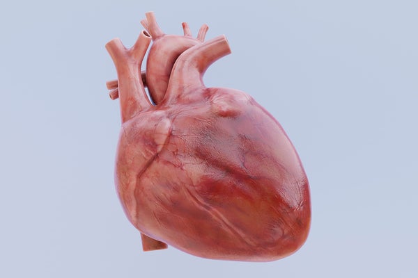
Realistic 3D Render of Human Heart

From artificial heart valves to cellular transplants, new treatments for cardiovascular ailments are being developed every day. To model how they work, researchers need a reliable way to observe the heart in action. Animal studies, computer models and various laboratory simulators made with dead heart tissue can all provide different views, but these approaches can be expensive, lacking in complexity or limited in their shelf life.

So to tinker with the heart, scientists have now developed a beating, biorobotic replica that can simulate the workings of both a healthy organ and a diseased one. This simulator combines pig heart tissue and soft robotic muscles and was described in two recent studies.

“Imagine a beating heart on a lab bench,” says Ellen Roche, a biomedical engineer at the Massachusetts Institute of Technology and the studies’ senior author. The simulator, which pumps a clear fluid instead of blood, is hooked up to instruments that measure blood flow, blood pressure, and more. It’s also customizable: the user can change the heart rate, blood pressure and other parameters, then watch how these changes affect the heart’s function in real time through an internal camera.

The simulator accurately replicates how blood flows through the heart—something that existing benchtop simulators using dead heart tissue could not do. Using live heart tissue from a pig instead, animated by robotics, granted Roche’s team far more control. (Pig hearts are similar in size and layout to human hearts and are often used in research.) The new hybrid simulator can also last longer than a live organ that is used on its own: whereas a pig heart hooked up to a pump in the lab would only continue beating for a few hours, Roche’s team was able to keep the simulator’s synthetic muscles going for months. The researchers haven’t measured the simulator’s exact limits yet. “We need to do robust shelf-life fatigue testing to see exactly how many cycles these things can do,” Roche says.

When it comes to modeling blood flow through the heart, the left and right sides of the organ are each their own challenge. “They require very customized models,” Roche says. The researchers tackled the left side first by focusing on the mitral valve, which controls the flow between the left atrium and ventricle (the heart’s upper and lower chambers). They re-created the healthy motion of this system before modeling a condition in which the valve becomes leaky, called mitral regurgitation. To demonstrate that the model could be used as an accurate simulation, the team had cardiac surgeons correct the valve with three different surgical interventions. These results were described in one of the two recent studies, which was published on Wednesday in Device.

“It’s a very complex pumping motion that you have to create in order for the blood to pump to your body at a really high pressure and flow,” says Clara Park, who co-authored both studies as a Ph.D. student at Roche’s lab at M.I.T.

Next the team modeled the mechanics on the right side of the heart. “The right heart is kind of the thinner, weaker muscle,” Park says, and it “doesn’t pump as hard.” The right heart simulator can replicate both healthy and abnormal functioning. These results were described in the team’s other recent study, which was published last month in Nature Cardiovascular Research.

Sarah Vigmostad, a biomedical engineer at the University of Iowa, who was not involved in the papers, believes that these simulators would be valuable for surgical planning, training or educational purposes. “I can also imagine their value in testing new interventions … designed to treat mitral regurgitation or other valve diseases,” she says. The ability to “tune” the heart to replicate various diseases could be very useful in research as well, she adds.

The biorobotic approach relies on live animal tissue, but Roche dreams of a completely 3-D-printed synthetic heart. Such an organ could be far more customizable. It could even be used to create patient-specific models that would allow people in treatment to observe a replica of their own beating heart in action and that would guide their doctors’ decisions. “We are moving to fully synthetic models [and] multi-material prints,” she says, which will require replicating heart tissue itself in the lab. “There’s a lot of ongoing work.”

Novel embalming solution for neurosurgical simulation in cadavers Laboratory investigation.


Surgical simulation using postmortem human heads is one of the most valid strategies for neurosurgical research and training. The authors customized an embalming formula that provides an optimal retraction profile and lifelike physical properties while preventing microorganism growth and brain decay for neurosurgical simulations in cadavers. They studied the properties of the customized formula and compared its use with the standard postmortem processing techniques: cryopreservation and formaldehyde-based embalming.

METHODS

Eighteen specimens were prepared for neurosurgical simulation: 6 formaldehyde embalmed, 6 cryopreserved, and 6 custom embalmed. The customized formula is a mixture of ethanol 62.4%, glycerol 17%, phenol 10.2%, formaldehyde 2.3%, and water 8.1%. After a standard pterional craniotomy, retraction profiles and brain stiffness were studied using an intracranial pressure transducer and monitor. Preservation time—that is, time that tissue remained in optimal condition—between specimen groups was also compared through periodical reports during a 48-hour simulation.

RESULTS

The mean (± standard deviation) retraction pressures were highest in the formaldehyde group and lowest in the cryopreserved group. The customized formula provided a mean retraction pressure almost 3 times lower than formaldehyde (36 ± 3 vs 103 ± 14 mm Hg, p < 0.01) and very similar to cryopreservation (24 ± 6 mm Hg, p < 0.01). For research purposes, preservation time in the cryopreserved group was limited to 4 hours and was unlimited for the customized and formaldehyde groups for the duration of the experiment.

CONCLUSIONS

The customized embalming solution described herein is optimal for allowing retraction and surgical maneuverability while preventing decay. The authors were able to significantly lower the formaldehyde content as compared with that in standard formulas. The custom embalming solution has the benefits from both cryopreservation (for example, biological brain tissue properties) and formaldehyde embalming (for example, preservation time and microorganism growth prevention) and minimizes their drawbacks, that is, rapid decay in the former and stiffness in the latter. The presented embalming formula provides an important advance for neurosurgical simulations in research and teaching.

Foramen ovale puncture, lesioning accuracy, and avoiding complications: microsurgical anatomy study with clinical implications.


Abstract

OBJECT

Foramen ovale (FO) puncture allows for trigeminal neuralgia treatment, FO electrode placement, and selected biopsy studies. The goals of this study were to demonstrate the anatomical basis of complications related to FO puncture, and provide anatomical landmarks for improvement of safety, selective lesioning of the trigeminal nerve (TN), and optimal placement of electrodes.

METHODS

Both sides of 50 dry skulls were studied to obtain the distances from the FO to relevant cranial base references. A total of 36 sides from 18 formalin-fixed specimens were dissected for Meckel cave and TN measurements. The best radiographic projection for FO visualization was assessed in 40 skulls, and the optimal trajectory angles, insertion depths, and topographies of the lesions were evaluated in 17 specimens. In addition, the differences in postoperative pain relief after the radiofrequency procedure among different branches of the TN were statistically assessed in 49 patients to determine if there was any TN branch less efficiently targeted.

RESULTS

Most severe complications during FO puncture are related to incorrect needle placement intracranially or extracranially. The needle should be inserted 25 mm lateral to the oral commissure, forming an approximately 45° angle with the hard palate in the lateral radiographic view, directed 20° medially in the anteroposterior view. Once the needle reaches the FO, it can be advanced by 20 mm, on average, up to the petrous ridge. If the needle/radiofrequency electrode tip remains more than 18 mm away from the midline, injury to the cavernous carotid artery is minimized. Anatomically there is less potential for complications when the needle/radiofrequency electrode is advanced no more than 2 mm away from the clival line in the lateral view, when the needle pierces the medial part of the FO toward the medial part of the trigeminal impression in the petrous ridge, and no more than 4 mm in the lateral part. The 40°/45° inferior transfacial–20° oblique radiographic projection visualized 96.2% of the FOs in dry skulls, and the remainder were not visualized in any other projection of the radiograph. Patients with V1 involvement experienced postoperative pain more frequently than did patients with V2 or V3 involvement. Anatomical targeting of V1 in specimens was more efficiently achieved by inserting the needle in the medial third of the FO; for V2 targeting, in the middle of the FO; and for V3 targeting, in the lateral third of the FO.

CONCLUSIONS

Knowledge of the extracranial and intracranial anatomical relationships of the FO is essential to understanding and avoiding complications during FO puncture. These data suggest that better radiographic visualization of the FO can improve lesioning accuracy depending on the part of the FO to be punctured. The angles and safety distances obtained may help the neurosurgeon minimize complications during FO puncture and TN lesioning.

Source: JNS


World’s leggiest millipede put under microscope.


The anatomical secrets of the world’s leggiest creature, a millipede with 750 legs, have been revealed by scientists.

The species, called Illacme plenipes, was first seen 80 years ago but was recently rediscovered in California.

Now researchers have found that as well as bearing an extreme number of legs, the creature may have more in common with millipedes that lived millions of years ago than today’s species.

The study is published in the journal ZooKeys.

“It’s a kind of mythical creature in the millipede world,” said Dr Paul Marek, an entomologist from the University of Arizona and the lead author of the paper.

Record breaker

In 2005, Dr Marek and his brother discovered some of the leggy arthropods lurking under boulders in the mountains of California. Until then, I.plenipes had not been glimpsed since 1926.

A paper published in the journal Nature outlined the rediscovery and described the creature’s basic biology, but the new research looked at the creature’s anatomy in much more detail.

Despite the name – most millipedes have far fewer than 1,000 feet. Most species belonging to the most common order Polydesmida have an average of just 62.

But Dr Marek confirmed that I.plenipes safely holds the record for the leggiest creature: females can have up to 750 legs, while males have up to 562.

“It seems like these legs evolved for their subterranean lifestyle,” he explained.

“They live deep underground: we found them about 10-15cm (4-6in) below the soil’s surface.

“They are typically found clinging onto sandstone boulders. Based on functional morphology of closely related species, it seems like all of these legs evolved to burrow under the ground and to cling onto these large boulders.”

Though they have many limbs, the creatures are small, measuring about about 3cm-long (1in).

Open wide

Close examination of the creature revealed that it had some ancient features.

 “Start Quote

Its anatomy retains a number of primitive characteristics”

Dr Paul MarekUniversity of Arizona

Most millipedes chew leaves and decaying vegetation with grinding mouth parts.

But the scientists found that this species had a more rudimentary anatomy. Its jaws are fused to its head, and Dr Marek believes that it pierces and then sucks up plant and fungal tissues to satisfy its appetite.

The creature’s body segments were also more similar to ancient millipedes than to most other species found today.

Dr Marek said that millions of years ago creatures like I.plenipes would have been widespread, but now it was one of the last of its kind.

He explained: “It is a relict species. Its most closely related lineages are in South Africa and there is nothing related to this species in the entire North America region. Its anatomy retains a number of primitive characteristics.”

The animal, which also has a number of other unusual features such as body hairs that secrete silk, is thought to be extremely rare and only found in a small area close to San Francisco.

Dr Marek said: “Based on our search of the area, it seem like it is known in three spots – and these spots are about 4.5km (3 miles) away from one and other.

“It does seem that this creature is restricted both in terms of geography and also evolutionarily.”

Dr Marek, a self-confessed millipede enthusiast, said that rediscovering the record breaking creature was a staggering experience, but that it would be “awesome” if someone unearthed an even leggier creature.

He said: “The name millipede would no longer be a misnomer – it would only need to add a few more segments to get an even 1,000 (legs), which would be fantastic.”

Source:BBC