9/11 Responders More Likely to Carry Genetic Mutations Tied to Cancer


Clonal hematopoiesis seen in 10% of firefighters, medical personnel on the scene after WTC attack

A photo of New York City Firefighters and first responders surveying the rubble of the World Trade Center on 9/11/2001.

Firefighters and emergency medical personnel who were among the first to respond to the World Trade Center (WTC) attack on Sept. 11, 2001 harbor significantly higher rates of genetic mutations associated with blood cancers and other inflammatory diseases, researchers have found.

A full 10% of WTC responders exposed to particulate matter from the burning twin towers had evidence of clonal hematopoiesis (CH) on deep targeting sequencing, as compared with 6.7% of non-WTC-exposed firefighters, reported Amit Verma, MBBS, of Albert Einstein College of Medicine in New York City, and colleagues.

The difference amounted to a threefold higher likelihood of CH among the WTC responders after controlling for age, sex, and race/ethnicity (OR 3.14, 95% CI 1.64-6.03, P=0.0006), according to the findings in Nature Medicine.

The data “demonstrate that environmental exposure to the WTC disaster site is associated with a higher burden of CH, exceeding that expected in normal aging, and establish a rationale for mutational testing of the larger WTC-exposed population,” the group wrote.

“These blood mutations are not only associated with an increased risk of blood cancers, but also associated with increased risk of heart disease, lung disease, and other inflammatory diseases,” Verma told MedPage Today in an email. “Finding these mutations can lead to preventive measures such as heart checkups, controlling cholesterol, checkups for early signs of cancer — the best way to cure cancer is to catch it early.”

“These findings suggest that sequencing tests can be used for not just firefighters, but also police force, EMT workers, and others affected by the WTC exposure,” Verma added.

In a previous study, investigators demonstrated that WTC exposure among first responders was associated with an increased risk of monoclonal gammopathy of unknown significance — a precursor of multiple myeloma. This suggests that “sufficient time has now elapsed after exposure for the manifestation of other premalignant conditions,” wrote Verma and Colleagues.

In their current study, the authors collected blood samples from 481 deidentified WTC-exposed New York City firefighters (n=429) and WTC-exposed emergency medical personnel (n=52) from 2013 to 2015. These samples were assessed for DNA isolation and sequencing. Control peripheral blood samples were obtained from 255 firefighters from the Nashville, Tennessee area who had comparable baseline demographics.

In the WTC-exposed group of responders, the authors identified 48 individuals with 57 CH-associated mutations, compared with 17 individuals in the non-WTC-exposed firefighter cohort.

The association between WTC exposure and CH on multivariate analyses were maintained when the researchers removed the WTC-exposed emergency medical personnel and only compared the exposed to non-exposed firefighters (OR 2.93, 95% CI 1.52-5.65, P=0.0014).

It was also maintained when they restricting the analysis to those whose smoking status was available (smoking status was not linked with CH):

  • All exposed responders: OR 3.05 (95% CI 1.54-6.06, P=0.0015)
  • Exposed firefighters: OR 2.78 (95% CI 1.39-5.59, P=0.004)

The frequency of somatic mutations in WTC-exposed first responders showed an age-related increase, and predominantly affected the DNMT3A (16 of 57) and TET2 (7 of 57) genes.

In order to determine the effects of WTC particulate matter exposure on hematopoietic stem cells in vivo, Verma and colleagues administered wild-type mice with doses of WTC particulate matter considered to be equivalent to the exposure experienced by first responders. They observed a significant expansion of hematopoietic stem cells in the treated mice 30 days after exposures, with no significant change in other populations.

A Never-Before-Seen Virus Was Just Found in the Sea, and It Kills Bacteria


When you think of a virus, you likely think of the microscopic agents that make life on land feel like a snot-infused hell. There are 219 virus species known to infect humans — rhinovirus, the poliovirus, influenza strains, and the like, which invade living cells, rapidly multiply, and make you sick.

In the ocean, there are approximately 10 million viruses in every milliliter of water. On Wednesday, scientists announced they discovered a new, never-before-seen family of viruses to add to the mix: abundant, bacteria-killing non-tailed viruses possibly related to bacterial viruses that invade the human gut. This discovery was published in Nature and made by scientists from MIT and the Albert Einstein College of Medicine.

This group of viruses — named Autolykiviridae after Autolycus, a particularly elusive character from Greek mythology — was never analyzed before because previous tests haven’t been able to detect them. While most viruses on land andin the ocean are double-stranded DNA viruses equipped with a “tail” that infects bacteria, this new group belongs to the non-tailed family, which have been historically much harder to analyze.

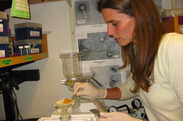
The Autolykivirdae are thought to play an important ecological role in the ocean as major bacteria-killers. In the study, the researchers collected samples of water off the coast of Massachusetts, and the viruses found in the samples were then incubated alongside a family of marine bacteria called Vibrionaceae. The researchers then analyzed the genomes of the viruses that successfully infected the bacteria. Of the 200 viruses collected, 18 were previously unrecognized and were non-tailed — the Autolykivirdae.

bacteria samples
Co-author Kathryn Kauffman processes seawater samples.

They then turned the new group of viruses loose on 300 strains of marine bacteria and compared how much damage the new group inflicted in contrast to tailed viruses. While the new group made up just 10 percent of the viruses observed, they did 40 percent of bacterial killing. Since these microscopic, oceanic predators were able to infect marine bacteria taken from outside of Massachusetts waters, the researchers were led to believe this group could potentially thrive throughout the ocean.

“When we found that, we were surprised,” co-author Martin Polz, Ph.D., announced in a statement released Wednesday.

Polz and his team also suspect that this new group of viruses are not ocean-specific and could be common within the human biome as well. The viruses, related to an ancient lineage that has been found previously in animals and eukaryotic organisms like algae, could have an effect on the human gut microbiome, say the researchers.

While we don’t typically think of viruses as good, some viruses can protecthumans against bacteria, instigating events like the creation of mucus, which can drive out germs. We don’t have a great sense of what exactly they do, but the gastrointestinal tracts of mammals are full of viruses — suggesting they likely play an important job in maintaining our health.

If you want to learn more about this new group of viruses, check out this explainer from study co-author Libusha Kelly, Ph.D., an assistant professor at the Albert Einstein College of Medicine.

Adult Stature and Risk of Cancer at Different Anatomic Sites in a Cohort of Postmenopausal Women.


Abstract

Background: Prospective studies in Western and Asian populations suggest that height is a risk factor for various cancers. However, few studies have explored potential confounding or effect modification of the association by other factors.

Methods: We examined the association between height measured at enrollment in 144,701 women participating in the Women’s Health Initiative and risk of all cancers combined and cancer at 19 specific sites. Over a median follow-up of 12.0 years, 20,928 incident cancers were identified. We used Cox proportional hazards models to estimate HR and 95% confidence intervals (CI) per 10 cm increase in height, with adjustment for established risk factors. We also examined potential effect modification of the association with all cancer and specific cancers.

Results: Height was significantly positively associated with risk of all cancers (HR = 1.13; 95% CI, 1.11–1.16), as well as with cancers of the thyroid, rectum, kidney, endometrium, colorectum, colon, ovary, and breast, and with multiple myeloma and melanoma (range of HRs: 1.13 for breast cancer to 1.29 for multiple myeloma and thyroid cancer). These associations were generally insensitive to adjustment for confounders, and there was little evidence of effect modification.

Conclusions: This study confirms the positive association of height with risk of all cancers and a substantial number of cancer sites.

Source: http://cebp.aacrjournals.org

The Taller the Woman, the Greater the Cancer Risk?


Taller postmenopausal women face higher risks for 10 types of cancer, according to a study in Cancer Epidemiology, Biomarkers and Prevention.

Researchers examined the association between height and cancer risk among some 145,000 Women’s Health Initiative participants. During roughly 12 years of follow-up, 14% received diagnoses of invasive cancer.

After multivariable adjustment, the risk for all cancers increased significantly, by 13%, with each 10-cm (4-inch) increase in height. In particular, risks for the following types of cancer were increased: breast, colon, colorectal, endometrial, melanoma, multiple melanoma, ovarian, rectal, renal, and thyroid. Additional adjustment for cancer screening did not alter the results.

The researchers say height should be considered “a marker for one or more exposures that influence cancer risk rather than a risk factor itself.”

Source:Cancer Epidemiology, Biomarkers & Prevention 


New theory uncovers cancer’s deep evolutionary roots.


A new way to look at cancer – by tracing its deep evolutionary roots to the dawn of multicellularity more than a billion years ago – has been proposed by Paul Davies of Arizona State University’s Beyond Center for Fundamental Concepts in Science in collaboration with Charles Lineweaver of the Australian National University. If their theory is correct, it promises to transform the approach to cancer therapy, and to link the origin of cancer to the origin of life and the developmental processes of embryos.

Paul Davies

Davies and Lineweaver are both theoretical physicists and cosmologists with experience in the field of astrobiology – the search for life beyond Earth. They turned to cancer research only recently, in part because of the creation at Arizona State University of the Center for the Convergence of Physical Science and Cancer Biology. The center is one of twelve established by the National Cancer Institute to encourage physical scientists to lend their insights into tackling cancer.

The new theory challenges the orthodox view that cancer develops anew in each host by a series of chance mutational accidents. Davies and Lineweaver claim that cancer is actually an organized and systematic response to some sort of stress or physical challenge. It might be triggered by a random accident, they say, but thereafter it more or less predictably unfolds.

Their view of cancer is outlined in the article “Exposing cancer’s deep evolutionary roots,” written by Davies. It appears in a special July issue of Physics World devoted to the physics of cancer.

“We envisage cancer as the execution of an ancient program pre-loaded into the genomes of all cells,” says Davies, an Arizona State University Regents’ Professor in ASU’s College of Liberal Arts and Sciences. “It is rather like Windows defaulting to ‘safe mode’ after suffering an insult of some sort.” As such, he describes cancer as a throwback to an ancestral phenotype.

The new theory predicts that as cancer progresses through more and more malignant stages, it will express genes that are more deeply conserved among multicellular organisms, and so are in some sense more ancient. Davies and Lineweaver are currently testing this prediction by comparing gene expression data from cancer biopsies with phylogenetic trees going back 1.6 billion years, with the help of Luis Cisneros, a postdoctoral researcher with ASU’s Beyond Center.

But if this is the case, then why hasn’t evolution eliminated the ancient cancer subroutine?

“Because it fulfills absolutely crucial functions during the early stages of embryo development,” Davies explains. “Genes that are active in the embryo and normally dormant thereafter are found to be switched back on in cancer. These same genes are the ‘ancient’ ones, deep in the tree of multicellular life.”

The link with embryo development has been known to cancer biologists for a long time, says Davies, but the significance of this fact is rarely appreciated. If the new theory is correct, researchers should find that the more malignant stages of cancer will re-express genes from the earliest stages of embryogenesis. Davies adds that there is already some evidence for this in several experimental studies, including recent research at Harvard University and the Albert Einstein College of Medicine in New York.

“As cancer progresses through its various stages within a single organism, it should be like running the evolutionary and developmental arrows of time backward at high speed,” says Davies.

This could provide clues to future treatments. For example, when life took the momentous step from single cells to multicellular assemblages, Earth had low levels of oxygen. Sure enough, cancer reverts to an ancient form of metabolism called fermentation, which can supply energy with little need for oxygen, although it requires lots of sugar.

Davies and Lineweaver predict that if cancer cells are saturated with oxygen but deprived of sugar, they will become more stressed than healthy cells, slowing them down or even killing them. ASU’s Center for the Convergence of Physical Science and Cancer Biology, of which Davies is principal investigator, is planning a workshop in November to examine the clinical evidence for this.

“It is clear that some radically new thinking is needed,” Davies states. “Like aging, cancer seems to be a deeply embedded part of the life process. Also like aging, cancer generally cannot be cured but its effects can certainly be mitigated, for example, by delaying onset and extending periods of dormancy. But we will learn to do this effectively only when we better understand cancer, including its place in the great sweep of evolutionary history.”

Source: asunews.asu.edu