One maternal death occurs every 2 minutes


A pregnancy- or childbirth-related death occurred once every 2 minutes in 2020, marking significant shortcomings in women’s health over the last several years, according to a new report developed by United Nations agencies.

“While pregnancy should be a time of immense hope and a positive experience for all women, it is tragically still a shockingly dangerous experience for millions around the world who lack access to high quality, respectful health care,” WHO Director-General Tedros Adhanom Ghebreyesus, PhD, MSc, said in a press release. “These new statistics reveal the urgent need to ensure every woman and girl has access to critical health services before, during and after childbirth, and that they can fully exercise their reproductive rights.”

Maternal mortality
Data derived from: Trends in maternal mortality 2000 to 2020.

The report analyzed national, regional and global trends in maternal mortality from 2000 to 2020, with the latter year registering a total of 287,000 maternal mortalities worldwide. According to WHO, this is only a slight decrease from 309,000 maternal deaths in 2016.

The organization noted that some improvements in reducing maternal mortality were made from 2000 to 2015; however, the rates in most regions either stagnated or risen beyond that time. From 2016 to 2020, maternal mortality rates increased in two of the eight U.N. regions: Europe and North America by 17% and Latin America and the Caribbean by 15%. Meanwhile, declines were observed in Australia and New Zealand, where deaths declined 35%, and Central and Southern Asia, which experienced a 16% decline.

Maternal deaths were most heavily concentrated in the poorest areas and countries impacted by conflict, according to WHO. In 2020, approximately 70% of maternal deaths occurred in sub-Saharan Africa.

The leading causes of maternal mortality included:

  • severe bleeding;
  • complications from unsafe abortions;
  • high BP;
  • pregnancy-related infections; and
  • underlying conditions that may have been aggravated by pregnancy, including HIV.

Although most conditions and ensuing maternal deaths are preventable, WHO noted that global lapses in women’s health care and resources have contributed to rising mortality. According to the organization, about one-third of women do not receive even half of the recommended eight antenatal check-ups or receive postnatal care, while about 270 million women lack access to family planning methods.

“Reducing maternal mortality remains one of the most pressing global health challenges,” John Wilmoth, the director of the U.N.’s Population Division of the Department of Economic and Social Affairs, said in the release. “With immediate action, more investments in primary health care and stronger, more resilient health systems, we can save lives, improve health and well-being, and advance the rights of and opportunities for women and adolescents.”

Nature’s Best Remedies for Aging Skin


Skip the expensive creams and chemical peels until you’ve nurtured your skin naturally

We all get old, but nature offers some plant compounds that can help us weather the years with more youthful skin.(Kzenon/Shutterstock)

We all get old, but nature offers some plant compounds that can help us weather the years with more youthful skin.(Kzenon/Shutterstock)

Even those who may have won the genetic lottery or have unlimited amounts of money to spend on skincare aren’t exempt from the natural effects of aging.

But while the beauty industry is bent on spending a fortune trying to convince people of the supreme wonders of chemical peels, Botox injections, and serums, there’s an entire world of natural solutions to slow the effects of aging.

Ditch the expensive creams and peels for now and explore these natural remedies that may help you delay or manage the signs of aging skin.

Aloe Vera

Aloe vera is an established topical skin remedy but is also often added to fruit juice or smoothies in gel form. A 2009 study found that taking aloe orally reduced facial wrinkles.Thirty healthy women over age 45 took aloe vera gel as a supplement, with one group taking a low dose of 1,200 milligrams (mg) a day and another taking a high dose of 3,600 mg a day.

The researchers concluded that aloe gel significantly improved wrinkles in both groups after just 90 days. Additionally, the lower-dose group had improved skin elasticity. How? Aloe increased collagen production, leading to enhanced structural support of skin and fewer wrinkles.

A separate study suggested that continued intake of aloe sterol contributed to maintaining healthy skin, as shown in “statistical differences” in areas such as skin moisture, skin elasticity, and collagen score.

Red Ginseng

Red ginseng contains bioactive compounds that include antioxidants and anti-aging agents. It’s available in various forms such as tinctures, powders, liquid extracts, and capsules.

In Korea, 82 healthy women over age 40 took part in a double-blind, placebo-controlled study. Every day, they received either a placebo or 3 grams (g) of an herbal mixture with red ginseng extract.

After measuring facial wrinkles, elasticity, water content, red patches, and pigmentation, the researchers found that facial wrinkles significantly improved in the red ginseng extract group. A number of biochemical markers of wrinkle damage also improved. “These results substantiate the alleged beneficial effects of red ginseng on photoaging and support its use as an effective ‘beauty food,’” the authors wrote.

Korean red ginseng also proved to be an excellent antiaging product in a separate study. On human skin, ginseng cream increased skin resilience and skin moisture as well as enhanced skin tone.

Soy

Soy extract appears to rejuvenate the structure of mature skin. In a placebo-controlled in vivo study, topically applying an isoflavone-containing emulsion significantly flattened the dermal-epidermal junction, considered the most reproducible structural change in aged skin.

Data from separate research seemed to confirm the findings above, indicating that orally taking 40 mg of soy isoflavone aglycones per day improved the aged skin of middle-aged women.

In a 2007 double-blind, 12-week study, a moisturizer containing compounds found in soy was found to be safe and effective in preventing photoaging. The moisturizer with stabilized soy extracts can be used to ameliorate overall skin tone and texture attributed to photoaging, the authors noted.

Pine Bark

A 2012 study comprising 112 women found pine bark extract to be safe and effective in substantially improving skin color and decreasing pigmentation of age spots caused by mild to moderate photoaging.

Japanese researchers divided the subjects into two groups, the first receiving 100 mg of pine bark extract each day and the second getting only 40 mg. The subjects received the standardized extracts known as Pycnogenol, found to significantly improve hydration and elasticity of the participants’ skin.

Pycnogenol also significantly increased the activity of an enzyme that’s important in the synthesis of hyaluronic acid, which then increases skin moisture and minimizes the appearance of wrinkles. It also affected genes involved in creating new collagen.

Astaxanthin

Astaxanthin is a carotenoid derived from microalgae, found in studies to mitigate skin photoaging and age-related skin diseases through its antioxidant and anti-inflammatory properties. A meta-analysis found that ingesting or topically using astaxanthin may be effective in reducing skin aging and improving moisture and elasticity, therefore providing promising cosmetic applications.

In a 2018 review, the carotenoid was also found to prevent UV-induced inflammation, wrinkling, and skin pigmentation after exposure to ultraviolet radiation, which can cause significant damage to skin tissue.

A Simple, Free Way to Heal Your Gut—With No Side Effects


(Magic mine/Shutterstock)

The gut is our main digestive organ and “second brain.” An imbalanced gut can negatively affect our body’s absorption of nutrients, blood sugar regulation, immunity, and even our mood. And there is a free method to help heal your gut: meditation.

Dr. Sunjya K. Schweig is an expert in complex chronic illnesses and a practitioner of integrative and functional medicine for almost three decades. According to his clinical experience, meditation can reduce stress, which in turn has a positive effect on the gut via several pathways. Very importantly, meditation can modulate the gut immune system and systemic inflammation, thus facilitating recovery from gut conditions.

Meditation Can Boost Good Gut Microbiome

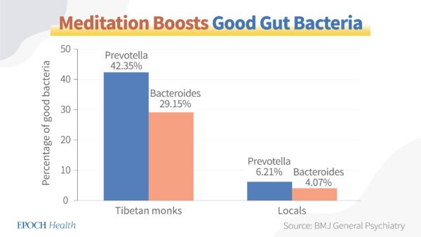
A study recently published in BMJ General Psychiatry analyzed the gut microbiome compositions of 56 Tibetan Buddhist monks and some residents who don’t regularly meditate. 

Researchers discovered that  the monks’ long-term deep meditation practice had significantly enriched the good bacteria in their intestinal systems, yielding “a reduced risk of anxiety, depression, and cardiovascular disease.” 

Epoch Times Photo
Differences between the microbiota of monks and locals.

Specifically, the proportions of good bacteria Prevotella and Bacteroides among the Tibetan monks were 42.35 percent and 29.15 percent, respectively, whereas their proportions were 6.21 percent and 4.07 percent in the locals. The monks also had a larger quantity of the good gut bacteria of Megamonas and Faecalibacterium than the local residents. 

Stress and depression facilitate dysbiosis of the gut microbiota, and meditation can reduce stress and the risk of depression, in turn promoting a better gut microbiome

Additionally, per a meta-analysis, meditation can help regulate the stress response and maintain a healthy gut-barrier function. The researchers recommended integrating meditation into existing health care models for improved effects.

Scientists also recommend using meditation as an effective tool in regulating gut health, as meditation could significantly improve the gastrointestinal quality of life index (GIQLI). 

Furthermore, according to a Harvard blog, meditation can activate the parasympathetic nervous system, which in turn promotes gut motility

When gastrointestinal motility is slow, bacteria and yeast cannot be removed from the body on time, leading to a build-up of bacteria and/or yeast in the small intestines, called small intestinal bacterial overgrowth (SIBO). SIBO can lead to chronic diarrhea and malabsorption of nutrients, causing depletion of certain fat-soluble vitamins such as A, D, E, and K. 

Meditation Can Improve Gut Diseases and Other Digestive Diseases

When suffering from stress, our digestive system simply cannot function well, according to Dr. Tawny Kross, a pain specialist and physical therapist at Kross Centered Care who specializes in chronic pain. 

Due to significant lifestyle changes in recent decades, the number of people with gastrointestinal disorders worldwide has been on the rise. According to the National Institute of Diabetes and Digestive and Kidney Diseases, 60 to 70 million Americans are affected by digestive diseases. Nevertheless, meditation can be an effective tool in relieving several common gut and other digestive diseases. 

Kross discovered in her practice that when the issue of stress is addressed by interventions such as meditation, her clients’ gut health would improve drastically. “When mindfulness (meditation) is combined with dietary changes, there is also a more powerful and greater impact,” she said.

Oftentimes patients with gastrointestinal symptoms improve when they engage in a meditation program, “because meditation has been shown to decrease stress levels,” and it also alters their nervous system, thus changing their gut for the better, according to Dr. Daniel A. Monti, chair of the Department of Integrative Medicine & Nutritional Sciences at Thomas Jefferson University and CEO of Marcus Institute of Integrative Health.

Irritable Bowel Syndrome (IBS)

One of Kross’ clients suffered from stomach pain and IBS symptoms for over a decade, but after he started practicing meditation, his stomach pain and IBS soon improved.

And combined with the proper nutrition, he started having regular bowel movements, and his gut issues later disappeared. 

Irritable bowel syndrome is a disorder of gut-brain interaction and a group of symptoms, including abdominal pain, bloating, diarrhea, and constipation. Ten to 15 percent of American adults suffer from IBS symptoms.

According to Dr. Kenneth Brown, a gastroenterologist, recent research showed that the practice of meditation can reduce IBS symptoms and the amount of time patients spend in the bathroom with digestive issues. Meditation can also decrease patients’ stress and anxiety levels, while the amount of serotonin in their bodies increases. Serotonin is a key hormone in regulating our digestive systems.

In a study published in the American Journal of Gastroenterology, 75 female IBS patients were randomly assigned to eight weekly half-day meditation sessions. The patients who participated in these sessions experienced a substantial reduction in IBS symptom severity and distress, as well as improved quality of life. 

In addition, such beneficial therapeutic effects lasted for at least three months after the sessions.

In a different follow-up study, 10 IBS patients who had been practicing relaxation response meditation for one year experienced significant reductions in IBS symptoms in comparison with their pretreatment conditions, including abdominal pain, diarrhea, flatulence, and bloating. The researchers suggested that continued use of meditation is an effective treatment for IBS in the long term. 

Crohn’s Disease

Crohn’s disease is a type of inflammatory bowel disease that causes inflammation of the digestive tract. Its major symptoms include abdominal pain, severe diarrhea, fever, fatigue, bowel obstruction, and weight loss. Patients with Crohn’s disease are at a higher risk of colon cancer.

Evidence shows that meditation can support Crohn’s disease patients’ subjective well-being homeostasis, which is the systematic management of positive feelings about ourselves. This may be due to meditation’s ability to help patients regulate their emotions.

In another 2022 German study, patients with Crohn’s disease were randomly assigned to two groups. One group received one weekly six-hour meditation session for 10 weeks. The study participants then took questionnaires on inflammatory bowel disease symptoms at week 12, and then at week 36. 

The patients who participated in the meditation sessions regularly were later extremely satisfied with their experience. At the end of the study, the researchers recommended meditation as a comprehensive approach to Crohn’s disease patients’ treatment.

Gastroesophageal Reflux Disease (GERD)

GERD is a type of upper gastrointestinal disease, where stomach acid regularly flows up into the esophagus. Its main symptoms include heartburn, difficulty swallowing, painful swallowing, and noncardiac chest pain.

Symptoms of GERD can be exacerbated by stress and depression, and meditation can help reduce both, thus positively improving GERD symptoms. In a longitudinal study in the official publication of the Indian Society of Gastroenterology, the group of participants who practiced meditation showed a greater decrease in their levels of depression than the controls. As a result, the researchers perceive meditation as an effective therapy for GERD patients.

Chronic Gastritis

Gastritis is an inflammation, irritation, or erosion of the lining of the stomach. Its symptoms may include upper abdominal pain, nausea, vomiting, bloating, and heartburn.

In a 2021 study, 146 chronic gastritis and gastric ulcer patients were assigned to two groups. One group received the standardized nursing intervention, while the other group received meditation training on top of the standardized care. The analysis showed that the nursing efficacy rate, nursing satisfaction rate, and general self-efficacy scale score of the group that meditated were higher.

Moreover, the first group’s self-rating anxiety and depressive scale scores were lower. The researchers concluded that the combination of standardized nursing intervention and meditation had curative effects on these patients.

Meditation Provides Pain Relief 

Monti says meditation can offer pain relief to patients with digestive diseases, as well. 

Since meditation can calm the sympathetic nervous system, according to the Harvard blog, it can reduce chronic pain, since the sympathetic nervous system tends to exacerbate physical symptoms of diseases such as pain. Therefore, meditation can be used as an effective and safe tool for pain control.

Patients with digestive diseases, including celiac disease, inflammatory bowel diseases, hemorrhoids, and anal fissures can benefit from the pain relief offered by meditation. 

‘No Side Effects’ 

Pharmaceuticals can be necessary for treating gut diseases, but inevitably, they also bring side effects. Meditation can be a good alternative treatment to incorporate.

“Often, meditation can reduce the need for medication, allowing the patient more natural relief without the side effects of pharmaceutical drugs,” Dr. Sony Sherpa, a holistic physician from organic wellness company Nature’s Rise, said.

It also doesn’t interfere with or offset the effects of medication. Paul Harrison, a corporate personal meditation coach based in Ontario, Canada, concurred. “There’s really no evidence to suggest that meditation will interfere with any kind of medication or with any other (medical) intervention.”

Other Ways to Maintain a Healthy Gut

In addition to meditation, there are many other ways to keep a healthy gastrointestinal system.

For example, maintaining a balanced diet with lots of fiber is important to our gut health. We need to consume 25 to 35 grams of fiber per day, and they can be from whole grains, fruits, vegetables, legumes, and nuts. To help our body better digest fiber and other nutrients, we also need to drink an adequate amount of water, which is four to six cups of water per day for most people. 

We also need to work out regularly to supply more blood flow to the gut. 

And finally, the intake of probiotic foods such as yogurt, sauerkraut (shredded fermented cabbages), kimchi (Korean-style fermented vegetables), miso (Japanese-style seasoning made with fermented soybeans), kombucha (fermented black or green tea), pickles, sourdough bread, and certain cheeses can also help with the health of our gut microbiota.

How Eating and Drinking Sugar Can Cause Diabetes


Very exciting research from Princeton University explains how taking in sugared drinks and any sugar added to foods (not in whole fruits and vegetables) can cause diabetes (Cell Metabolism, Feb 6, 2018;27(2):351–361).

The most common sugar source in foods contains two sugars, glucose and fructose. Glucose is the only sugar that your body allows to circulate in your bloodstream. The authors of this study show that fructose is converted to glucose primarily in the intestines, and not in the liver as scientists thought previously. However, if you overload your intestines with fructose, the unchanged fructose can pass through your intestines into your bloodstream and into your liver where it causes fat to accumulate in your liver to cause high blood sugar levels that are characteristic of diabetes.

This exciting new study also shows that fructose that is not converted to glucose in the small intestine can pass to the colon where it fosters the growth of harmful bacteria. These harmful types of bacteria try to invade the cells in your colon, which turns on your immunity to cause inflammation that increases risk for obesity, heart attacks and some cancers. On the other hand, adding high-fiber foods to a person’s diet helps to prevent diabetes by fostering the growth of the good colon bacteria that do not invade the colon cells and help to reduce inflammation (Cell, January 2014). See Gut Bacteria Linked to Diabetes

Why Sugar in Fruits and Vegetables Is Safer
Virtually all studies on the subject show that eating fruits and vegetables is associated with the prevention and control of diabetes (PLoS Medicine, April 11, 2017), even though they contain sugar. The most likely explanation is that:
• Sugar added to foods and in all sugared drinks (including fruit juices) causes an immediate overload of fructose that allows some fructose to pass unchanged from the intestines into the bloodstream and then directly into the liver, where fructose is converted to fatty triglycerides, which fill up the liver cells with fat. Fat in the liver prevents the liver from lowering high blood sugar levels the way it is supposed to do.
• The sugar in fruits and vegetables is prevented from causing a high rise in blood fructose by the soluble fiber and anti-oxidants in them (Cell. January 9, 2014). The added sugars used in beverages and foods are the same as the sugars in fruits and vegetables, but when the sugars are extracted from their plant sources (sugar beets, sugar cane, maple trees, corn, flowers, grapes, apples and so forth), the beneficial soluble fiber and numerous antioxidants are removed.

Why Sugar in Drinks is Absorbed Most Quickly
Solid food is not allowed to pass directly into your intestines because it could block the intestines and kill you. When you eat food, your pyloric sphincter at the end of your stomach closes to prevent solid food from entering your intestines. Then your stomach muscles contract and push only a liquid soup through the closed sphincter muscle into your intestines. However, when you take sugar in a liquid, it is not stopped by the pyloric sphincter and enters the intestines immediately to allow the fructose to enter the liver and turn into fat.

How a Fatty Liver Can Cause Diabetes
A high rise in blood sugar can damage cells throughout your body. To prevent blood sugar levels from rising too high after eating, your pancreas releases insulin that is supposed to lower blood sugar by driving sugar from the bloodstream into the liver. However, if your liver is full of fat, your liver cannot accept the sugar and blood sugar levels rise even higher. High blood sugar levels cause sugar to stick to the outer membranes of every type of cell in your body. Once attached, sugar can never get off and eventually destroys cells to cause all the harmful side effects of diabetes.

My Recommendations
Almost 50 percent of North Americans will develop diabetes. To reduce your risk for becoming diabetic:
• Avoid being overweight.
• Prevent fat from accumulating in your liver by losing excess weight until you cannot pinch more than three inches of fat under the skin near your belly button. I recommend intermittent fasting for weight loss.
• Restrict or avoid drinks with sugar (including fruit juices) and foods with added sugars.
• If you are overweight or have high blood sugar levels after eating (>140 mg/dl one hour after a meal) you should also restrict all refined carbohydrates (bakery products, pastas, most dry breakfast cereals and so forth), processed meats and red meat (which increase risk for not being able to respond to insulin), and fried foods.
• Try to exercise every day. Contracting muscles draw sugar from your bloodstream without needing insulin.
• Avoid vitamin D deficiency as it can prevent your cells from responding to insulin. Hydroxy vitamin D levels should be >20 ng/ml.

How Sugar-Added Foods and Drinks Increase Risk for Heart Disease


Researchers followed more than 110,000 people for nine years and found that the more free sugar a person takes in, the greater the risk for heart disease (BMC Medicine, Feb 14, 2023;21(34)). Each five percent increase in free sugar intake in a participant’s daily diet resulted in a six percent higher risk of heart disease and a 10 percent higher risk of stroke. Furthermore, a higher fiber intake and replacing refined grain starch and free sugars with whole grains and non-free sugars appeared to help protect against heart attacks. The authors explained how increased intake of sugar-added foods and drinks can cause heart disease: Those who took in more sugar-added foods and drinks had higher blood triglycerides and greater waist circumferences.

Note: Free sugars are defined by these researchers as any sugars added to foods by the manufacturer, cook or consumer, plus sugars present in honey, syrups, and fruit juices. (Non-free sugars are those naturally occurring in fruit, vegetables, and dairy products). See How Eating and Drinking Sugar Can Cause Diabetes

This study agrees with many other studies that show that heart damage is associated with refined sugar intake, not the total amount of carbohydrates (Lancet, 2017;390:2050–62). An earlier review of studies on sugar-added foods shows that people who take in 10-25 percent of their calories from sugared beverages and foods suffer a 30 percent higher risk for heart attacks, compared with people who take less than ten percent of calories from added sugars (British Medical Journal: Open Heart, Dec. 11, 2014). The review also found that:
• higher added-sugar intake was associated with increases in systolic and diastolic blood pressure of 6.9 and 5.6 mm Hg
• the more sugared foods you eat, the higher your bad LDL cholesterol
Sugars occurring naturally in foods, such as fruit, did not appear to increase risk for high blood pressure or heart attacks. Another review of twelve scientifically-dependable studies involving 409,707 participants showed that sugar-sweetened beverages were associated with increased risk for high blood pressure, a major risk factor for diabetes and heart attacks (The American J of Cardiol, February 2014).

How Intake of Added Sugars Increases Heart Attack Risk
• Taking in sugar added foods and drinks causes a high rise in blood sugar.
• To prevent blood sugar from rising higher, the pancreas release insulin which lowers blood sugar by driving sugar from the bloodstream into the liver.
• As soon as the liver fills up with sugar, it can store no more sugar, so the extra sugar is converted by your liver into fatty triglycerides, so blood levels of triglycerides rise too high (>150).
• To prevent blood triglyceride levels from rising too high and causing clots, your good HDL cholesterol carries the triglycerides into the liver, so your HDL cholesterol goes down and you store extra fat in your liver to cause a fatty liver.
• As the liver collects extra fat, it cannot respond to insulin that lowers blood sugar by driving sugar from the bloodstream into the liver, so your blood sugar rises even higher and you have insulin-resistant diabetes.
• As the cycle continues, extra fat is deposited in the liver and the belly, which gives you a big belly.
• Then your liver takes large numbers of triglyceride molecules and combines them with lesser numbers of cholesterol molecules to make the bad LDL cholesterol, which causes plaques to form in arteries (arteriosclerosis), which can break off to lead to a heart attack.

How Can You Tell If You Are Insulin Resistant?
People who are insulin resistant usually have what is called metabolic syndrome. You probably have metabolic syndrome if you have three or more of:
• storing fat primarily in your belly
• small hips compared to your stomach
• being overweight
• blood triglycerides >150
• HDL cholesterol <40
• high blood pressure
• a fatty liver
• fasting blood sugar >100
• HbA1c >5.7
• high insulin levels

Treat Insulin Resistance with Lifestyle Changes
• Restrict sugared drinks and foods with added sugars.
• Limit refined carbohydrates such as foods made from flour
• Eat plenty of fruits, vegetables and seeds. Unprocessed vegetables, whole grains, nuts, other seeds and most fruits contain complex carbohydrates and fats that are not released rapidly into the bloodstream. These nutrient-rich foods do not cause high rises in blood sugar and insulin.
• Exercise. Resting muscles draw no sugar from the bloodstream, while contracting muscles draw sugar rapidly from the bloodstream and don’t even need insulin to do so. The more intensely you exercise, the less insulin is needed by muscles to draw sugar from the blood. This effect lasts for up to 17 hours after you finish exercising.
• Avoid being overweight. Your liver controls blood sugar levels. When blood sugar levels rise, insulin drives sugar from the bloodstream into the liver. However, the more fat you have stored in your liver, the harder it is for sugar to enter liver cells in response to insulin. A fatty liver will raise blood sugar levels even higher by releasing stored sugar from its cells into the bloodstream.

Definition of Sugar-Added Foods and Drinks
Added sugars are those put in food during processing or food preparation:
• packaged table sugar and all other high-calorie sweeteners
• all sugars in liquid form such as sugar in fruit juice, syrups, honey, vegetable juice, purees, pastes and similar products in which the cellular structure of the food has been broken down
• Other refined carbohydrates such as ground-up grains are similar to “added sugars” because grinding breaks up the seed capsules of whole grains to markedly raise blood sugar levels in foods made with flour (bread, pasta, pretzels, bagels, cookies, crackers and many dry breakfast cereals). A person who is insulin resistant should try to limit all sources of refined carbohydrates, not just added sugars.
• Sugars in dairy products or in fruits or vegetables are not considered “added sugars”.

Wilt Chamberlain’s Myocarditis


Wilt Chamberlain was possibly the greatest basketball player and the greatest athlete ever. The 63-year-old Chamberlain was reported to have died of heart damage called myocarditis, but how could arguably the world’s greatest and fittest athlete die of heart damage? A possible explanation would be venereal diseases, which are a common cause of myocarditis (Case Rep Infect Dis, 2015; 2015: 385126). Chamberlain wrote a book, A View from Above (published in 1991) in which he claimed that had had sex with more than 20,000 women. He received a tremendous amount of criticism for this claim, but he never backed down in defending it. Every casual sexual contact puts a person at high risk of acquiring a venereal disease, since more than 67.6 million North American adults have venereal diseases such as chlamydia, mycoplasma, ureaplasma, trichomoniasis, genital herpes, human papillomavirus (HPV) and many more (Sexually Transmitted Diseases, April 2021;48(4):208-214).

Success in Every Sport He Tried
Wilton Norman Chamberlain was born in Philadelphia in 1936. He was 6’11” when he entered Philadelphia’s Overbrook High School, and led them to three public school championships and two all-city titles.  In high school, he:
• ran the quarter mile in less than 48 seconds, to set the U.S. high school record, and ran the half mile in 1:58.3
• high-jumped 6’6″ and broad jumped 22 feet
• was the state of Pennsylvania shot put champion at 53’4″

He was the most recruited high school player in the country, with more than 200 colleges interested in him.  He went to the University of Kansas, where he:
• was the best basketball player in the country
• won the Big 8 high jump championship his junior year
• threw the shot put more than 47 feet
• was 4th in the  1956 Kansas Relays’ hop-step-jump

In the National Basketball Association, he scored more than 100 points in a single game and averaged more than 30 points per game throughout his professional career.  He bench pressed 500 pounds, was timed at 4.6 seconds in the 40-yard dash and had an incredible vertical jump of 48 inches.  After his amazing NBA career, he became arguably the best volleyball player in the world. At age 60, he ran in the Honolulu marathon and competed in a 50-mile race in Canada.

The Great Lover
Long after his athletic career ended, Chamberlain made news with his claim that he had had sex with 20,000 women. That comes to 500 women per year, or 10 different women per week in his 40-year career of sharing his sperm. This would probably make the world’s greatest athlete the most prolific lover of all time.

Diagnosis: Cardiomyopathy
Chamberlain’s health first became an issue in the 1960s, when a former coach told the news media that the star player might have had a heart attack before the 1964 season. But Chamberlain denied it.

In 1992, when Chamberlain gathered with former teammates for a halftime ceremony marking the anniversary of their 1971-72 NBA championship, he had to leave early because he was having trouble breathing. He was admitted to a hospital and found to have an irregular heartbeat. He was released from the hospital after three days, wearing a heart monitoring device.

During his last years, he was diagnosed as having cardiomyopathy which means that his heart was too weak to pump blood through his body. Cardiomyopathy can be caused by:
• arteriosclerosis (Chamberlain did not have high cholesterol and was still able to do amazing athletic feats in his 60s)
• damaged heart valves (he did not have this)
• genetic conditions (nobody in his family was reported to have a similar condition),
• long-term high blood pressure (his doctors never would have missed this)
• metabolic disorders such as obesity, thyroid disease or diabetes (no evidence of any of these)
• nutritional deficiencies of vitamins or minerals (his doctors would have made that diagnosis)
• drinking too much alcohol over many years (he was not an alcoholic)
• recreational drugs such as cocaine, amphetamines or anabolic steroids (no evidence of this)
• chemotherapy or radiation to treat cancer (does not apply to him)
• iron buildup called hemochromatosis (an easy diagnosis that would not have been missed),
• sarcoidosis or amyloidosis (his doctors would not have missed these)
• autoimmune diseases such as lupus (no evidence that he had any of these diseases) 
• infections

Infections that Cause Cardiomyopathy
Chamberlain’s heart failure came from an infection that caused heart muscle damage, called myocarditis:
• He was hospitalized seven years before his death with an irregular heartbeat
• He was unable to go anywhere in the last months of his life
• He lost up to 50 pounds in the few weeks before he died. Heart failure caused large amounts of fluid to collect in his legs and doctors had to give him drugs to get rid of this massive accumulation of fluid.

The most likely cause of Chamberlain’s cardiomyopathy was an infection in his heart with bacteria such as chlamydia, mycoplasma or ureaplasma (N Am J Med Sci, 2013 Mar; 5(3): 169–181).  Regular bacterial and viral cultures will not grow these germs. A doctor would have had to order special cultures and sometimes even these cultures fail to grow them. It was only in 1999, after Chamberlain’s death, that researchers proved that chlamydia and mycoplasma can cause the inflammation that forms plaques in arteries (arteriosclerosis), damages the heart and causes heart attacks (Science, 1999;283:1335-9).  The first dependable test for the sexually transmitted Mycoplasma genitalium was approved by the FDA on February 1, 2019.

The fact that he lost 50 pounds and was unable to go anywhere in the last months of his life points to a diagnosis of heart failure, caused by cardiomyopathy (heart muscle damage), possibly caused by an infection such as chlamydia or mycoplasma.  He could easily have acquired these infections from making love to considerably fewer than the 20,000 women that he claimed. These infections may be cured by taking antibiotics, such as minocycline, doxycycline or clarithromycin, for several weeks or months.  His body was cremated, so we will never know for sure how he died.

Efanesoctocog Alfa Prophylaxis for Patients with Severe Hemophilia A


Abstract

Background

Efanesoctocog alfa provides high sustained factor VIII activity by overcoming the von Willebrand factor–imposed half-life ceiling. The efficacy, safety, and pharmacokinetics of efanesoctocog alfa for prophylaxis and treatment of bleeding episodes in previously treated patients with severe hemophilia A are unclear.

Methods

We conducted a phase 3 study involving patients 12 years of age or older with severe hemophilia A. In group A, patients received once-weekly prophylaxis with efanesoctocog alfa (50 IU per kilogram of body weight) for 52 weeks. In group B, patients received on-demand treatment with efanesoctocog alfa for 26 weeks, followed by once-weekly prophylaxis with efanesoctocog alfa for 26 weeks. The primary end point was the mean annualized bleeding rate in group A; the key secondary end point was an intrapatient comparison of the annualized bleeding rate during prophylaxis in group A with the rate during prestudy factor VIII prophylaxis. Additional end points included treatment of bleeding episodes, safety, pharmacokinetics, and changes in physical health, pain, and joint health.

Results

In group A (133 patients), the median annualized bleeding rate was 0 (interquartile range, 0 to 1.04), and the estimated mean annualized bleeding rate was 0.71 (95% confidence interval [CI], 0.52 to 0.97). The mean annualized bleeding rate decreased from 2.96 (95% CI, 2.00 to 4.37) to 0.69 (95% CI, 0.43 to 1.11), a finding that showed superiority over prestudy factor VIII prophylaxis (P<0.001). A total of 26 patients were enrolled in group B. In the overall population, nearly all bleeding episodes (97%) resolved with one injection of efanesoctocog alfa. Weekly prophylaxis with efanesoctocog alfa provided mean factor VIII activity of more than 40 IU per deciliter for the majority of the week and of 15 IU per deciliter at day 7. Prophylaxis with efanesoctocog alfa for 52 weeks (group A) improved physical health (P<0.001), pain intensity (P=0.03), and joint health (P=0.01). In the overall study population, efanesoctocog alfa had an acceptable side-effect profile, and the development of inhibitors to factor VIII was not detected.

Conclusions

In patients with severe hemophilia A, once-weekly efanesoctocog alfa provided superior bleeding prevention to prestudy prophylaxis, normal to near-normal factor VIII activity, and improvements in physical health, pain, and joint health. (Funded by Sanofi and Sobi; XTEND-1 ClinicalTrials.gov number, NCT04161495. opens in new tab.)

QUICK TAKEEfanesoctocog Alfa for Severe Hemophilia A 02:20

Regular prophylaxis to prevent spontaneous bleeding and joint damage is the standard treatment for severe hemophilia A.1 A trough factor VIII level of 1 IU per deciliter (or 1%) was considered to be an adequate goal for prophylaxis for many years, but recently revised World Federation of Hemophilia guidelines acknowledge that many clinicians now prefer to target trough factor VIII activity of 3 to 5 IU per deciliter or higher for their patients.1 However, both life-threatening bleeding and bleeding into joints still occur, with the latter contributing to substantial morbidity as a result of chronic pain and hemophilic arthropathy.2-6 Normalization of factor VIII levels may increase protection from both spontaneous and traumatic (including life-threatening) bleeding and preserve joint health, particularly for patients at higher risk, such as those with an active lifestyle or with recurrent hemarthrosis.7,8 The interaction between factor VIII and endogenous von Willebrand factor (VWF) imposes a ceiling of 8 to 19 hours on the half-life of current factor VIII replacement products.9-17 Therefore, maintaining factor VIII levels in the normal range (50 to 150 IU per deciliter) or levels that are close to normal (>40 to <50 IU per deciliter; defined herein as “near-normal”) with currently available factor VIII therapies requires frequent administration, which confers a substantial treatment burden on people with hemophilia and their caregivers.1

Efanesoctocog alfa (formerly BIVV001) is a new class of factor VIII replacement therapy designed to decouple recombinant factor VIII from endogenous VWF and overcome the VWF-imposed half-life ceiling.18,19 Efanesoctocog alfa is composed of a single recombinant factor VIII protein and three additional components that contribute to its increased half-life: an Fc domain that facilitates recycling through the neonatal Fc receptor pathway, covalent linkage to a VWF D′D3 factor VIII binding domain to decouple recombinant factor VIII from endogenous VWF, and two XTEN polypeptides to shield efanesoctocog alfa from proteolytic degradation and clearance.18,19 A schematic diagram of the molecule is provided in the Supplementary Appendix, available with the full text of this article at NEJM.org. The pharmacokinetics and safety of single- and repeat-dose efanesoctocog alfa have been evaluated in phase 1 and 1–2a studies.12,13,20 In the sequential single-dose study, single-dose efanesoctocog alfa (50 IU per kilogram of body weight) had a mean terminal half-life of 43.3 hours, which was approximately three to four times as long as those of rurioctocog alfa (15.4 hours) and octocog alfa (11.0 hours).13 Here, we report the results of XTEND-1, a pivotal phase 3 study to determine the efficacy, safety, and pharmacokinetics of efanesoctocog alfa for routine prophylaxis, treatment of bleeding episodes, and perioperative management in previously treated adults and adolescents with severe hemophilia A.

Methods

Population and Study Design

XTEND-1 was an open-label, multicenter study involving previously treated patients 12 years of age or older with endogenous factor VIII activity of less than 1 IU per deciliter (<1%) or a documented genotype known to produce severe hemophilia A. Patients were required to have had at least 150 previous exposure days to recombinant or plasma-derived factor VIII concentrates or cryoprecipitate. Patients were assigned to receive efanesoctocog alfa as prophylaxis (group A) or as on-demand treatment, followed by prophylaxis (group B). To be included in group A, patients were required to have been receiving a prophylactic regimen; to be included in group B, patients were required to have been receiving on-demand treatment with factor VIII replacement and to have had at least 6 bleeding episodes in the previous 6 months or at least 12 bleeding episodes in the previous 12 months.

Patients were excluded if they had a positive test for factor VIII inhibitor (≥0.6 Bethesda units [BU] per milliliter) at screening or a history of a positive inhibitor test, clinical signs or symptoms of a decreased response to factor VIII, other known coagulation disorders, a history of hypersensitivity or anaphylaxis to factor VIII therapies, or major surgery within 8 weeks before screening. Patients with human immunodeficiency virus (HIV) infection were excluded if they had a CD4 lymphocyte count of 200 cells or less per microliter or a viral load of 400 copies or more per milliliter. A full list of enrollment criteria is provided in Table S1 in the Supplementary Appendix.

Study Treatment

Patients in group A were assigned to receive once-weekly prophylactic doses of 50 IU per kilogram of intravenous efanesoctocog alfa for 52 weeks. Patients in group B were assigned to receive on-demand treatment with 50 IU per kilogram of intravenous efanesoctocog alfa for 26 weeks, followed by once-weekly prophylactic doses of 50 IU per kilogram of intravenous efanesoctocog alfa for 26 weeks (Fig. S1).

Bleeding episodes were treated with a single dose of 50 IU per kilogram of efanesoctocog alfa. If the bleeding episode did not resolve, additional doses of 30 or 50 IU per kilogram could be administered every 2 or 3 days, as needed. Patients who underwent major surgery were included in the surgery subgroup. These patients received a preoperative loading dose of 50 IU per kilogram of efanesoctocog alfa, followed by additional doses of 30 or 50 IU per kilogram every 2 or 3 days, as needed.

End Points

The primary end point was the mean annualized bleeding rate in group A. The key secondary efficacy end point was an intrapatient comparison of the annualized bleeding rate in group A and the rate during prestudy prophylaxis in a subgroup of patients who participated in a prospective, observational prestudy (242HA201/OBS16221) and who had at least 6 months of available efficacy data from both the prestudy and XTEND-1 (see the Methods section in the Supplementary Appendix). In the observational prestudy, data were collected on bleeding episodes and treatment with standard-care factor VIII prophylaxis. Additional secondary end points included the annualized bleeding rate according to bleeding type (all bleeding and spontaneous bleeding) and location (joint), an intrapatient comparison of the annualized bleeding rate during the prophylaxis period with the rate during the on-demand treatment period in group B, bleeding-treatment outcomes, perioperative management of major surgery, pharmacokinetics, quality of life (including physical health and pain intensity), joint health, and safety.

Quality of life was assessed with the use of the Hemophilia Quality of Life Questionnaire for Adults (Haem-A-QoL; administered to patients ≥17 years of age) and the Patient-Reported Outcomes Measurement Information System (PROMIS) Pain Intensity–Short Form 3a, which measures the worst pain intensity within the past 7 days. Response to treatment was evaluated by means of the Physician’s Global Assessment. Response to treatment of bleeding episodes was measured with the use of the 4-point International Society on Thrombosis and Haemostasis scale, and the hemostatic response to surgery was assessed with the use of the 4-point surgical procedures scale. Joint health was assessed with the use of the Hemophilia Joint Health Score (HJHS) (see the Methods section in the Supplementary Appendix). All bleeding rates refer to treated bleeding events unless otherwise stated. Target-joint resolution at week 52 was assessed. A target joint was defined as a major joint with at least three spontaneous bleeding episodes in a consecutive 6-month period before study entry.1 Target-joint resolution was defined as no more than two episodes of bleeding into that joint during 12 months of continuous exposure to efanesoctocog alfa. Exit interviews were conducted in a subgroup of patients to provide a qualitative assessment of the clinical meaningfulness of the patient-reported end points for physical health and pain.

Other Assessments

A subgroup of patients in group A underwent sequential blood sampling for pharmacokinetic assessment (up to 336 hours) at week 1 and a reassessment at week 26. All other patients underwent abbreviated sampling (up to 168 hours) for factor VIII activity during week 1. Sampling for peak and trough factor VIII activity occurred at weeks 4 and 13 (group A only) and weeks 26, 39, and 52 (both groups). Factor VIII activity was measured by means of the activated partial-thromboplastin time (aPTT)–based one-stage clotting assay with the use of the Actin FSL reagent and the World Health Organization 6th International Standard for factor VIII.

The development of factor VIII inhibitors was assessed with the use of a Nijmegen-modified Bethesda assay, and the development of antidrug antibodies was assessed with the use of an efanesoctocog alfa–specific antidrug-antibody assay.1,21 Samples were tested at screening, baseline, and weeks 4, 13, 26, 39, and 52. The development of inhibitors to factor VIII was defined as a result of 0.6 BU or more per milliliter that was confirmed by a second positive result from a separate sample drawn 2 to 4 weeks later. Both tests were performed by the central laboratory.

Study Oversight

The study was performed in accordance with the principles of the Declaration of Helsinki and local regulations. The study was funded by Sanofi and Sobi. All the authors had access to the primary clinical-trial data and vouch for the completeness and accuracy of the data and for the adherence of the study to the protocol (available at NEJM.org). The manuscript was developed with medical writing support funded by Sanofi and Sobi.

Statistical Analysis

The primary end point was estimated in the full analysis set (all patients in group A who received at least one dose of efanesoctocog alfa) with the use of a negative-binomial regression model. Annualized bleeding rates were calculated on the basis of the number of bleeding episodes during the efficacy period (see the Supplementary Appendix for a definition of the efficacy period). If the upper limit of the 97.5% confidence interval for the annualized bleeding rate in group A was 6 or less, the intervention was considered to be effective (see the Methods section in the Supplementary Appendix for rationale). The intrapatient comparison of the annualized bleeding rate during prophylaxis in group A and the rate during prestudy prophylaxis was assessed with the use of a negative-binomial regression model. Noninferiority and superiority of efanesoctocog alfa prophylaxis to prestudy prophylaxis were evaluated sequentially (see the Methods section in the Supplementary Appendix for criteria used to assess noninferiority and superiority). The adjusted mean change from baseline to week 52 in physical health (Haem-A-QoL physical-health score), pain (PROMIS pain-intensity score), and joint health (HJHS) were estimated by means of mixed-effects models with repeated measures, as part of a prespecified hierarchical testing framework.

Baseline factor VIII levels were assessed after a minimum washout period of 4 or 5 days, depending on previous therapy, and before administration of the first dose of efanesoctocog alfa. Pharmacokinetic variables were computed with the use of baseline-corrected values for factor VIII activity. Safety outcomes were analyzed with the use of descriptive statistics. The incidence of the development of inhibitors to factor VIII was determined with the use of an exact 95% confidence interval on the basis of the binomial distribution.

Results

Patient Characteristics

Figure 1. Enrollment, Randomization, and Follow-up.Table 1. Demographic and Clinical Characteristics of the Patients at Baseline.

A total of 159 patients from 48 centers in 19 countries were enrolled in the study, with 133 in group A and 26 in group B. Of the 159 patients, 149 (94%) completed the study and 10 (6%) discontinued the study prematurely (Figure 1). The most common reasons for discontinuation was withdrawal of consent and the use of prohibited concomitant medication (3 patients each). The demographic and clinical characteristics of the patients at baseline were representative of previously treated patients with severe hemophilia A (Table 1 and Table S4). One female patient with severe hemophilia A was enrolled in group A. Almost half the patients had a genotype associated with an increased risk of the development of inhibitors to factor VIII (36% had intron 22 inversions, and 9% had nonsense mutations) (Table S2).

The mean (±SD) number of exposure days to efanesoctocog alfa was 48.4±10.5 in the overall population, 50.9±9.1 in group A, 11.9±4.3 during the on-demand treatment period in group B, and 23.8±6.1 days during the prophylaxis period in group B. Combined dose and administration-interval adherence was 98% in group A and 88% in group B.

Efficacy

Table 2. Annualized Bleeding Rates.

In group A, the median annualized bleeding rate was 0.00 (interquartile range, 0.00 to 1.04), and the estimated mean annualized bleeding rate was 0.71 (95% confidence interval [CI], 0.52 to 0.97) (Table 2). The intervention was considered to be effective (i.e., the upper limit of the one-sided 97.5% CI was ≤6). Furthermore, all the patients were assessed by the physician as having an excellent or effective response to efanesoctocog alfa. In group A, 65% of the patients with evaluable data during the efficacy period had zero bleeding episodes and 93% had zero to two bleeding episodes. No episodes of spontaneous bleeding were reported in 107 of 133 patients (80%) in group A and in 22 of 26 patients (85%) during the prophylaxis period in group B (Table 2).

Switching from prestudy standard-care factor VIII prophylaxis to efanesoctocog alfa prophylaxis in group A decreased the mean annualized bleeding rate from 2.96 to 0.69, a reduction of 77% (key secondary end point), which showed superiority over prestudy prophylaxis (annualized bleeding rate ratio, 0.23; 95% CI, 0.13 to 0.42; P<0.001) (Table 2). The annualized bleeding rate also decreased when patients switched from on-demand efanesoctocog alfa to prophylaxis in group B (mean [±SD] annualized bleeding rate, 21.42±7.41 vs. 0.69±1.35).

A total of 362 bleeding events occurred during the study, with the majority (268 of 362, 74%) occurring in group B during the on-demand treatment period. A single injection of efanesoctocog alfa (50 IU per kilogram) was sufficient to resolve 97% of bleeding events in the study. A total of 317 of 334 evaluated injections (95%) were assessed as resulting in excellent or good responses.

A total of 13 patients underwent 14 major surgeries (defined in the Methods section in the Supplementary Appendix) during the study. Two major surgeries took place after the last dose of efanesoctocog alfa and were not assessed. For the 12 surgeries that could be evaluated, all hemostatic responses to efanesoctocog alfa were deemed by the investigator or surgeon to be excellent.

Pharmacokinetics

Figure 2. Factor VIII Activity over Time and Pharmacokinetic Variables.

Once-weekly prophylaxis with efanesoctocog alfa (50 IU per kilogram) provided mean factor VIII activity of more than 40 IU per deciliter for approximately 4 days after administration and of 15 IU per deciliter at day 7 (Figure 2 and Fig S2). The geometric mean half-life was 47.0 hours (95% CI, 42.3 to 52.2), the steady state clearance 0.439 ml per hour per kilogram (95% CI, 0.390 to 0.493), the maximum factor VIII activity 151 IU per deciliter (95% CI, 137 to 167), and the area under the activity–time curve from hour 0 to infinity 11,500 h×IU per deciliter (95% CI, 10,200 to 13,000). There was minimal accumulation of once-weekly efanesoctocog alfa.

Physical Health, Pain, and Joint Health

Table 3. Patient-Reported Physical Health and Pain Intensity (Group A).

For patients receiving prestudy standard-care factor FVIII prophylaxis who were enrolled in group A, weekly prophylaxis with efanesoctocog alfa resulted in significant improvements from baseline in the Haem-A-QoL physical-health score (P<0.001) and the PROMIS pain-intensity score (P=0.03) at week 52 (Table 3). The percentage of these patients in group A who reported no use of medication for hemophilia-related pain in the previous 14 days was higher at week 52 than at baseline (80% vs. 73%) (Fig. S3). In addition, the mean HJHS (scores range from 0 to 124, with higher scores indicating worse joint health) decreased from baseline to week 52 (mean change, −1.54; 95% CI, −2.70 to −0.37; P=0.01) in group A. All 45 target joints resolved among the 14 patients who had target joints identified at baseline and at least 12 months of on-study prophylaxis. Exit interviews support the improvements observed in physical health. All 29 patients who had exit interviews preferred weekly efanesoctocog alfa prophylaxis to their previous hemophilia treatment.

Immunogenicity and Safety

The development of inhibitors to factor VIII was not detected (overall incidence, 0%; 95% CI, 0.0 to 2.3), and no reports of serious allergic reactions, anaphylaxis, or vascular thrombotic events occurred. Of the 159 patients who received at least one dose of efanesoctocog alfa, 123 (77%) had at least one adverse event that developed or worsened during the treatment period, including 15 patients (9%) with at least one serious adverse event (Table S3). The most common adverse events overall (reported in >5% of patients) were headache (in 32 patients [20%]), arthralgia (in 26 [16%]), fall (in 10 [6%]), and back pain (in 9 [6%]). One patient in group B had metastatic pancreatic carcinoma that was fatal and was assessed by the investigator as not being related to efanesoctocog alfa. Two patients had adverse events leading to discontinuation of efanesoctocog alfa: decreased CD4 lymphocyte count in a patient with a history of HIV infection and combined tibia–fibula fracture in a patient who received treatment with another factor VIII product, which was prohibited during the study.

A total of 11 patients (7%) were positive for preexisting antidrug antibodies before receiving efanesoctocog alfa, with no discernible effect on any pharmacokinetic variable that was assessed in comparison with the antibody-negative population on day 1 of the study. During the study, transient antidrug antibodies developed in 4 patients (3%). The factor VIII activity–time profiles, calculated pharmacokinetic variables, and bleeding rates were similar in the patients in whom antidrug antibodies developed and those who were antibody-negative throughout the study.

Discussion

Once-weekly prophylaxis with efanesoctocog alfa (50 IU per kilogram) provided effective prevention of bleeding and superior bleeding protection as compared with prestudy standard-care factor VIII prophylaxis. Significant improvements in physical health, pain, and joint health were shown. Furthermore, 80% (group A) and 85% (group B) of patients had zero spontaneous bleeding episodes during the prophylaxis periods. When bleeding events did occur, a single injection of 50 IU per kilogram of efanesoctocog alfa provided effective resolution for 97% of such events. The majority of adverse events were mild in degree and did not result in discontinuation of efanesoctocog alfa. The development of inhibitors to factor VIII was not detected, and no reports of serious allergic reactions, anaphylaxis, or vascular thrombotic events were noted.

Up to 46% of people with hemophilia report living with chronic pain,22 which can negatively affect physical and psychological health.23 Prophylaxis with efanesoctocog alfa significantly improved physical health (Haem-A-QoL physical-health score) and pain (PROMIS pain-intensity score) in patients who were receiving prestudy standard-care factor VIII prophylaxis. In addition, the percentage of patients reporting no use of pain medication during the previous 14 days increased during the study. Collectively, these results suggest the prophylaxis with efanesoctocog alfa has the potential to provide meaningful improvements in physical health and pain for persons with severe hemophilia A. In addition, prophylaxis with efanesoctocog alfa positively affected joint health, as shown by the complete resolution of target joints, absence of bleeding into joints in 72% (group A) and 81% (group B) of the patients during prophylaxis, and a modest but significant improvement in the HJHS in patients receiving previous factor VIII prophylaxis. These end points are planned to be assessed over longer periods in the phase 3 extension study (XTEND-ed; ClinicalTrials.gov number, NCT04644575. opens in new tab).

Efanesoctocog alfa (50 IU per kilogram) had a long geometric mean half-life of 47.0 hours (95% CI, 42.3 to 52.2) and maintained mean factor VIII levels in the normal to near-normal range (>40 IU per deciliter) for 4 days and of 15 IU per deciliter at day 7, as determined by the aPTT-based one-stage clotting assay. In addition to maintaining high sustained factor VIII levels, uncoupling recombinant factor VIII from VWF may also reduce interpatient pharmacokinetic variability of efanesoctocog alfa and increase the predictability of the factor VIII profile over time, because circulating VWF levels vary widely among patients and within the same person.24 The reduced interpatient pharmacokinetic variability is supported by a recent phase 1 sequential pharmacokinetic study,13 which showed a coefficient of variation for clearance of 45% for octocog alfa, 33% for rurioctocog alfa pegol, and 19% for efanesoctocog alfa.13 The low variability in efanesoctocog alfa clearance allows the use of a standard weekly dose of 50 IU per kilogram for prophylaxis and reduces the need for monitoring of factor VIII activity levels for individualized pharmacokinetic-based prophylaxis. Decoupling recombinant factor VIII from VWF does not affect clot formation, as shown by the efficacy results in the current study and by previous in vitro and in vivo results.25

Collectively, these results show that by maintaining high sustained factor VIII activity, once-weekly efanesoctocog alfa provided substantial improvements in clinical outcomes and quality of life for patients with severe hemophilia A. Additional phase 3 studies involving patients with severe hemophilia A are ongoing to assess pediatric outcomes (XTEND-Kids, NCT04759131. opens in new tab) and the long-term safety and efficacy of prophylaxis with efanesoctocog alfa.

Source: NEJM

FDA approves Altuviiio as once-weekly treatment for hemophilia A


The FDA approved efanesoctocog alfa, a once-weekly treatment for children and adults with hemophilia A.

The approval applies to use of the factor VIII replacement therapy as routine prophylaxis to reduce the frequency of bleeding episodes, on-demand treatment for control of bleeds, and for perioperative management of bleeding. Efanesoctocog alfa (Altuviiio; Bioverativ Therapeutics, Sanofi) is not indicated for treatment of von Willebrand disease.

 The main entrance of FDA Building 1.

“This is both a treatment and a prophylaxis, whereas other novel therapies are only prevention drugs,” Margaret V. Ragni, MD, MPH, professor of medicine and clinical translational science at University of Pittsburgh School of Medicine, medical director at Hemophilia Center of Western Pennsylvania and expert with ASH, told Healio. “By all measures that we use, this treatment is safe, it is very effective and it is less burdensome.”

Efanesoctocog alfa, a novel fusion protein, is designed to be given as once-weekly recombinant factor VIII replacement therapy to prevent bleeding. The FDA previously granted the treatment priority review, in addition to breakthrough therapy designation for the treatment of hemophilia A.

Margaret V. Ragni, MD, MPH

Margaret V. Ragni

The FDA based approval on results of the phase 3 XTEND-1 trial, in which efanesoctocog alfa met the primary endpoint by providing significant bleed protection with a mean annualized bleeding rate of 0.7 (95% CI, 0.5-1) and median annualized bleeding rate of 0 (Q1, Q3: 0, 1). The treatment also conferred a significant 77% (95% CI, 58-87) reduction in intrapatient annualized bleed rate compared with factor VIII prophylaxis therapy.

The impact efanesoctocog alfa will have on clinical care is unclear at this point because it works differently than emicizumab-kxwh (Hemlibra, Genentech), another factor VIII replacement therapy, according to Ragni. It’s too early to tell if a drug that is both preventive and a treatment produces a “medically less impressive outcome,” Ragni said.

Nevertheless, if efanesoctocog alfa continues to provide significant bleed protection, “this can be a game-changer in the sense that it’s a single go-to drug,” she added.

Studies leading to its approval showed no significant treatment-related safety issues, Ragni said.

“It caused minor side effects that we have come to expect from drugs of this nature — mostly headaches,” added Ragni, who was not involved in the studies that led to the drug’s approval.

Knee Pain: Treat With Lifestyle Changes


(RossHelen/Shutterstock)

A review of 47 studies on 22,037 patients with knee osteoarthritis treated for at least 12 months showed no clear difference in controlling long-term pain between medications and placebos (JAMA, 2018;320(24):2564-2579). There was a slight time-limited pain control with a non-steroidal (celecoxib) and glucosamine.

With the exception of immune suppressants that have lots of serious side effects, medications, and health supplements do not prevent progressive damage to joints and are used only to help lessen pain. The list of medications for osteoarthritis is huge, including:

  • Nonsteroidal anti-inflammatory drugs (NSAIDs) such as ibuprofen and other pain medicines.
  • Antioxidants.
  • Bone strengtheners such as bisphosphonates.
  • Joint injections such as hyaluronic acid and corticosteroids.
  • Chemicals found in cartilage such as glucosamine and chondroitin sulfate.
  • Disease-modifying agents such as cindunistat or sprifermin.

Know the Cause of Your Joint Pain

If you do not already have a diagnosis, check with a doctor to see if you have rheumatoid arthritis, psoriatic arthritis, gout, reactive arthritis from an infection, or some other known cause of joint pain. If you have sudden locking of your joint that gets better and then recurs, you may have “joint mice,” loose pieces of cartilage that slip between your cartilage to cause horrible pain. This can usually be cured by removing the loose pieces with arthroscopic surgery.

Osteoarthritis is the most common cause of chronic and progressive joint pain. It can eventually destroy the cartilage in joints and is among the most prevalent chronic diseases and a leading cause of disability worldwide (JAMA, 2018;319(14):1444-1472). Eighty percent of North Americans have X-ray evidence of osteoarthritis by age 65, and sixty percent have significant joint pain. More than 700,000 people in North America have their knees replaced each year, mostly for this condition.

What Causes Osteoarthritis?

A diagnosis of osteoarthritis used to mean that your doctor had ruled out other known causes of knee pain and had no idea what was causing your joint pain. Now we know that people with osteoarthritis have high blood levels of galectins that turn on a person’s immune system to cause inflammation, just as in rheumatoid arthritis or reactive arthritis (Journal of Immunology, Feb 15, 2016;196(4):1910-1921). If your immune system stays overactive, the same chemicals and cells that are used to attack germs can attack and destroy the cartilage in your joints.

A review of 68 studies showed that osteoarthritis is associated with everything that increases inflammation, such as obesity, high blood pressure, high cholesterol, diabetes, and metabolic syndrome (Rheumatology, May 1, 2018;57(suppl_4):iv61–iv74; Rheumatology, Jan 1, 2016;55(1):16–24), and that this joint pain is reduced temporarily by anything associated with the control of inflammation:

  • Omega-3 oils in fish.
  • Leafy green foods such as kale, spinach, and parsley.
  • Weight reduction in people who are overweight (but not in thin people).
  • Strengthening and flexibility exercises (Arthritis Care & Research, Dec 5, 2017;69(12) and aerobic exercise (The Knee, January 18, 2018).
  • Avoidance of smoke.
  • Restriction of alcohol.

Several studies have shown that exercise is more effective than surgery (arthroscopic partial meniscectomy) in treating people with knee pain and degenerative meniscal tears (BMJ, July 20, 2016; N Engl J Med, 2013;368:1675-84). See: Arthroscopic Knee Surgery is Usually Useless.

Anti-Inflammatory Lifestyle to Treat Osteoarthritis

  • Eat an anti-inflammatory diet that includes lots of fruits, vegetables, whole grains, beans, nuts, and other seeds, and restricts red meat, processed meats, foods with added sugar, all sugared drinks including fruit juices, and fried foods. People who eat an anti-inflammatory Mediterranean-style diet are at reduced likelihood to get osteoarthritis (Clin Nutr, Oct 8, 2016).
  • Lose weight if overweight. Excess weight causes inflammation, and obesity is a major risk factor for osteoarthritis (Int J Obes Relat Metab Disord, 2001;25(5):622-627); Osteoarthritis Cartilage, Oct 27, 2015. S1063-4584(15)01364-3; Arthritis and Rheumatism, Sept 15, 2008;59(9):1207-13). Losing as little as 11 pounds reduced risk of developing knee osteoarthritis among women by 50 percent (Arthritis and Rheumatism, August 1998;41(8):1343-55).
  • Move more. Osteoarthritis almost always worsens with inactivity. Exercise increases cartilage quality in osteoarthritis (Med and Sci in Sprts and Ex. Mar 23, 2017), and a review of 55 studies showed that weight-bearing exercise reduced pain and improved joint function (British Journal of Sports Medicine, September 24, 2015). Aerobic and strength training for 20 weeks markedly decreased knee pain and increased mobility (Arthritis Care & Research, Aug. 30, 2016). A review of six studies of 656 men and women with knee osteoarthritis found that exercise improves symptoms of knee pain in osteoarthritis and that it didn’t make much difference whether the knee exercise program was of low or high intensity (Cochrane Database Syst Rev, 2015 Oct 29;(10):CD010203). However, you need to be guided by pain and always stop if the pain worsens.
  • Avoid impact sports. The force of your feet hitting the ground can break off cartilage in your knees. Do not run, jump, or participate in sports that involve the strong impact of your foot hitting the ground. Non-impact sports include cycling, swimming, water aerobics, and use of machines such as ellipticals or stair-steppers where your feet are supported by the equipment as you move.

Use Medications As Needed to Control Pain

It is acceptable to try to control your pain with NSAIDs [non-steroidal anti-inflammatory drugs] or other pain medications, but realize that they do nothing to stop progressive joint destruction and do not cure the pain. Take the lowest dose that helps to relieve your pain and use them only when needed.

If the pain becomes so unbearable that it keeps you awake at night, it may be time to consider a knee replacement. However, joint replacements do not last forever; at this time most replacements last about 15 years, so put off the surgery as long as you can. Your surgeon should caution you that replacement joints increase risk for infections because any infection in your body can settle in the replaced joint since it has no local immunity.

Why We Shouldn’t Multitask


Focusing on one thing at a time is a healthier way to live, from both Eastern and Western perspective

Overwhelming our brains usually means overwhelming our body also, especially our spleen.(Elnur/Shutterstock)

Overwhelming our brains usually means overwhelming our body also, especially our spleen

How many of us were raised singing the praises of multitasking? That shining beacon of productivity we are all supposed to aspire to? But the ugly truth is that multitasking is not only bad for our brains, it turns out that we are simply not designed for it.

Most of us spend a large part of every day multitasking. Whether it is listening to the radio while making breakfast, reading the newspaper while we eat, or scrolling through our Instagram feed while at work, we are routinely doing several things at the same time.

Multitasking seems like a great way to be productive by doing many things at once. But, what is actually happening is that we are switching our focus from one thing to another because the brain simply can’t do more than one thing at a time. In fact, research shows that multitasking reduces our ability to focus, increases levels of stress, and causes us to make more mistakes.

Multitasking and the Brain

Recently, there has been a lot of research investigating the functioning and processing limitations of the brain. An article published in the journal Cerebrum in 2019 summarizes several interesting findings, including that we tend to overestimate our ability to multitask effectively when, in reality, there is almost no correlation to our actual abilities. Research has shown that the brain is suited to only do one thing at a time and multitasking places a burden on several important systems. Research shows that multitaskers complete tasks more slowly with less efficiency and are more easily distracted.

It’s also interesting to note that some correlations were found between chronic multitaskers and certain personality traits, an example of which is that chronic multitaskers tend to be more impulsive, although it’s still unclear whether people with certain traits tend to multitask more or if heavy multitaskers are actually rewiring their brains.

In a study conducted at the Massachusetts Institute of Technology, scientists present the dangers of multitasking, particularly in the context of driving. When the brain switches from one task to another, it is using what neurologists refer to as executive function. These are cognitive processes used to determine, how, when, and in what order tasks are performed.

This happens in two parts. Goal shifting and rule activation.

  1. Goal shifting is what happens when we decide to switch to doing something else.
  2. Rule activation is when the brain moves its focus from the information needed to accomplish the present task to the information needed to accomplish the new one.

These are what psychologists call “task switch costs,” which are the negative effects associated with switching from one task to another. They result in a decrease in accuracy and an increase in the time it takes to complete a task. All these processes take fractions of a second, but that time can add up when we are continually switching back and forth, and can become potentially dangerous when we are doing something where our undivided attention is critical, such as driving.

The MIT study’s findings explain that our eyes are only able to see clearly at the center of our vision and the brain “fills in” the rest of the information in the visual field. The average adult is only able to perceive and process three or four things simultaneously, and cognitive performance decreases the more we try to process. Multitasking may be benign when we are doing something such as folding laundry while watching TV but it can be catastrophic amid tasks where fractions of a second matter, like driving. The study estimates that distracted driving accounts for 50 percent of all accidents on the road.

An Eastern View of Multitasking

In Eastern medicine, the harmful effects of multitasking are well known, but their source is perhaps unusual. Multitasking is seen not to affect the brain per se, but the spleen. Yes, you read that right. The spleen gets little attention in Western medicine but is of vital importance in Eastern medicine. The spleen is located in the upper left quadrant of the abdomen, is an important part of the immune system, and is the largest lymphatic organ in the body.

In Eastern medicine, the spleen and stomach are the main organs of digestion. But, this isn’t digestion in the traditional sense. The spleen and stomach digest and process not only food and drink but all of the stimulus that comes in through our sensory organs.

In this view, the spleen is directly related to our capacity for processing information. How well we manage our thoughts, concentrate, exercise discernment, and form intentions are dependent on the strength of the spleen.

This idea may seem odd, but the more we learn about the impossible complexity of the human body, the more we learn that it functions as a highly integrated whole. It’s now well recognized that what we eat affects mood, and how we move affects cognition and our chances of developing Alzheimer’s.

So while Western science describes multitasking based solely on its effects in the brain, it’s important to recognize that what happens in the mind has cascading effects on the body. In the Eastern medical view, the spleen has a significant role within this process.

Worry and overthinking both are associated with the spleen, and overindulgence in either impedes its ability to perform important functions in the body. This isn’t unlike the discovery that stress shifts the body’s biochemistry in profound ways, rousing different hormones and activating or shutting down different processes. Similarly, a spleen weakened from other factors, such as too much external stimulation, makes a person more susceptible to worry which can develop into things like anxiety and depression, commonplace in the modern world.

And just as nutrition or exercise can affect our emotions and neurology, in the Eastern view, cold is an important factor that impairs the spleen’s ability to digest and process properly. Cold slows, hardens, and constricts various processes in the body and is also seen to “extinguish the digestive fire.” Putting ice in drinks and eating cold foods, especially ice cream, weakens the spleen. The spleen also has important responsibilities in creating qi, which is the energetic force that powers all biological processes, so keeping it functioning optimally is important for the health of the entire body. Qi is largely created from the food we eat and the air we breathe.

In the Eastern view, one of the best ways to support the spleen and ensure that it can process various stimuli properly is by doing one thing at a time, and doing it with mindful intention. This allows the spleen to focus all of its energy on the task at hand efficiently, without waste.

One way to support this is by reducing other loads on the spleen. For example, you can reduce the digestive load by eating soups. These are warming and take little energy to digest. That’s also the reason they are prescribed when we are sick, as our bodies need to conserve energy for fighting off invading pathogens.

We can also help the spleen conserve precious energy by chewing our food well. This, along with eating slowly, and mindfully—while not doing anything else—are simple but highly effective ways to keep the spleen in good working order.

With this in mind, the spleen is a very overworked organ in the context of our busy modern lifestyles. Our over-busy lives, complex processed foods, environmental toxins, and cacophony of media leave us overwhelmed. That takes a toll not only on the brain, but also on the spleen’s finite capacity for digesting and processing. Doing one thing at a time and taking frequent breaks is important for a healthy body and mind.

Breaking the Habit

Scientists suggest some ways that we can break the multitasking habit.

The first thing to do is evaluate all the things you are trying to accomplish and prioritize them. Then simply do the most important one first and try to allot a specific amount of time to it, like an hour or two, before moving to the next task on your list.

Schedule a specific time of day to do things like checking your email, looking at social media, and anything else that you feel has a strong pull on your attention. This will allow your mind to relax, knowing it will get to do those things, but also allow it to focus on the task at hand.

Putting cellphones and other devices that cause distractions in another room can also help break the habit of multitasking. Removing the temptation to check email, or look at notifications will help your brain focus, your body relax, and make you more productive.

Multitasking seems to be a natural byproduct of living in a fast-paced environment and having a wealth of information at our fingertips. While this is a wonderful benefit of technology and living in the information age, balancing how much we take in at any one time can prove a little more challenging.

The fact that science is discovering that this simply isn’t good for us and that the benefits we may feel from doing many things at once simply don’t exist, may encourage us to get back to just doing one thing at a time with attention and focus. After all, it seems that this is the way we were designed to be.