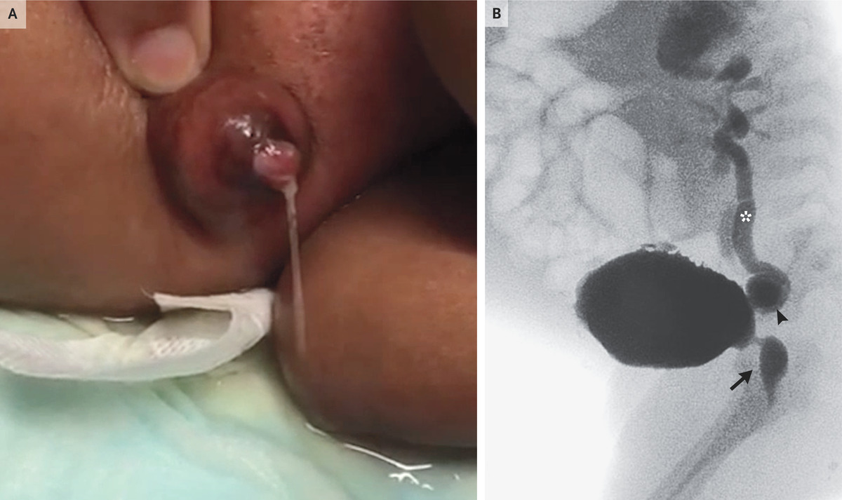
A 1-month-old boy was transferred to a tertiary care hospital for the management of obstructive uropathy. He had initially been brought to a local hospital for progressive abdominal distention. At the local hospital, laboratory studies had shown kidney injury, and ultrasonography of the abdomen showed a thickened bladder wall, ascites, hydronephrosis of both kidneys, and a perinephric collection around the left kidney. He was subsequently transferred for further care. On arrival at the tertiary care hospital, the physical examination was notable for a distended abdomen, reducible umbilical hernia, and rash on the lower abdomen (Panel A). When the abdomen was palpated, urine flowed from the umbilicus (Panel A and video). A voiding cystourethrogram (Panel B, lateral view) showed a dilated posterior urethra (arrow), bladder diverticula (arrowhead), and vesicoureteral reflux (asterisk) on both sides. A diagnosis of posterior urethral valves was made. Posterior urethral valves — congenital obstructing membranes in the lumen of the urethra — are a common cause of urinary tract obstruction in male infants. In severe cases, retrograde accumulation of urine can result in vesicoureteral reflux, bladder diverticula, urinomas, urinary ascites, and a patent urachus — all of which were seen in this case. Urachus ligation, urinoma drainage, urethral valve ablation, and a vesicostomy with later closure were performed. During 2 years of subsequent follow-up, the patient’s development and kidney function remained normal.
