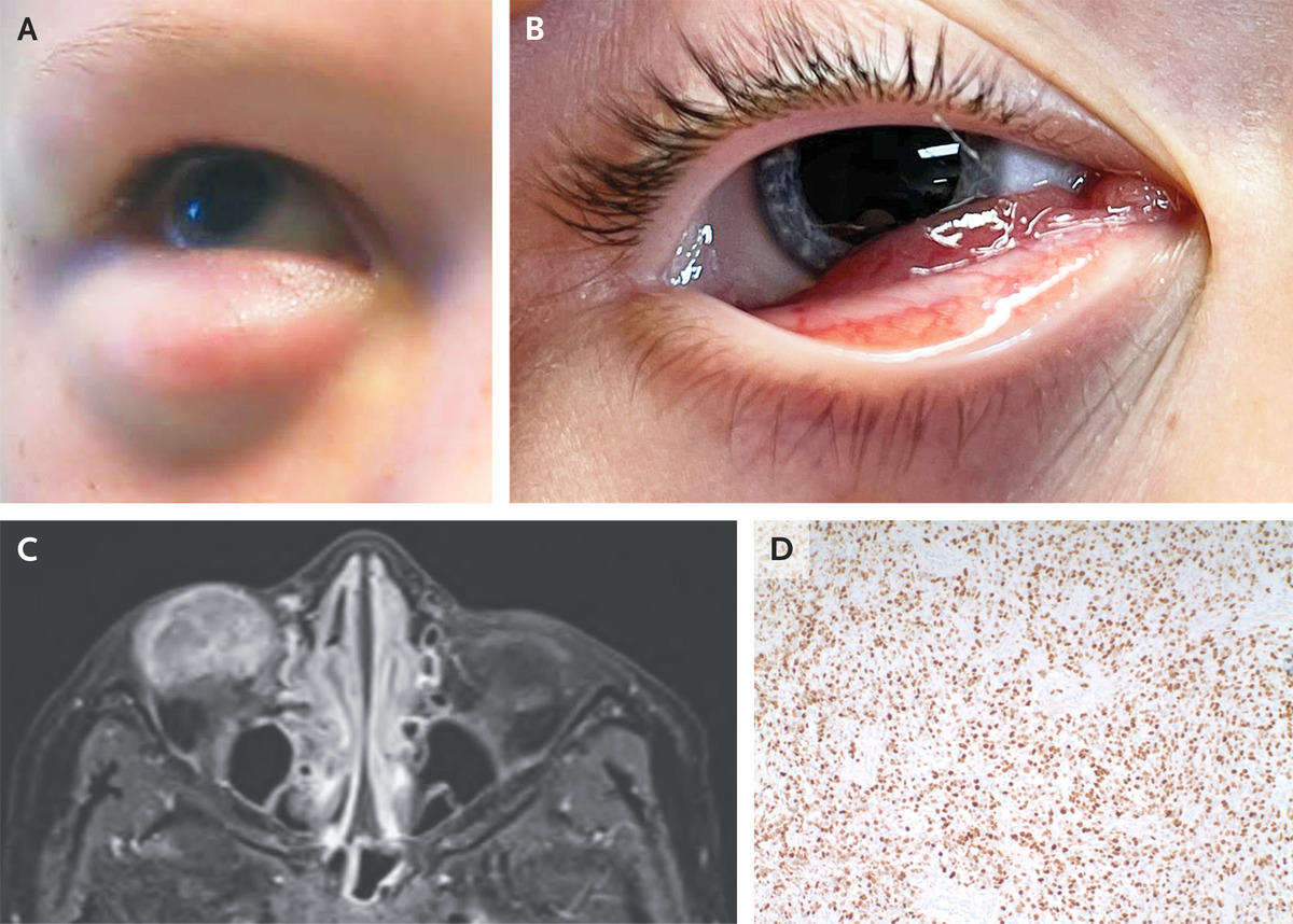
A previously healthy 8-year-old boy was brought to the emergency department with a 2-week history of painless swelling of the right lower eyelid. On physical examination, the swelling was found to be present during primary gaze (Panel A) but reduced during downward gaze . A fixed mass measuring 1.5 cm by 2.5 cm was palpable within the eyelid. When the right eye was opened, erythematous subconjunctival tissue was seen protruding from under the lower eyelid (Panel B). Visual acuity and extraocular movements were normal in both eyes. Magnetic resonance imaging of the orbits showed a heterogeneous, circumscribed, T1-hypointense, T2-hyperintense mass along the anteroinferior portion of the right globe (Panel C, T1 fat-suppression sequence with contrast medium). An incisional biopsy was performed, and histopathological examination showed dense sheets of small, round, blue cells that stained positive for myogenin (Panel D). A diagnosis of embryonal rhabdomyosarcoma — the most common primary malignant orbital tumor in children — was made. A staging evaluation indicated stage 1 involvement, and the patient underwent mass debulking, adjuvant chemotherapy, and radiotherapy. At follow-up 3 months later, imaging showed no evidence of disease and the patient’s vision remained normal. Eyelid swelling that varies with eye movements should arouse suspicion for an extraocular muscle condition, including malignant tumors, as in this case.
SOURCE: NEJM
