https://diabetesjournals.org/care/search-results?f_AllAuthors=Raveendhara+R.+Bannuru
Day: 12/28/2022
Aducanumab Approved for Treatment of Alzheimer’s.
Scientists develop blood test to detect Alzheimer’s disease
Scientists develop blood test to detect Alzheimer’s disease https://www.wionews.com/science/scientists-develop-blood-test-to-detect-alzheimers-disease-547445.
NASA Gets Unusually Close Glimpse of Black Hole Snacking on Star

A disk of hot gas swirls around a black hole in this illustration. The stream of gas stretching to the right is what remains of a star that was pulled apart by the black hole. A cloud of hot plasma (gas atoms with their electrons stripped away) above the black hole is known as a corona.
Recent observations of a black hole devouring a wandering star may help scientists understand more complex black hole feeding behaviors
Multiple NASA telescopes recently observed a massive black hole tearing apart an unlucky star that wandered too close. Located about 250 million light-years from Earth in the center of another galaxy, it was the fifth-closest example of a black hole destroying a star ever observed.
Once the star had been thoroughly ruptured by the black hole’s gravity, astronomers saw a dramatic rise in high-energy X-ray light around the black hole. This indicated that as the stellar material was pulled toward its doom, it formed an extremely hot structure above the black hole called a corona. NASA’s NuSTAR (Nuclear Spectroscopic Telescopic Array) satellite is the most sensitive space telescope capable of observing these wavelengths of light, and the event’s proximity provided an unprecedented view of the corona’s formation and evolution, according to a new study published in the Astrophysical Journal.
The work demonstrates how the destruction of a star by a black hole – a process formally known as a tidal disruption event – could be used to better understand what happens to material that’s captured by one of these behemoths before it’s fully devoured.
Most black holes that scientists can study are surrounded by hot gas that has accumulated over many years, sometimes millennia, and formed disks billions of miles wide. In some cases, these disks shine brighter than entire galaxies. Even around these bright sources, but especially around much less active black holes, a single star being torn apart and consumed stands out. And from start to finish, the process often takes only a matter of weeks or months. The observability and short duration of tidal disruption events make them especially attractive to astronomers, who can tease apart how the black hole’s gravity manipulates the material around it, creating incredible light shows and new physical features.
“Tidal disruption events are a sort of cosmic laboratory,” said study co-author Suvi Gezari, an astronomer at the Space Telescope Science Institute in Baltimore. “They’re our window into the real-time feeding of a massive black hole lurking in the center of a galaxy.”
When a star wanders too close to a black hole, the intense gravity will stretch the star out until it becomes a long river of hot gas, as shown in this animation. The gas is then whipped around the black hole and is gradually pulled into orbit, forming a bright disk.
A Surprising Signal
The focus of the new study is an event called AT2021ehb, which took place in a galaxy with a central black hole about 10 million times the mass of our Sun (about the difference between a bowling ball and the Titanic). During this tidal disruption event, the side of the star nearest the black hole was pulled harder than the far side of the star, stretching the entire thing apart and leaving nothing but a long noodle of hot gas.
Scientists think that the stream of gas gets whipped around a black hole during such events, colliding with itself. This is thought to create shock waves and outward flows of gas that generate visible light, as well as wavelengths not visible to the human eye, such as ultraviolet light and X-rays. The material then starts to settle into a disk rotating around the black hole like water circling a drain, with friction generating low-energy X-rays. In the case of AT2021ehb, this series of events took place over just 100 days.
The event was first spotted on March 1, 2021, by the Zwicky Transient Facility (ZTF), located at the Palomar Observatory in Southern California. It was subsequently studied by NASA’s Neil Gehrels Swift Observatory and Neutron star Interior Composition Explorer (NICER) telescope (which observes longer X-ray wavelengths than Swift).
Then, around 300 days after the event was first spotted, NASA’s NuSTAR began observing the system. Scientists were surprised when NuSTAR detected a corona – a cloud of hot plasma, or gas atoms with their electrons stripped away – since coronae usually appear with jets of gas that flow in opposite directions from a black hole. However, with the AT2021ehb tidal event, there were no jets, which made the corona observation unexpected. Coronae emit higher-energy X-rays than any other part of a black hole, but scientists don’t know where the plasma comes from or exactly how it gets so hot.
“We’ve never seen a tidal disruption event with X-ray emission like this without a jet present, and that’s really spectacular because it means we can potentially disentangle what causes jets and what causes coronae,” said Yuhan Yao, a graduate student at Caltech in Pasadena, California, and lead author of the new study. “Our observations of AT2021ehb are in agreement with the idea that magnetic fields have something to do with how the corona forms, and we want to know what’s causing that magnetic field to get so strong.”
Yao is also leading an effort to look for more tidal disruption events identified by ZTF and to then observe them with telescopes like Swift, NICER, and NuSTAR. Each new observation offers the potential for new insights or opportunities to confirm what has been observed in AT2021ehb and other tidal disruption events. “We want to find as many as we can,” Yao said.
Genetically Modified Mosquitoes Stunt Malaria Parasite Growth, Prevent Transmission
Scientists led by researchers from the Transmission:Zero team at Imperial College London have engineered mosquitoes that slow the growth of malaria-causing parasites in their guts, and prevent transmission of the disease to humans. The mosquitoes carry a genetic modification that causes them to produce compounds in the gut that stunt the growth of the malaria parasites, meaning that the parasites are unlikely to reach the mosquitoes’ salivary glands and be passed on to a human in a bite before the insects die.
The research team showed that the strategy can dramatically reduce the possibility of malaria spreading, in a lab setting. If proven safe and effective in real-world settings it could offer a powerful new tool to help eliminate malaria. Collaborators from the Institute for Disease Modeling at the Bill and Melinda Gates Foundation also developed a model that, for the first time, can assess the impact of such modifications if used in a variety of African settings. They found that the modification developed by the Transmission:Zero team could be a powerful tool for bringing down cases of malaria even where transmission is high.
The innovation is designed so it can be coupled with existing “gene drive” technology to promote the spread of the modification and drastically cut malaria transmission. The researchers aim to test whether their approach can block the transmission of parasites that have infected humans, as well as those that have been lab-reared. The safety of the new modification will be tested thoroughly before combining it with a gene drive for real-world tests.
If all goes well, field trials are anticipated within 2–3 years. George Christophides, PhD, a professor in the department of life sciences at Imperial, said, “History has taught us that there is no silver bullet when it comes to malaria control, thus we will have to use all the weapons we have at our disposal and generate even more. Gene drive is one such very powerful weapon that in combination with drugs, vaccines, and mosquito control can help stop the spread of malaria and save human lives.”
Christophides is co-lead author of the team’s published paper in Science Advances, which is titled, “Gene drive mosquitoes can aid malaria elimination by retarding Plasmodium sporogonic development.”
Malaria remains one of the world’s most devastating diseases, putting at risk about half of the world’s population. In 2021 alone, the disease infected 241 million and killed 627,000 people, mostly children aged below five years, in sub-Saharan Africa.
The authors wrote, “Despite the availability of the first World Health Organization–approved malaria vaccine the necessity to develop alternative intervention strategies remains pressing, particularly if malaria elimination is to remain the goal.” Co-first author Tibebu Habtewold, PhD, at the department of life sciences at Imperial, further explained, “Since 2015, the progress in tackling malaria has stalled. Mosquitoes and the parasites they carry are becoming resistant to available interventions such as insecticides and treatments, and funding has plateaued. We need to develop innovative new tools.”
Malaria is transmitted between people after a female mosquito bites someone infected with the malaria parasite. The parasite then develops into its next stage in the mosquito’s gut and travels to its salivary glands, ready to infect the next person the mosquito bites.
However, only around 10% of mosquitoes live long enough for the parasite to develop far enough to be infectious. The team aimed to lengthen the odds even further, by extending the time it takes for the parasite to develop in the gut.
The Transmission:Zero team genetically modified Anopheles gambiae, the primary malaria-carrying species of mosquito in sub-Saharan Africa. On taking a blood meal, mosquitoes with the modification produce two antimicrobial peptides (AMPs) in the gut, which impair the malaria parasite’s development. The two peptides are magainin 2, which was first found within skin secretions of the African claw frog Xenopus laevis, and melittin, a primary toxin component of the European honey bee Apis mellifera.
Inhibiting the parasite’s development causes a few days’ delay before the next parasite stage could reach the mosquito salivary glands, by which time most mosquitoes in nature are expected to die. The peptides work by interfering with the energy metabolism of the parasite, which also has some effect on the mosquito, causing them to have a shorter lifespan and further decreasing their ability to pass on the parasite. “As the modification additionally reduces female mosquito life span, the possibility of infectious sporozoites to be transmitted to a new host is reduced markedly,” the team stated in the published report.

Co-first author of the study Astrid Hoermann, PhD, from the department of life sciences at Imperial, said: “For many years, we have been trying to no avail to make mosquitoes that cannot be infected by the parasite or ones that can clear all the parasites with their immune system. Delaying a parasite’s development inside the mosquito is a conceptual shift that has opened many more opportunities to block malaria transmission from mosquitoes to humans.”
If the genetic modification is to help prevent malaria spread in the real world, it will need to be spread from lab-bred mosquitoes to wild populations. Normal interbreeding would achieve this to a certain degree, but because the modification has a “fitness cost” in the form of reduced lifespan, it would likely be quickly eliminated as a result of natural selection.
Gene drive is an additional genetic trick that could be added to the mosquitoes and cause preferential inheritance of the antiparasite genetic modification, making it spread more widely among any natural populations. And as the researchers further noted, “Modeling suggests that propagation of this modification via gene drive promises to break the malaria transmission cycle across a range of epidemiological scenarios in sub-Saharan Africa even if the effector itself is eventually replaced by resistant alleles because of the fitness cost that it imposes.”
For the reported study, the researchers demonstrated that the small genetic modifications to the malaria mosquitoes successfully impeded transmission of two different malaria parasite Plasmodium species, the deadliest human parasite, P. falciparum, and the rodent parasite P. berghei. “It achieves this by hampering parasite sporogonic development that occurs in the oocyst, markedly delaying the emergence of infectious sporozoites, and we attribute this effect to the known propensity of these AMPs for interfering with mitochondrial function,” they wrote.
The authors acknowledged that their strategy would require careful planning to minimize any risks before field trials can be considered. The Transmission:Zero team is creating two separate, but compatible strains of modified mosquitoes—one with the antiparasite modification and one with the gene drive.
They can then test the antiparasite modification on its own first, only adding in the gene drive once it has been shown to be effective. Co-lead author Nikolai Windbichler, PhD, from the department of life sciences at Imperial, said, “We are now aiming to test whether this modification can block malaria transmission not just using parasites we have reared in the lab but also from parasites that have infected humans. If this proves to be true, then we will be ready to take this to field trials within the next two to three years.”
With partners in Tanzania, the team has set up a facility to generate and handle genetically modified mosquitoes and conduct some initial tests. These include collecting parasites from locally infected schoolchildren, to ensure that the modification works against the parasites circulating in relevant communities.
They are also fully risk-assessing any potential releases of modified mosquitoes, taking into account any potential hazards and making sure they have acceptance from the local community. But they are hopeful that their intervention can ultimately help to eradicate malaria.
The investigators concluded in their paper, “This modification is already designed for gene drive and requires no further adjustment before deployment, while, at the same time, it is inert on its own and thus can be safely tested in an endemic setting under standard containment protocols. It thus enables the next step for testing antimalarial effectors, i.e., to evaluate their transmission blocking modifications against parasites directly sampled from patients in malaria-endemic countries.”
Tick-Bite Induced Mammalian Meat Allergy Unraveled
Scientists at the Garvan Institute of Medical Research in Australia have gained mechanistic insights on a life-threatening mammalian meat allergy brought on by tick bites that could pave the way for therapeutics to treat the rare anaphylactic response.
The study takes a close look at how human antibodies interact with a sugar molecule called alpha-gal that is found on gut bacteria, malarial parasites, the cancer drug cetuximab, and tick proteins, but not in humans, old world monkeys, and other great apes.
The findings were published on July 4, 2022, in an article in the Proceedings of the National Academy of Sciences (PNAS) titled, “Genetic and structural basis of the human anti-alpha-galactosyl antibody response.” The study confirms the role of alpha-gal as the key molecule underlying this unique allergy and may lead to strategies toward antibody therapeutics for alpha-gal syndrome in mammalian meat allergy, malaria, and xenotransplantation.
Northern Sydney has the highest prevalence of tick-induced mammalian-meat allergy, with more than 1800 cases reported. The Sunshine Coast hinterland around Maleny in Queensland is another hot spot. Sheryl van Nunen, MBBS, MM, FRACP, an allergy specialist at Sydney’s Northern Beaches Hospital, and a co-author on the paper, was the first clinician to link tick bites with mammalian-meat allergy. “Not a week goes by that I don’t see two people with this allergy,” she said.
“Our findings outline common binding modes and germline usage underpinning the alpha-gal response, providing molecular insights and guidance for drug development efforts,” the authors noted. The study is led by Daniel Christ, PhD, professor and head of antibody therapeutics and director of the Centre for Targeted Therapy at Garvan, and Joanne Reed, PhD, an associate professor at the Westmead Institute.
Human serum contains high levels (nearly 1%) of antibodies against alpha-gal. Therefore, when humans are exposed to alpha-gal—such as, through bites of the paralysis-inducing tick Ixodes holocyclus that is endemic to Eastern Australia—the human immune system mounts a massive anaphylactic response.
In the current study, the researchers investigated the basis of anti-alpha gal antibody gene restriction to the germline. They analyzed the structural and genetic mechanisms of alpha-gal recognition by solving the crystal structure of the mouse immunoglobulin M (M86) bound to alpha-gal. They also characterized the affinity and structures of human anti-alpha-gal antibodies in alpha-gal binding B cells isolated from healthy individuals and patients of mammalian meat allergy.
Analyzing blood from patients with tick bite-induced mammalian-meat allergy, the researchers frequently found the antibody type IGHV3-7 in response to alpha-gal. Molecular analysis showed that alpha-gal fits snugly into a pocket in this antibody. “We have more than 70 types of antibodies and this one is significantly overrepresented with alpha-gal recognition. We seem to be genetically predisposed to being sensitive to this sugar,” Christ said.
Alpha-gal is not found in humans and higher primates because a gene for the enzyme that adds the final galactose in alpha-gal was inactivated in the hominid lineage some 20 to 30 million years ago. This inactivation potentially occurred as a survival mechanism when pathogens with alpha-gal emerged. The high level of alpha-gal antibody levels in humans is likely driven by the constant exposure to resident gut bacteria that also express alpha-gal on their surfaces. The current study points to the evolutionary benefit of having an antibody response against alpha-gal.
“Humans lost the capacity to produce α-gal throughout evolution, but we don’t know why,” said Reed. “The suspicion is that it has to do with protection against infectious disease.”
Christ noted a rapid immune response to alpha-gal could destroy the malarial parasite Plasmodium that bears alpha-gal on its coat, protecting a person from malaria.
The reason why some people develop anaphylaxis and others don’t, remains unknown. It could be related to the number of tick bites, the amount of saliva injected during a tick bite or genetic sensitivity, says van Nunen. About a third of the population with sensitivity to alpha-gal exhibit symptoms of an allergy to mammalian meat. A second tick bite can more than double this allergic response.
Potential Inverse DNA Vaccine for Multiple Sclerosis
Conventional vaccines enhance the body’s immune response against a foreign protein. In an autoimmune disease, where the body’s misdirected immune system attacks its own cells, could an “inverse vaccine” turn down, or better still turn off, a pathological immune response? This question intrigued Lawrence Steinman, MD, PhD, a professor of neurology, pediatrics, and genetics at Stanford University and chairman of Pasithea Therapeutics.

Pasithea, dedicated to developing drugs for neurological disorders, recently announced encouraging preclinical results that support the efficacy of a tolerizing, inverse DNA vaccine for multiple sclerosis (MS). Based on experiments conducted with Hooke Laboratories, Pasithea reported that intramuscular injections of the candidate vaccine (PAS002) delayed the onset of paralysis, and reduced severity of peak disease. Prophylactic administration also reduced the incidence and severity of relapse in the mouse model.
“The results of this study show that this technology has the potential to tolerize to GlialCAM, a myelin molecule that has molecular similarity to the Epstein Barr Virus that triggers MS,” said Steinman.

Viral connections
The definitive cause for MS is unclear, however, many studies have found B lymphocytes infected with EBV (Epstein Barr Virus) in the brains of MS patients, indicating EBV could trigger MS. Yet nearly 95% of all adults carry EBV but do not develop MS.
“Although EBV is the trigger and is necessary, fortunately, it’s not sufficient to trigger MS in most of us,” explained Steinman.
The direct cause is difficult to demonstrate for a disease like MS, but an epidemiological study led by Albert Ascherio, PhD, professor of epidemiology and nutrition at the Harvard School of Public Health, demonstrated that the risk for MS increases 32-fold upon infection with EBV and not upon infection with other similar viruses. Of 10 million U.S. military veterans included in the study, 801 had MS and 800 of them were positive for EBV. Moreover, 35 individuals who did not have any evidence of EBV infection when they entered the military, showed the presence of EBV in blood before they were diagnosed with MS, further supporting EBV’s causative role in MS.
An editorial on Asherio’s study in Science that Steinman co-wrote mentions, “These findings provide compelling data that implicate EBV as the trigger for the development of MS.”
A few days after Ascherio’s article in Science, Steinman’s group published an article in Nature (“Clonally expanded B cells in multiple sclerosis bind EBV EBNA1 and GlialCAM“) that showed a human cell adhesion protein found in myelin called GlialCAM is attacked in MS. The study provided structural and functional evidence that a domain in GlialCAM that resembles an EBV transcription factor called EBNA1 (EBV nuclear antigen 1) plays a pivotal role in MS pathogenesis. The rationale underlying the development of Pasithea’s tolerizing vaccine for MS, PAS002, is based on these findings.
“The tolerizing approach advanced by Pasithea represents a new paradigm in the treatment of MS, which has application to other autoimmune, neuroinflammatory, and neurodegenerative diseases. Their advance was made possible by the discovery that EBNA1 is a mimic of GlialCAM and serves as an immune target in MS,” said Scott Zamvil, PhD, a professor of neurology and immunology at the University of California, San Francisco, and chair in MS research. “DNA vaccinations targeting GlialCAM are unique and provide a robust clinical benefit. I can see this approach could be beneficial to patients both within the early inflammatory and secondary neurodegenerative phases of MS.” (Zamvil was not involved in this study).
Tracking pathogenesis
A hallmark of MS is the production of antibodies by the clonal expansion of B cell-derived plasmablasts in the brain.
“There are only a few diseases where if you do a spinal tap and look in the cerebrospinal fluid, you find high levels of immunoglobulins that are clonal,” Steinman said. “That was the map we used to home in on clonal antibodies that are directed to the piece of EBV that mimics GlialCAM.”
These antibodies bind GlialCAM that is expressed on astrocytes and oligodendrocytes in the white matter, resulting in the destruction of myelin that envelops axons, and eventually the disintegration of the neuronal axons.
“The majority of MS patients have a clonal antibody in the spinal fluid that cross-reacts between GlialCAM and EBNA1, and 25 to 30% of patients have that marker in the blood,” said Steinman. “We don’t know if that’s a snapshot in time, or if longitudinally studies would reveal increases at some points. Those studies are ongoing.”
B cells and their plasmablast progeny express a cell surface molecule called integrin a4, that enables these cells to travel from the bone marrow into the systemic circulation and finally cross the blood-brain barrier into the brain.
“One of our remarkable discoveries is that the main homing molecule alpha4 integrin that allows plasmablasts that make the clonal antibody to enter into the cerebrospinal fluid (CSF), is lost once the plasmablasts are in the brain,” said Steinman. “This means the plasmablasts can enter [the brain], but they cannot escape!”

Steinman’s team collaborated with a team led by William Robinson, MD, PhD, a professor of immunology and rheumatology at Stanford University. “Robinson’s patented technology (US Patent 2013:WO2012148497 A2012148493) for deconvoluting heavy and light chain clonal antibody expansions was critical in identifying the monoclonal antibody that binds EBNA-1 and GlialCAM,” said Steinman.
The team identified the cross-reactive antibody through single-cell sequencing of plasmablasts in blood and CSF collected from MS patients, and protein microarray assays of CSF-derived antibodies against MS-associated viruses. Using sequence analysis, interferometry affinity measures, and X-ray crystallography, the team determined the crystal structure of the EBNA1–peptide epitope bound to a fragment of the autoreactive antibody. This enabled the team to track the development of the GlialCAM cross-reactive antibody.
Molecular mimicry and its boosters

When parts of a host’s own protein resemble parts of a viral protein, the “molecular mimicry” can induce immune cells into attacking cells bearing such molecular mimics. Ordinarily, the body’s T lymphocytes are adept at recognizing foreign proteins (antigens), parts of which are presented by cell surface HLA (human leucocyte antigen) that act as molecular billboards on the cellular landscape.
“People with a type of HLA molecule called HLADRB*1501 or HLRDR2 are more susceptible to the consequences of molecular mimicry,” said Steinman.
Post-translational modifications of the shared domain enhance the mimicry between GlialCAM and EBNA1. The molecular mimic includes serine residues which when phosphorylated causes the cross-reactive antibody to bind more intensely.

Steinman said, “One of the genetic risk factors for susceptibility to MS are kinases that can phosphorylate proteins. That may be one of the reasons that although most of us have antibody to EBV in our blood, we do not have antibody in our CSF that is cross-reactive between EBNA-1 and GlialCAM.” The research community is looking at modulating kinases that control disease susceptibility by determining whether the protein is phosphorylated.
Modeling the disease
Steinman’s team used the EAE (experimental autoimmune encephalomyelitis) mouse model in their preclinical studies. This model was generated by Thomas Rivers nearly 90 years ago to understand why the smallpox vaccine was causing disseminated encephalomyelitis—a rare autoimmune disease marked by inflammation in the brain and spinal cord—in a small percentage of individuals vaccinated for smallpox.
EAE is the most common experimental model for MS. Studies in the EAE model have led to the development of three blockbuster drugs for MS: Copaxone made by Teva, Tysabri (natalizumab), a monoclonal antibody against integrin-alpha4 from Steinman’s lab, and Mayzent (siponimod), an oral drug for progress MS from Novartis.
“If I scored three holes in one, I would be considered a pretty good golfer and not just lucky,” Steinman said, emphasizing the importance of the EAE model in MS. “No one’s going to invest in a drug for MS unless it works in EAE.”
Inspired by Koch’s postulates in establishing the cause of a disease, Steinman’s team injected the EAE model with EBNA1 and found this increased the severity of paralysis associated with MS.
“The EBNA1 was associated with noncoding CpG sequences of DNA that activate the innate immune system. The CpG given at the dose in Lanz’s paper worsened paralysis in EAE, compared to giving the same peptide mixed with CpG that was scrambled. This in some ways is a fulfillment of Koch’s postulates,” explained Steinman.
Treating MS without shutting down the immune system
The therapies that are currently approved for MS are drugs that seriously modulate the immune system. Steinman said, “Tysabri blocks the ability of the immune system to traffic into the brain. It’s very good in shutting down MS, but if your brain has to fight an infection and your immune system is blocked, you’re susceptible.” Antigen-specific tolerance is the holy grail of an immune therapy—it is the therapeutic gap that Pasithea seeks to fill with its tolerizing vaccines.
“If we know what one of the main triggers of the disease is, and we could shut down the response to it, we may have one of the long sought after goals that you would want to see for an immune therapy: blocking the cause of the disease and leaving the rest of your immune system free to fight infections,” said Steinman. “We don’t want to shut down the immune system, if we can avoid it.”
To achieve such antigen-specific tolerance, Steinman’s team obtained promising results with an inverse DNA vaccine for MS that encoded the full-length myelin basic protein (MBP), in an earlier study. The first-generation vaccine reduced levels of autoantibodies against myelin and showed improved MRI brain lesions in a Phase II trial on 267 MS patients. However, with stronger therapies like Tysabri available, investors were reluctant to invest in an inverse MBP DNA vaccine, and the trials were discontinued.
“Now with GlialCAM linked to the EBNA1, which is a definitive trigger of MS, investigators like myself, are ready to push ahead again with DNA vaccines to GlialCAM, and perhaps with RNA vaccines like the one developed by BioNTech,” said Steinman.
In addition, the earlier vaccine encoding MBP included immune-stimulatory CpG sequences in the plasmid vector that posed a hindrance. “Testing the newer versions of the DNA plasmid with no CpG and with the coding region targeting GlialCAM promises to be more efficacious than tolerizing to MBP,” Steinman added.
A problem with both RNA and DNA vaccines is that they can stimulate the immune system via toll-like receptors. Recent work from BioNTech shows changing uridine to pseudouridine prevents RNA from stimulating the immune system. Eliminating CpG sequences from the vector backbone accomplishes a similar effect for DNA vaccines.
“There are some very interesting comparisons in what we’re trying to do with DNA and what BioNTech is doing with RNA. So far with traditional immunizing vaccines, RNA beat DNA. We have Moderna, Pfizer, BioNtech producing RNA vaccines, but we don’t have too much traction with DNA vaccines,” said Steinman. “We’ll have to see if DNA vaccines are better at tolerizing than RNA vaccines.”
Pasithea’s inverse DNA vaccine
Pasithea’s tolerizing vaccine PAS002 aims to suppress encephalomyelitis and MS. Its CpG-less plasmid vector encodes the EBNA1 peptide that mimics GlialCAM. Preclinical results in the EAE model show that PAS002 significantly lowers GlialCAM antibodies.
“The data show a reduction in the intensity of the initial attack and subsequent relapse in the EAE model. Initial results are telling us PAS002 does have foundations for moving this ahead,” said Steinman. “We’re somewhat comfortable as are the regulatory agencies with the idea of a vaccine where the DNA is modified and we’re encoding an antigen. It’s very bold and we want to make sure it’s safe and efficacious.”
“The tolerizing vaccine developed by Steinman based on his GlialCAM discovery is a major advance in our attempts to develop an immunologically specific, nontoxic therapy for multiple sclerosis,” said Howard Weiner, PhD, a professor of neurology at Harvard Medical School. (Weiner was not involved in Steinman’s study). “The beauty of using an inverse vaccine encoding a protein already present, is that it takes advantage of the body’s own regulatory system to fight disease.”
The vaccine can be administered subcutaneously or intramuscularly and does need to be designed to cross the blood-brain barrier. Unlike biologics such as monoclonal antibodies, tolerized immune cells cross the blood-brain barrier efficiently. “We want to tolerize the immune system outside the brain. If they see inflammation, tolerized immune cells can get into the brain and broadcast tolerance rather than inflammation.”
Steinman does not expect PAS002 to be a one-shot wonder. “One thing we’re learning on the other side of the equation with nucleic acid vaccines is that the durability of the effects isn’t as long as we had hope for,” said Steinman. Preclinical titers indicate the vaccine would need to be administered periodically but the regimen remains to be worked out in future studies.
In a serendipitous turn of events, Steinman found PAS002 could potentially play a role in vaccine development efforts against monkeypox. Pox viruses, including monkeypox, contain the same GlialCAM sequence that is present in EBNA1. Although the first line of defense against an infection such as monkeypox is still a conventional vaccine that boosts immune responses against the pathogen, Pasithea’s tolerizing vaccine could reverse uncontrolled immune responses in individuals who develop encephalomyelitis following monkey pox infection or vaccination. “But this will need further studies,” said Steinman.
Bolstered by the promising preclinical results, Steinman is determined to see PAS002 advance to the clinic for MS. His team is also developing biomarker assays to study the effect on anti-GlialCAM antibodies that they plan to deploy in the upcoming clinical trials.
mRNA Vaccine Provides Broad Protection against All Known Influenza Subtypes
Researchers at the Perelman School of Medicine at the University of Pennsylvania have developed an experimental multivalent mRNA-based vaccine against all 20 known subtypes of influenza virus. Their approach differs from previous attempts to craft a universal flu vaccine, by including antigens specific to each subtype, rather than just a smaller set of antigens shared among subtypes. This strategy harnesses the same mRNA technology as that employed in the Pfizer and Moderna SARS-CoV-2 vaccines. The mRNA technology that enabled those COVID-19 vaccines was pioneered at Penn.

Tests in animal models showed that the vaccine dramatically reduced signs of illness and protected from death, even when the animals were exposed to flu strains different from those used in making the vaccine.
The team suggests that their technology could lead to the development of a universal flu vaccine that protects against potential future pandemics. “The idea here is to have a vaccine that will give people a baseline level of immune memory to diverse flu strains, so that there will be far less disease and death when the next flu pandemic occurs,” said study senior author Scott Hensley, PhD, a professor of microbiology at in the Perelman School of Medicine. Hensley and colleagues reported on the development of their mRNA vaccine in Science, in a report titled, “A multivalent nucleoside-modified mRNA vaccine against all known influenza virus subtypes.” Hensley and his laboratory collaborated in the study with the laboratory of mRNA vaccine pioneer Drew Weissman, MD, PhD, the Roberts Family professor in vaccine research and director of vaccine research at Penn Medicine.
Influenza viruses periodically cause pandemics with enormous death tolls. The best known of these was the 1918–19 “Spanish flu” pandemic, which killed tens of millions of people worldwide. Flu viruses can circulate in birds, pigs, and other animals, and pandemics can start when one of these strains jumps to humans and acquires mutations that adapt it better for spreading among humans. The authors explained, “There are at least 18 different influenza A virus (IAV) subtypes that circulate in animal reservoirs, and these viruses occasionally enter the human population and cause a pandemic.”
Current flu vaccines are “seasonal” vaccines that protect against recently circulating strains, but would not be expected to protect against new, pandemic strains. And even with increased global surveillance, it is difficult to predict which flu strain will cause the next flu pandemic, making a universal vaccine important. As the investigators noted, “Although surveillance programs and modeling studies have increased our knowledge of pandemic risk, we cannot accurately predict which influenza subtype will cause the next pandemic.”
There are several universal influenza vaccines in development to provide protection against diverse influenza virus subtypes, the team continued. Most of these vaccine candidates include a limited number of antigens that have epitopes that are conserved across different influenza virus subtypes.
In contrast, the strategy employed by the Penn Medicine researchers is to vaccinate using immunogens—a type of antigen that stimulates immune responses—from all known influenza A and influenza B virus (IBV) subtypes in order to elicit broad protection. This vaccination strategy is not expected to provide “sterilizing” immunity that completely prevents viral infections. “Instead of focusing on immunogens to elicit antibodies against epitopes that are conserved among many different influenza virus strains, we designed a vaccine that encodes separate immunogens from all known IAV subtypes and IBV lineages,” the team explained.
Their newly reported study confirmed that the vaccine elicited a memory immune response that can be quickly recalled and adapted to new pandemic viral strains, significantly reducing severe illness and death from infections. “It would be comparable to first-generation SARS-CoV-2 mRNA vaccines, which were targeted to the original Wuhan strain of the coronavirus,” Hensley said. “Against later variants such as Omicron, these original vaccines did not fully block viral infections, but they continue to provide durable protection against severe disease and death.”
For their flu vaccine, the researchers prepared 20 different nanoparticle encapsulated mRNAs, each encoding a different hemagglutinin antigen. The experimental mRNA-lipid nanoparticle vaccine developed by Hensley and colleagues encoded hemagglutinin (HA) antigens from all 20 known influenza A and B virus subtypes.
When injected and taken up by the cells of recipients, the vaccine then resulted in production of copies of the key flu virus hemagglutinin protein, for all 20 influenza hemagglutinin subtypes—H1 through H18 for influenza A viruses, and two more for influenza B viruses. “For a conventional vaccine, immunizing against all these subtypes would be a major challenge, but with mRNA technology, it’s relatively easy,” Hensley said.
Tested in mice, the mRNA vaccine elicited high levels of antibodies, which stayed elevated for at least four months, and reacted strongly to all 20 flu subtypes. Multivalent protein vaccines produced using more traditional methods elicited fewer antibodies and were less protective compared to the multivalent mRNA vaccine in the animals.
Moreover, the new vaccine seemed relatively unaffected by prior influenza virus exposures, which can skew immune responses to conventional influenza vaccines. The researchers observed that the antibody response in the mice was strong and broad whether or not the animals had been exposed to flu virus before. The team also carried out tests in ferrets that were vaccinated using a prime-boost approach, and challenged with an avian H1N1 virus, to mimic a pandemic caused by an unknown viral strain. The results of these experiments confirmed that compared with unvaccinated animals challenged by the same virus, the vaccinated ferrets lost less weight, and all survived, whereas two of the four unvaccinated ferrets died. Unvaccinated animals also displayed more clinical signs of disease relative to vaccinated animals after infection.
“Further studies will be required to fully elucidate the mechanisms by which the 20-HA mRNA vaccine provides protection,” the authors acknowledged. Their reported findings suggested that protection against antigenically matched strains is mediated by neutralizing antibodies, whereas protection against mismatched viral strains may occur through non-neutralizing mechanisms, such as antibody-dependent cellular cytotoxicity (ADCC).
Hensley and his colleagues currently are designing human clinical trials. The researchers envision that, if those trials are successful, the vaccine may be useful for eliciting long-term immune memory against all influenza subtypes in people of all age groups, including young children. “We think this vaccine could significantly reduce the chances of ever getting a severe flu infection,” Hensley said. As the team noted in their report, “It is likely that mRNA influenza vaccines that are imperfectly matched to novel pandemic influenza virus strains will not provide sterilizing immunity but will instead limit disease severity and protect against death through non-neutralizing mechanisms.”
In an accompanying perspective, Alyson A Kelvin, PhD, and Darryl Fallzarano, PhD, at the University of Saskatchewan, noted that the strengths of the mRNA platform for pandemic vaccine production include flexibility of antigen design, increased numbers of potential viral targets, speed of production, and inexpensive, scalable manufacturing. “These strengths are important when designing and producing vaccines for a highly diverse, unpredictable family of viruses that can easily spread globally in a matter of weeks,” they noted. However, they commented, “questions remain regarding the regulation and approval pathway of such a vaccine that targets viruses of pandemic potential but are not currently in human circulation.”
In principle, Hensley added, the same multivalent mRNA strategy could be used for other viruses with pandemic potential, including coronaviruses. “Multivalent mRNA-LNP vaccines may be applied against other variable pathogens, such as coronaviruses and rhinoviruses,” the scientists concluded. “For example, SARS-CoV-2mRNA vaccines are being updated to include multiple spike components to combat antigenically distinct strains. Additional studies will be required to determine the maximum number of antigens that can be simultaneously delivered through mRNA-LNP vaccines and the underlying immunological mechanisms that allow for the induction of responses against multiple antigens.”
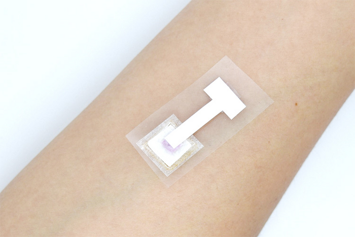
Skin Patch Test Detects COVID-19 in Under Three Minutes
A rapid and reliable skin-patch test can now detect the COVID-19 virus, and potentially other infectious agents in under three minutes, without the need to draw blood. This convenience overcomes a current challenge in identifying infected individuals who are averse to blood tests and could help restrict the spread of the pandemic.
The details of the new test were published on July 1, 2022, in an article in the journal Scientific Reports titled, “Anti‑SARS‑CoV‑2 IgM/IgG antibodies detection using a patch sensor containing porous microneedles and a paper‑based immunoassay.”
Asymptomatic individuals constitute 16–38% of the SARS-CoV-2 infected population which increases the difficulty of identifying infected individuals. The lack of convenient and sensitive tests to detect the virus in all individuals is continuing to limit global response to the pandemic.
Predominantly, SARS-CoV-2 is detected through RT-PCR (real-time reverse transcription polymerase chain reaction) on swab samples collected from the nose and throat. However, these tests require long detection times, high costs, specialized equipment and medical personnel, and are not feasible in areas where resources are limited.
Alternatively, COVID-19 infection is detected through antibody tests (immunoassays) using blood samples collected from finger pricks by a lancing device. These popular point-of-care options require gold nanoparticle-based testing strips and involve risks of cross contamination and biohazard.
Immunochromatographic tests that detect anti-SARS-CoV-2 immunoglobulin M (IgM) and immunoglobulin G (IgG) can provide clinically relevant information regarding the course of COVID-19 infection, but invasive blood sampling poses a major obstacle.
“To develop a minimally invasive detection assay that would avoid these drawbacks, we explored the idea of sampling and testing interstitial fluid, which is located in the epidermis and dermis layers of human skin,” said first author Leilei Bao, PhD, researcher at the Institute of Industrial Science, at The University of Tokyo. “Although the antibody levels in the interstitial fluid are approximately15–25% of those in blood, it was still feasible that anti-SARS-CoV-2 IgM/IgG antibodies could be detected and that interstitial fluid could act as a direct substitute for blood sampling.”

“We developed biodegradable porous microneedles made of polylactic acid that draws up the interstitial fluid from human skin,” said Beomjoon Kim, PhD, professor at the department of mechanical and biofunctional systems at The University of Tokyo, and the senior author of the paper. “Then, we constructed a paper-based immunoassay biosensor for the detection of SARS-CoV-2-specific antibodies.”
Dermal interstitial fluid is a rich and accessible source of protein biomarkers, including antibodies. The authors prepared polylactic acid microspheres from a single emulsion to form continuous micropores, which they then heated for half an hour at 180°C to bind them together. They demonstrated these microneedles fabricated with emulsion droplets could effectively penetrate and extract interstitial fluid by capillary effect from rat and pig skin, used to model human skin.
The extracted interstitial fluid flows vertically into the attached nitrocellulose paper biosensor, where virus-specific antibodies are detected visually through a colored-based reaction (colorimetry).
The researchers show that anti-SARS-CoV-2 IgM and IgG levels as low as 3 and 7 ng/mL, respectively, can be detected using the skin patch test which shows the advantage of this new method over current commercially available lateral-flow immunochromatographic assays (LFIA). The patch, 1.5 cm by 3.5 cm in size, detects anti-SARS-CoV-2 antibodies in the interstitial fluid in a painless and convenient test, in under three minutes.
The authors believe that the speed, safety, simplicity, convenience, and minimally invasive nature of the compact anti-SARS-CoV-2 IgM/IgG biosensor device will lead to its widespread use. In addition to COVID-19, the authors claim that the device can be customized to rapidly screen various infectious agents and provide a complementary diagnostic test.
Monkeypox Symptoms Described in Large International Study
An international collaboration of clinicians has identified the clinical symptoms in people infected with monkeypox in the largest case study series to date. Their findings will improve future diagnosis, help to slow the spread of infection, and help the international community prioritize the limited global supply of monkeypox vaccines and treatments to communities most at risk.
The number of monkeypox cases in the United States has surpassed 2,500 (the number of cases was 2,891 on July 22). The states with the highest case counts are New York, California, and Florida (900, 356, and 247, respectively.) The latest numbers (as of July 20) from the World Health Organization (WHO) are 14,000 cases, from more than 70 countries and territories, including 5 deaths—all in Africa.
Just a few months ago, before April, monkeypox virus infection in humans was not typically reported outside African regions—where it is endemic. Basic information that will aid the public health response, like transmission, risk factors, clinical presentation, and outcomes of infection, are not well understood.
Now, a study—which is the result of an international collaboration across 16 countries—is adding some useful information to the current understanding. The study identifies new clinical symptoms of monkeypox infection, which will aid the ability to diagnose the infection in the future and help to slow the spread of infection. It is the largest case series to date, reporting on 528 confirmed infections (from 43 sites in 16 countries) between April 27 and June 24, 2022.
The report is published in The New England Journal of Medicine (NEJM) in the paper, “Monkeypox Virus Infection in Humans across 16 Countries—April–June 2022.”
Many of the infected individuals reviewed in the study presented with symptoms not recognized in current medical definitions of monkeypox. These symptoms include single genital lesions and sores on the mouth or anus. The clinical symptoms are similar to those of sexually transmitted infections (STIs) and can easily lead to misdiagnosis. In some people, anal and oral symptoms have led to people being admitted to the hospital for management of pain and difficulties swallowing. This points to the importance that these new clinical symptoms be recognized and healthcare professionals be educated on how to identify and manage the disease. Misdiagnosis can slow detection and thus hinder efforts to control the spread of the virus. The study will therefore lead to increased rates of diagnosis when persons from at-risk groups present with traditional STI symptoms.
The authors noted that transmission was suspected to have occurred through sexual activity in 95% of the persons with infection. In this case series, 95% of the persons presented with a rash (with 64% having less than 10 lesions), 73% had anogenital lesions, and 41% had mucosal lesions (with 54 having a single genital lesion).
The study includes data on common systemic features preceding the rash: they included fever (62%), lethargy (41%), myalgia (31%), and headache (27%); the swelling of lymph nodes was also common (reported in 56%).
Among the 23 persons with a clear exposure history, the median incubation period was 7 days (with a range of 3 to 20). Monkeypox virus DNA was detected in a large proportion (29 of the 32 persons) in whom seminal fluid was analyzed. However, “this may be incidental,” said John Thornhill, PhD, consultant physician in sexual health and HIV and clinical senior lecturer at Barts NHS Health Trust and Queen Mary University of London. “We do not know that it is present at a high enough level to facilitate sexual transmission. More work is needed to understand this better.”
“This truly global case series has enabled doctors from 16 countries to share their extensive clinical experience and many clinical photographs to help other doctors in places with fewer cases,” noted Chloe Orkin, MBChB, FRCP, MD, professor of HIV medicine at Queen Mary University of London and director of the SHARE collaborative. “We have shown that the current international case definitions need to be expanded to add symptoms that are not currently included, such as sores in the mouth, on the anal mucosa, and single ulcers. These particular symptoms can be severe and have led to hospital admissions so it is important to make a diagnosis. Expanding the case definition will help doctors more easily recognize the infection and so prevent people from passing it on. Given the global constraints on vaccine and anti-viral supply for this chronically underfunded, neglected tropical infection, prevention remains a key tool in limiting the global spread of human monkeypox infection.”
In addition, noted Thornhill, “we identified new clinical presentations in people with monkeypox. While we expected various skin problems and rashes, we also found that one in ten people had only a single skin lesion in the genital area, and 15% had anal and/or rectal pain. These different presentations highlight that monkeypox infections could be missed or easily confused with common sexually transmitted infections such as syphilis or herpes. We, therefore, suggest broadening the current case definitions.”
The findings of this study, including the identification of those most at risk of infection, will help to aid the global response to the virus. There is a global shortage of both vaccines and treatments for human monkeypox infection. Public health interventions aimed at the high-risk group could help to detect and slow the spread of the virus. Although sexual closeness is the most likely route of transmission in most cases, researchers stress that the virus can be transmitted by any close physical contact, through large respiratory droplets and potentially through clothing and other surfaces. The findings from this study will help public health measures—such as enhanced testing and education—to be developed and implemented, working with at-risk groups to ensure that they are appropriate and non-stigmatizing.
