HAWKING SOLVED HOW INFORMATION COULD ESCAPE BLACK HOLES


Stephen Hawking announced during at the KTH Royal Institute of Technology in Stockholm, in Sweden, that he has solved how you could escape a black hole.

The presentation was made at the Hawking Radiation conference, which was co-hosted by the theoretical physics institute, Nordita, and the University of North Carolina, on the campus of KTH Royal Institute of Technology in Stockholm.

Hawking, said”

“I propose that the information is stored not in the interior of the black hole as one might expect, but on its boundary, the event horizon. The message of this lecture is that black holes ain’t as black as they are painted. They are not the eternal prisons they were once thought. Things can get out of a black hole both on the outside and possibly come out in another universe.”

Watch the vide. URL: https://youtu.be/DkRDmJpthXg

Pregnancy Test That Can Predict Down Syndrome, Miscarriage, And Twins May Be Available In Just 2 Years


pregnancy test
The MAP Test hopes to provide expectant parents with accurate information faster.

The days of wondering what to expect when you’re expecting may soon be over. Researchers in the UK are working on eliminating much of the mystery surrounding pregnancies by developing a mail-in test that can predict everything from whether or not a couple is having twins to if an unborn child may have Down syndrome, using nothing more than a urine sample.

MAP Diagnostics is the company behind the first-of-its-kind pregnancy test that would reveal intricate details of both a mother and child’s health months before the actual due date, Smithsonian reported. The test, known as the MAP test, was originally developed to analyze proteins in a mother’s urine to predict chances of having an aneuploid pregnancy, or a pregnancy in which the child has an abnormal number of chromosomes in his cells (the cause of Down syndrome ). The Telegraph reported that the technique is similar to that used in IVF when screening for the best embryo to implant.

However, according to the company’s website, the researchers soon realized that the same technology used to predict Down syndrome could be used to predict other conditions, including: preeclampsia, hyperemesis gravidarum, ectopic pregnancies, gestational diabetes, assisted reproduction, intrauterine growth restriction, and gestational cancer.

According to MAP’s founder and chief scientific officer, Stephen Butler, this ultimate pregnancy test would provide expectant parents with more accurate test results than those currently available. “What we’re trying to do is create more information so people can make better decisions,” Butler explained, as reported by Smithsonian.

The pregnancy test we’re familiar with today was patented in 1972, but humans have recorded their efforts to predict pregnancies for nearly a thousand years. In the 1920s, scientists discovered the pregnancy hormone, human chorionic gonadotropin (hCG), and pregnancy tests started to become a little more accurate. The MAP test works off a similar concept as traditional pregnancy tests but goes one step further by analyzing the hCG for proteins that can indicate other health conditions.

Unlike at-home pregnancy tests, the MAP test would need to be sent to a lab for proper analysis. Also, a woman would need to be at least eight weeks into her pregnancy to get an accurate result. Currently, an amniocentesis procedure, where a doctor draws amniotic fluid from the mother’s uterus using a needle, is the most common way to predict Down syndrome in unborn children. The new test would theoretically provide a non-invasive way to arrive at the same answer.

The test for Down syndrome may take up to two years before becoming available to the public, but MAP Diagnostics developers hope their simpler tests, such as those for preeclampsia, will become available to the global market within six months. According toSmithsonian, the most recent developments on the MAP test were presented at the European Society of Human Reproduction and Embryology in Lisbon in June. Researchers are expected to release further information on their developments at the American Society for Reproductive Medicine conference in Baltimore this fall.

What Is Okra (Lady’s Finger) And 6 Benefits Of Adding The Medicinal Vegetable To Your Diet


Pieces of okra
Sink your teeth into okra’s health benefits, which range from treating diabetes to preventing kidney disease. Rebecca Wilson, CC BY 2.0

The beginning of September signals that fall is on its way, along with leaves, and… okra? The exotic medicinal and culinary vegetable (available year-round), is best to get in early fall when crops in Southern states reach their peak. Typically, okra is used as a thickening agent in soups like gumbo because of its ooey-gooey texture, but it can double as a nutritional powerhouse filled with vitamins, minerals, and other nutrients that provide an array of health benefits from treating diabetes to preventing kidney disease.

A single cup of raw okra has a little over 30 calories, about 3 grams of dietary fiber, 2 grams of protein, 7.6 grams carbohydrates, 0.1 grams of fat, 21 milligrams of vitamin C, around 88 micrograms of folate, and 57 milligrams of magnesium. This makes okra a nutrition hero and a very available food when it comes to our health.

Whether you consume okra stewed, boiled, fried, or even in pickled form, you can reap the health benefits of this little green vegetable any time of the year. Here’s how:

Alleviates Asthma

Consuming even small amounts of fruits or vegetables rich in vitamin C, like okra (21 milligrams per cup), can alleviate asthma symptoms. A 2000 study published in the journalThorax found the intake of citrus or kiwi fruits conferred a highly protective effect against wheezing symptoms in childhood. The protective effect was seen even among children who ate fruit only one to two times per week. The researchers found this to be especially true among already susceptible patients.

Lowers Cholesterol

Okra not only promotes good digestive health, but also good cholesterol levels due to its high fiber content. Soluble fiber can be dissolved in water, which means that it breaks down in the digestive tract. There, it also binds to cholesterol in other foods so that it can be excreted along with other wastes. In turn, total cholesterol levels plummet, according to the Harvard Health Publications. Okra also helps to lower cholesterol by replacing all the foods you eat with high fat and cholesterol levels — okra contains no cholesterol and very little fat.

Manages Diabetes

Soluble fiber can help diabetics because of its ability to keep blood glucose levels stable — it affects how sugar is absorbed in the intestines. In a 2011 study published in the journalISRN Pharmaceutics, researchers soaked sliced okra pods in water and then gave rats the solution through a gastric feeding tube — a control group wasn’t fed this solution. The researchers found okra helped reduce the absorption rate of glucose and in turn reduced blood sugar levels in the treated rats.

Boosts Immune System

Okra’s rich vitamin C content and antioxidant components also double as decent immune boosters against unsafe free radicals, while also supporting the immune system. Vitamin C stimulates the immune system to create more white blood cells, which can help battle other foreign pathogens and materials in the body.

Prevents Kidney Disease

Regularly eating okra can be helpful for preventing kidney disease. A 2005 study published in the Jilin Medical Journal found patients who ate okra daily reduced clinical signs of kidney damage more than those who were on a diabetic diet. This is helpful since nearly half of kidney disease cases develop from diabetes.

Promotes Healthy Pregnancy

Okra’s high levels of vitamin A, B vitamins (B1, B2, B6), and vitamin C, and traces of zinc and calcium, make it an ideal vegetable to eat during pregnancy. Okra also serves as a supplement for fiber and folic acid. This helps prevent birth defects like spina bifida and can even stop constipation during pregnancy.

The Eko Core Is A Digital Upgrade For The Centuries-Old Stethoscope


In a few months, the stethoscope will celebrate its 200th birthday. A medical breakthrough in 1816, it’s still a part of nearly every doctor’s visit today and a symbol of medicine itself.

Yet the stethoscope hasn’t changed much in the past 200 years. No different than in the 17th century, listening to a heartbeat has been a manual process that relies entirely on a doctor’s ear to detect irregularities.

That is, until today. And it’s all thanks to Eko Devices, a Berkeley-based startup that just became the youngest team to secure FDA clearance for a Class II medical device. Co-founders Connor Landgraf, Jason Bellet, and Tyler Crouch, all rather recent graduates of UC Berkeley, have added a digital dimension to the centuries-old tool.

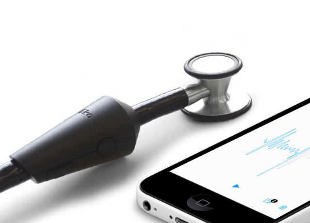
Dubbed the Eko Core, their solution is an adapter that attaches to the typical stethoscope and streams the heartbeat data to the cloud, providing doctors with an entirely new layer of information to analyze.

“It’s incredibly challenging to hear a minute heart murmur, especially in patients with high heart rates,” says Landgraf. “Cardiologists say it’s almost like a musical ear, it’s something that you have to learn over five or ten years of practice.”

With the Eko Core, the physician can see the heartbeat in wave form on a mobile device as well as hear the sound at an amplified level. Both the visible and audible data can be recorded and easily shared between physicians and hospitals.

For doctors, this takes a lot of the guesswork out of detecting murmurs, valve problems, and blockages in the arteries.

And for the 70 percent of pediatric patients with suspected heart murmurs who are unnecessarily referred to a cardiologist, Eko will be able to save thousands of dollars in avoidable echocardiograms.

“Physicians don’t have confidence in their ability to use the stethoscope in a lot of situations so they frequently refer people to cardiologists when it’s not necessary,” Landgraf says.

Eko is running a pilot with Stanford Hospital, where all residents will be using the Eko Core device as a training tool. Today, the company is releasing the device to the public for $199 on its own, or $299 with a stethoscope included.

Over the next few months, the team will be continuing to develop an algorithm that analyzes the data collected by all Eko devices in order to match heartbeats to conditions in real-time. Kind of like the “Shazam for heartbeats,” as Landgraf says doctors like to call it.

“Connecting patients to physicians with noninvasive tools to understand what’s going on in peoples’ hearts is going to be really powerful,” Landgraf says. “Right now you can catheterize a patient to find out what the pressures are like inside of the heart, but it’s very invasive and inefficient.”

The condition-detecting feature is slated to launch in Q1 of next year, and Landgraf hints that Eko will eventually roll out additional products to give physicians a better understanding of the heart in a noninvasive manner.

Since Eko was founded in 2013, the company has raised $2.8 million from Stanford’s StartX Fund, FOUNDER.org founder Michael Baum, and the co-founders of Shazam, among others.

Mutation Status Guides Advanced NSCLC Therapy


The presence or absence of mutations in advanced non-small cell lung cancer (NSCLC) should guide selection of first-line systemic therapy, according to an updated clinical guideline from the American Society of Clinical Oncology.

Patients with squamous-cell tumors that have no gene alterations should begin treatment with combination platinum-based cytotoxic chemotherapy, so long as they have good performance status (0 or 1). Optionally, bevacizumab (Avastin) may be added when the platinum agent is carboplatin. For patients with performance status 2, either chemotherapy or palliative care alone is an acceptable option.

In the presence of sensitizing EGFR mutations, appropriate first-line therapy is afatinib (Gilotrif), erlotinib (Tarceva), or gefitinib (Iressa). Treatment should begin with crizotinib (Xalkori) when patients have tumors with ALK or ROS1rearrangements, as published online in the Journal of Clinical Oncology.

Patients who respond to treatment have the option of continuing the same regimen as maintenance therapy, switching to a different therapy, or taking a break from therapy.

“Although there is no cure for patients with stage IV non-small cell lung cancer, various treatment options are available that can help patients control their cancer longer,” guideline panel co-chair Gregory Masters, MD, of Christiana Care Health System in Newark, Del., said in a statement. “This guideline will help doctors choose the most appropriate therapies, depending on the biology of the tumor and the patient’s general well-being.”

For first-line conventional chemotherapy for nonsquamous NSCLC, also favors platinum-containing regimens. The panel noted that use of nonplatinum doublets has weak supporting evidence. Additionally, “there are no FDA-approved nonplatinum regimens,” the authors pointed out.

The guideline addresses treatment options for multiple combinations of tumor mutation status, performance status, and histologic subtype, as well as quality of response (if any) and the selection of maintenance therapy.

The panel decided to remove a previous recommendation that allowed clinicians to “consider addition of cetuximab (Erbitux) to cisplatin-vinorelbine in first-line therapy in patients with EGFR-positive tumor.” Cetuximab has no regulatory approval for NSCLC in the United States, Canada, or Europe, and a recent trial of a cetuximab-containing combination ended in a negative result, the panel noted.

Options for second-line therapy included docetaxel, erlotinib, and gefitinib, plus pemetrexed (Alimta) as an additional option for patients with nonsquamous tumors. Patients with EGFR-mutant tumors can receive chemotherapy or a different EGFR inhibitor, depending on response to initial therapy. Crizotinib or chemotherapy is appropriate for patients with ALKrearrangements.

For third-line therapy, the panel backed the existing ASCO recommendation of erlotinib for patients with performance status 0 to 3 and no prior exposure to erlotinib or gefitinib. The authors found data insufficient to recommend for or against cytotoxic chemotherapy in third line.

The ASCO panel supported early palliative care, alone or in addition to primary therapy, throughout the guideline.

“Early palliative care is associated with improved survival of patients with advanced lung cancer,” panel co-chair David H. Johnson, MD, of the University of Texas Southwestern Medical Center in Dallas, said in the ASCO statement. “Hospice care also improves patient quality of life and reduces caregiver distress.”

Food Cravings May Rewire Obese People’s Brains In Same Way As Drug Addiction: Should There Be Rehab For Food Addicts?


Food Addiction
Obese people at a buffet had their brains scanned, and what the images revealed shows addict-like circuitry.

Drug addicts can become physically dependent on a substance to the point of overdose, and according to a new study, food cravings take hold of the brain in an eerily similar way. The findings were presented at the European College of Neuropsychopharmacology’s annual conference in Amsterdam by a collaborative team of researchers from the University of Granada in Spain and Monash University in Australia.

“There is an ongoing controversy over whether obesity can be called a ‘food addiction,’ but in fact there is very little research which shows whether or not this might be true,” said the study’s lead author Oren Contreras-Rodríguez, a cognitive psychology researcher at the University of Granada, in a press release. “Reward processing following food stimuli in obesity is associated with neural changes similar to those found in substance addiction.”

The research team laid out a buffet of food for 39 obese and 42 normal-weight participants. They let them eat what they wanted before evaluating each person in a functional magnetic resonance imaging (f-MRI) machine. The brain scans revealed food cravings affected obese participants’ neural connections in the brain differently than those who were considered a normal weight.

Obese individuals had a stronger connection between their dorsal caudate (responsible for reward-based habits) and somatosensory cortex (responsible for measuring the energy value of food, such as calories).  Meanwhile, normal-weight individuals had a stronger connection between the ventral putamen (responsible for evaluating flavors) and the orbitofrontal cortex (responsible for decision making). With this data, researchers were able to predict which obese individuals would gain weight three months later based on how strong of a connection there was between their dorsal caudate and somatosensory cortex.

These are the same neural changes researchers have seen play out in a drug addict’s brain. Past research has shown how cocaine and sugar play out in nearly identical ways in the brain. The most recent study in 2013 found that lab rats became addicted to Oreos in an alarmingly similar way to cocaine, leading researchers to believe food addiction is one of the underlying causes of the obesity epidemic. According to the Centers for Disease Control and Prevention(CDC), more than one-third of adults in America are obese, with childhood obesity rates trailing closely behind; the CDC reported the number of obese children has tripled in the last 30 years.

Food addiction may pose a more dangerous threat to public health, because unlike heroin, a box of Oreos isn’t illegal. One of the study’s co-authors said in a statement, “Even though we associate significant health hazards in taking drugs like cocaine and morphine, high-fat/ high-sugar foods may present even more of a danger because of their accessibility and affordability.”

By measuring a person’s connective strength between two areas of the brain, researchers may be able to predict who is struggling with food addiction. However, Contreras-Rodríguez explains this phenomenon needs to be viewed as an association between food craving behavior and brain changes. Further research must be conducted before the research team can establish a cause and effect link.

“These findings provide potential brain biomarkers which we can use to help manage obesity,” Contreras-Rodríguez said. “For example, through pharmacotherapies and brain stimulation techniques that might help control food intake in clinical situations.”

How Much Does Sugar Contribute to Obesity?


Last week, the British Medical Journal published a review article titled “Dietary Sugars and Body Weight”, concluding that “free sugars” and sugar-sweetened beverages contribute to weight gain.  But what are “free sugars”, and why does the scientific literature suggest that the relationship between sugar intake and body weight isn’t as straightforward as it may initially appear?


In a new review paper (meta-analysis), Lisa Te Morenga and colleagues review the studies evaluating the link between certain types of sugar intake and body weight in adults and children.  These studies include both observational studies and randomized controlled trials.  They conclude that the intake of “free sugars” and sugar-sweetened beverages are linked with higher body weight in both groups (1).

This conclusion appears sound and I have no quibbles with it.  But what are “free sugars”?  And why does this conclusion seem to be at odds with an older literature suggesting that people who eat more sugar tend to be leaner?

What are “free sugars”?

Here is the definition of “free sugars” they provide in table 1 of the paper, which is a standard definition used by the World Health Organization and the Food and Agriculture Organization:

All monosaccharides and disaccharides added to foods by the manufacturer, cook, or consumer; sugars naturally present in honey, syrups, and fruit juices.

So the term  refers to added sugars and fruit juices, but excludes the sugar that occurs naturally in fruit.  Importantly, it doesn’t refer to total sugar intake, but rather to a major component of total sugar intake.

In plain language then, what the authors found is that added sugar and sweet beverage consumption are associated with a higher body weight in observational studies.  In controlled trials, these sugars increased body weight when calorie intake wasn’t held constant, and had no effect on body weight when calorie intake was held constant.  For me, this conclusion is consistent both with the scientific literature I’ve read, and with common sense.

They do state in the paper that their result applies to specific types of sugar intake, rather than total sugar intake, but at certain points it sounds as if they’re referring to total sugar intake.  For example, the title of the paper doesn’t specify that the paper is specifically about added sugars and sweet beverages.  This could easily lead to misunderstandings about what they actually found.

The relationship between sugar intake and body weight is more complex than you may realize

When I first skimmed through the paper, I thought it was about total sugar intake, and I was surprised to see that they found an association between sugar intake and a higher body weight.  Why?  Because most of the observational studies that have examined the association between total sugar intake and body weight have found that people who eat more total sugar weigh less.  And the remaining studies found no association.  There is virtually no observational evidence that people who eat more total sugar weigh more than people who eat less, or gain more weight over time.

The hypothesis that sugar intake could be linked to weight gain is a pretty obvious one, and it’s been around for a long time.  Consequently, many observational studies have evaluated it, beginning in the 1970s.  James Hill reviewed these studies back in 1995 (2), concluding:

Carbohydrates, particularly refined sugars, are still widely assumed to be fattening. However, there is now a substantial body of epidemiologic evidence refuting this view…  Almost all of the above studies support the contention that a high-carbohydrate, high-sugars diet is associated with lower body weight and that this association is by no means trivial.

Whether or not you agree with this hypothesis, it’s still pretty interesting to note the marked difference between this conclusion and the result recently published by Te Morenga and colleagues, particularly since many of the same studies were available to be included in both review papers.

A major difference between the two review studies is that Hill was concerned with total sugar intake, while Te Morega was concerned with added sugars and sweet beverages specifically.  Today, few diet-health observational studies focus on total sugar intake as an outcome; usually the outcomes are focused on sugar-sweetened beverages, fruit intake, or other subsets of sugar intake.  Maybe people got tired of seeing that total sugar intake is associated with leanness?  Maybe the finding isn’t novel enough anymore?  Or maybe it just makes the sugar-health story a little too complicated?  I find it a bit strange, personally.

These are observational studies we’re talking about here, so we do have to be cautious about interpreting them.  Who knows how well self-reported sugar intake, and components of sugar intake, actually correspond to real intake.  And who knows to what extent differences in body weight are caused by differences in sugar intake, rather than other things that are associated with sugar intake.

Still, I think when we look at all of the data together, including the two papers I discussed in this post, a fairly logical and consistent picture emerges: added sugars and sweet beverages tend to be fattening because they lead us to eat too many calories, but whole fresh fruit isn’t fattening and probably actually tends to be slimming.  When it comes to body weight, it’s not so much the sugar itself, but the way in which it’s packaged.

“Please empathize with me, Doctor!”


The doctor/patient relationship has been the central instrument of healing throughout the history of medicine. Specific treatments come and specific treatments go. Some help patients; some hurt patients; many have no impact at all. But the constant of 4000 years of modern medicine has been the healing impact of the relationship with a doctor, however ineffective or harmful the type of treatment provided.


In recent years, high tech medicine has undercut the value previously placed on the doctor/patient relationship. Doctors spend more and more time tending their powerful medical toys, less and less time getting to know their patients. They treat lab values, not people.

This would be OK if the new medicine lived up to its promise of razzle/dazzle, technically-based cures. But usually it doesn’t. Diseases are really complicated and we are much better at finding abnormalities than at making people better. And medical errors, often caused by doctors not knowing their patients, have become the third leading cause of death in the US.

We need to combine the science of medicine with its art and to get our doctors and our patients back in sync. Medical schools are finally beginning to recognize this and are revising their entrance test to place more emphasis on the social, not just the biological sciences. It is crucial that we make medicine more humane.

The “Empathize With Me, Doctor!” project is a promising initiative in this direction, developed by Vassilios Kiosses and Ioannis Dimoliatis of the Medical Education Unit at the University of Ioannina in Greece. They write:

“We provide an experiential training program aimed at improving health care professionals’ empathy, based on the Person-Centered Approach (PCA) founded by Carl Rogers. Unconditional positive regard, empathy, and congruence are elements that can create a safe climate where students develop alternative ways to relate with each other and with their patients.

The training in empathy lasts 60 hours, distributed in three 3-day intense workshops, occurring at 4 week intervals. There are three modules: theory, personal development, and skills development.

Empathy is not taught as a technique but as a philosophy and a new way of being and relating. This is why an experiential training program is needed.

The theory part of the training includes an introduction to communication skills and specifically the importance of non-verbal communication. The student is taught how to gather a medical history in a person-centered way, combining open ended questions that allow patients to take the lead with more structured medical interviewing. The clarification of what empathy is, how it is used in medicine and how it can be applied during doctor/patient relationship, constitutes a large part of the training.

Last but not least, medical undergraduates are introduced to the theory of bereavement in the medical context, and also how to break bad news empathetically.

It is important in creating an empathic climate to be aware of one’s own needs and boundaries during encounters with patients. That is why a personal development section is included, with experiential exercises in self-awareness, self-knowledge, and identification of others. To facilitate this process, we use artwork as triggers for introspection and increased awareness of their own and patient’s inner needs. Creating in clay or collage allows students to be more freely expressive. Through this section students seek to answer “what scares me during my practice?”, “what am I expecting from me?” or “how can I respect myself and my patient?”

Encounter groups provide an opportunity for verbal interaction and emotional expression. Students are encouraged to examine and explore their reactions and feelings about their relationship with their patients and others in the group.

The skills development section contains active listening exercises, role plays, non- verbal communication games and more practical implications of empathy. Medical undergraduates have the chance to try and develop such skills aiming at improving person-centered interviewing with a patient. Case studies are also used to help medical undergraduates to work on real, everyday problems in empathy.

This training aims at improving each student’s interaction in the training group with the hope it will then lead to more empathic encounters with patients. Trainers try to create a condition full of acceptance, genuineness, and empathy to help trainees try new ways of interacting and relating. This not only trains professionals in a more effective communication techniques, but also sensitizes them to act in a more genuine and humanistic way.

The experiential nature of the training, has elements far different from what is usual in medical school. No lectures or study at home is needed. Student often describe it as “a life-changing experience” and “this empathy training taught me a whole new way to relate.”

Hippocrates stated that patients often recover because they believe in their doctors. This training helps build that trust. Most doctors currently don’t even ask their patients what scares them and if they are anxious about their treatment and health outcomes. We need to make them more alive to the emotional reality of the medical contact.”

Amen.

On the first day of medical school 50 years ago, the spare, spectral chairman of medicine wished all of us students a life threatening illness that we would recover from. Only in this way, he said, would we fully understand what it was like to be a patient. When he retired soon after, he did an astounding thing- he entered medical school as a student and did everything all students were required to do over the four years. He wanted to experience first hand from both sides what was wrong and what was right about medical training.

I don’t have the dedication or endurance to repeat his heroic experiment. But I think there is no mystery in what is currently wrong with medical education. It has become far too technical and has lost its central focus on the relationship to the patient.

This is bad in emotional terms. But it is also bad in terms of medical outcomes. Doctors who don’t know their patients make easily avoidable technical mistakes that can have tragic consequences.

The best way “to do no harm” is to know and understand your patient.

– See more at: http://www.psychiatrictimes.com/blogs/couch-crisis/please-empathize-me-doctor#sthash.8QDKQqeP.dpuf
New reef discovered off Australia’s south coast rivals the Great Barrier Reef .


Absolutely incredible.

Scientists have discovered a never-before-seen coral reef off the southern coast of Australia, and they claim that its diversity of colourful coral, sponges, and abundant fish species could rival the UNESCO World Heritage-listed Great Barrier Reef.

The reef was found in the deep, chilly waters off the coast of Wilson’s Promontory National Park, which is 157 km south east of Melbourne and is already known for its beautiful terrestrial ecosystems. But up until now scientists hadn’t been able to explore the ocean terrain, which is a lot deeper than Queensland’s Great Barrier Reef.

The discovery was made by remotely operated vehicles, or ROVs, which were able to travel to depths of 100 metres and record footage of what they encountered with attached underwater cameras. Researchers from Parks Victoria had previously mapped the national park’s sea floor from above the surface and had seen evidence of some interesting underwater structures, but this is the first time they’d sent cameras down to explore. And they definitely weren’t expecting to find this.

During the three-day expedition, the ROVs also revealed massive coral fans, giant holes 90 m deep filled with schools of fish, boulders the size of houses, underwater caves, and 30-metre high sand dunes.

“The resulting footage shows that the deep reef habitats are teeming with life and are home to rich and abundant marine ecosystems that are comparable to Australia’s better-known tropical reef areas,” Parks Victoria marine science manager Steffan Howe said in a press release. “The extent and abundance of spectacular sponge gardens and corals is a particularly exciting find.”

In addition to the stunning coral life, the underwater robots captured footage of rare fish species, including the Australian barracuda and Longsnout Boarfish, which suggests that they could be thriving in the ecosystem. Howe also told The Huffington Post that he wouldn’t be surprised if they found some entirely new species in the area.

“We’re still analysing a lot of the video footage, which will take some time,” he said. “But given the diversity of the marine life we’ve seen, I wouldn’t be surprised if there were some species that we haven’t seen before.”

6717002-3x2-940x627Parks Victoria

The best part is that this new reef is protected by the Wilson’s Promontory Marine National Park, and is pretty inaccessible, so won’t be flooded by tourists any time soon. Howe admitted that for now the priority is to gain information to “minimise impact and threats to these sort of communities”.

But they do want to encourage diving once they’ve mapped out which parts of the reef are safe, in order to help explore the underwater ecosystem further. “We really hope that this will stimulate a lot of interest amongst divers and give them some appetite to explore this area,” said Howe.

In the meantime, we can enjoy this incredible footage and images of this underwater world, captured by the ROVs during the expedition.